1. The Clinical Approach
A single finding of "hydronephrosis" on ultrasound or CT does not answer anything by itself. A meaningful evaluation addresses:
- Is it unilateral or bilateral?
- Is it acute or chronic/gradual?
- Is there truly functionally significant obstruction, or just dilatation?
- What is the likely cause — local or lower in the urinary tract?
- Are there signs of infection?
- How is kidney function, overall and per kidney?
- Is there a solitary kidney?
- Does this need urgent decompression, or is planned investigation safe?
The answers to these questions determine whether this is a benign finding to be observed, or a condition that needs immediate intervention.
2. What Is Hydronephrosis
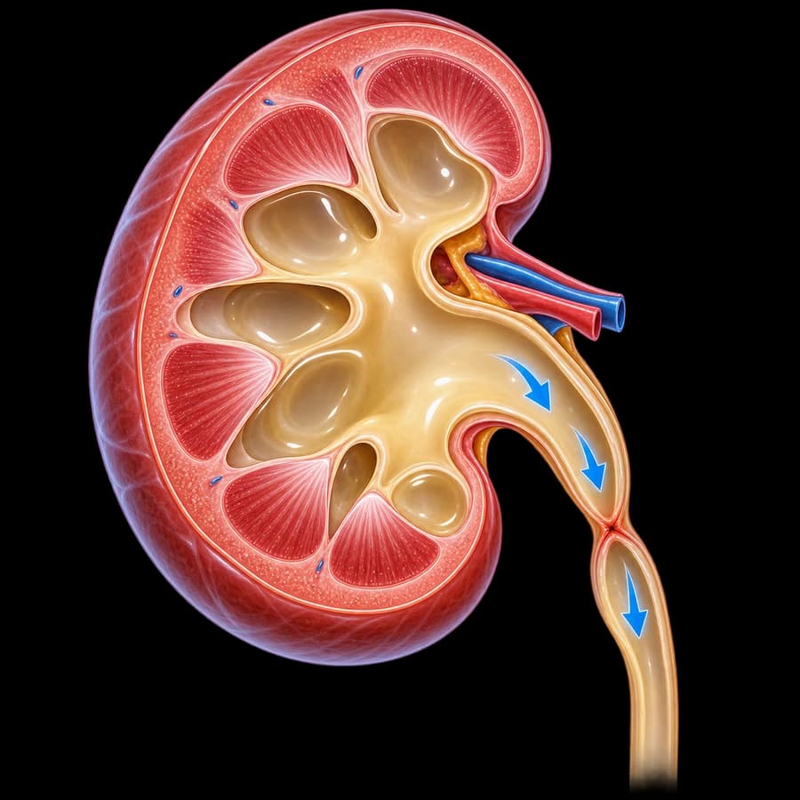
Hydronephrosis is dilatation of the renal pelvis (the funnel that collects urine inside the kidney) and calyces, due to urine accumulating because it is not draining at its normal rate toward the ureter and bladder. It is described on imaging — ultrasound, CT, or MRI.
The term "hydroureter" (or "hydroureteronephrosis") is used when the ureter itself is also dilated, which usually suggests that the cause of obstruction is lower down, in the ureter or bladder, rather than at the ureteropelvic junction.
3. Hydronephrosis ≠ Obstruction
This is the single most important distinction in this entire topic. Hydronephrosis is an imaging finding, while obstruction is a functional diagnosis. A dilated collecting system does not automatically mean there is a blockage harming kidney function.
Dilatation without significant obstruction
Can be seen in pregnancy, urinary reflux, an extrarenal pelvis, or as residual dilatation after previously successful treatment.
Dilatation with functionally significant obstruction
Requires documentation through clinical context, change over time, and, where needed, functional testing (e.g., a renal scan), before a treatment decision is made.
4. Dilatation Without Significant Obstruction
Not every dilatation needs treatment. Examples of benign or non-obstructive causes:
- Extrarenal pelvis — an anatomical variant without true obstruction.
- Parapelvic cysts — can mimic dilatation on ultrasound.
- Vesicoureteral reflux — urine flows back toward the kidney without a mechanical blockage.
- Residual dilatation after successful treatment of obstruction, without an active blockage.
- Normal physiological dilatation of pregnancy, more often on the right.
5. Unilateral vs Bilateral
The distribution of dilatation guides the differential diagnosis:
Unilateral
Usually a local cause in one kidney or ureter — a stone, UPJ obstruction, ureteral stricture, or external compression.
Bilateral
Raises suspicion of a cause lower down — prostate, bladder, pelvic mass — or a more generalized condition. It often needs faster, more complete evaluation, especially with worsening kidney function.
6. Acute vs Chronic Hydronephrosis
Acute obstruction (e.g. from a stone) usually causes intense flank pain because the renal capsule stretches suddenly. Chronic or gradual obstruction (e.g. from UPJ obstruction or progressive prostatic obstruction) can progress completely painlessly, while still causing progressive loss of kidney function.
Absence of pain does not mean absence of obstruction. This is why chronic hydronephrosis is often discovered incidentally, at an advanced stage.
7. Grade of Dilatation — What It Really Means
Descriptions such as "mild," "moderate," or "severe" hydronephrosis describe the grade of dilatation on imaging, not the severity of the underlying cause. Mild dilatation can accompany significant acute obstruction, while extensive chronic dilatation can correspond to a kidney with already minimal function and a relatively "quiet" clinical picture. Interpretation always combines the clinical context with, where needed, functional testing.
8. Common Causes
Ureteral Stone
The most common cause of acute unilateral hydronephrosis, with or without colicky pain.
Ureteropelvic Junction (UPJ) Obstruction
Congenital or acquired narrowing at the point where the pelvis meets the ureter, a common cause of chronic or recurrent dilatation.
Ureteral Stricture
Can be post-surgical, post-radiation, inflammatory, or idiopathic.
External Compression / Tumor
A urothelial tumor, pelvic or retroperitoneal mass, or infiltrative disease can compress the ureter from outside.
Obstruction Lower Down
Benign prostatic hyperplasia, chronic urinary retention, or neurogenic bladder can cause bilateral dilatation over time.
9. Ureteropelvic Junction (UPJ) Obstruction
This deserves special mention as one of the most common causes of persistent or recurrent hydronephrosis, especially in younger patients. The full clinical picture, functional evaluation with a renal scan, and current repair options (robotic or laparoscopic pyeloplasty, endopyelotomy) are covered in detail on the UPJ obstruction page.
10. Lower-Tract Causes: Prostate, Bladder, Reflux
When hydronephrosis is bilateral, the cause is often lower than the kidneys:
- Advanced benign prostatic hyperplasia with chronic bladder outlet obstruction.
- Chronic urinary retention with bladder overdistension.
- Neurogenic bladder, where high bladder pressure is transmitted upward.
- Vesicoureteral reflux, especially in children or congenital anomalies.
11. Hydronephrosis in Pregnancy
Mild dilatation, more often on the right side, is a common finding during pregnancy due to hormonal effects and mechanical pressure from the uterus, and usually needs no intervention. Ultrasound is the imaging test of choice. Severe pain, fever, or rapid worsening of symptoms always need immediate evaluation, regardless of pregnancy.
12. Children & Antenatal Dilatation
Antenatal hydronephrosis is one of the most common findings on prenatal ultrasound screening. In most cases it is mild and resolves on its own after birth, but it needs organized follow-up by a specialist to rule out UPJ obstruction, reflux, or another significant cause requiring treatment.
13. Does It Affect Kidney Function?
Persistent obstruction can gradually damage the kidney. However, serum creatinine reflects overall kidney function, not each kidney separately — so in unilateral obstruction with a normal contralateral kidney, creatinine can remain completely normal while the affected kidney loses a significant share of its function.
A normal creatinine does not rule out significant damage to one of the two kidneys. When per-kidney assessment is needed, a renal scan with split renal function is used.
14. Ultrasound: What It Shows & What It Doesn’t
Renal ultrasound is the first, radiation-free test and reliably detects dilatation of the pelvis and calyces. However, it cannot always determine the cause with certainty, or reliably distinguish functionally significant obstruction from mild, non-obstructive dilatation — which is why it is often combined with other tests depending on the clinical context.
15. CT & MR Urography
CT urography provides more precise anatomical localization of the cause (stone, stricture, external compression) and is used when detailed treatment planning is needed.
MR urography is chosen when radiation or iodinated contrast must be avoided — for example in pregnancy or kidney dysfunction — or when better soft-tissue detail around the urinary tract is needed.
16. MAG3 Renal Scan & Functional Testing
When imaging alone cannot clarify whether functionally significant obstruction is present, a diuretic MAG3 renal scan is used, assessing diuretic drainage of each kidney and relative (split) renal function.
The T½ half-time is not an absolute numerical cutoff that alone "decides" treatment. Intermediate results are common and are always interpreted together with the clinical picture, kidney function, and change over time.
17. Infected Obstructed Kidney — Emergency
When there is obstruction and infection behind the blockage, this is a urological emergency with a risk of urosepsis. The key mistake is attempting "definitive" treatment of the cause before the infection is stabilized.
The priority is immediate decompression (JJ stent or nephrostomy) and antibiotics — definitive treatment of the cause follows once stabilized.
18. JJ Stent vs Nephrostomy
Ureteral (JJ) stent
Internal drainage, from the kidney to the bladder through the ureter, usually placed endoscopically.
Percutaneous nephrostomy
External drainage, with a catheter placed through the skin directly into the kidney collecting system.
Neither removes the cause of obstruction — both achieve emergency decompression. The choice depends on the clinical picture, cause, and anatomy, not a general rule.
19. Solitary Kidney, Bilateral Obstruction & Anuria
Hydronephrosis in a solitary functioning kidney, bilateral obstruction, or complete cessation of urine output (anuria) are always treated as potentially urgent, regardless of how "mild" the dilatation looks on imaging, because the risk of irreversible damage is significantly higher.
20. When Observation Is Enough
Stable, painless, non-progressive dilatation, with a documented benign or non-obstructive cause and normal kidney function, can often be monitored with periodic imaging instead of immediate intervention. This decision must always be based on documented evaluation, not assumption.
21. Residual Dilatation After Treatment
Even after successfully relieving an obstruction, the collecting system can remain slightly dilated, without this meaning treatment failed. Success is judged by stabilization or improvement of kidney function and absence of progressive worsening over time, not just the diameter of the pelvis.
22. Treating the Underlying Cause
Treatment does not target "hydronephrosis" itself, but its underlying cause:
- Ureteral stone → see ureteral stones, endoscopic treatment where indicated.
- UPJ obstruction → robotic/laparoscopic pyeloplasty or endopyelotomy, see UPJ obstruction.
- Prostatic obstruction → see benign prostatic hyperplasia.
- External compression/tumor → individualized treatment depending on the cause.
23. The Patient Journey
- 1Hydronephrosis found on ultrasound or CT
- 2History + clinical examination
- 3Red flags? (fever / solitary kidney / bilateral obstruction / anuria)
- 4YES → urgent evaluation, lab work, and, if needed, immediate decompression + antibiotics
- 5NO → planned investigation of cause: lab work, CT/MR urography if needed, functional testing where indicated
- 6Documentation of cause and severity of obstruction
- 7Observation for a stable, non-obstructive picture • or targeted treatment of the cause
- 8Follow-up imaging ± functional testing to confirm stabilization
24. Myths to Forget
"Hydronephrosis is a diagnosis on its own"
No. It is an imaging finding; its significance depends on the underlying cause.
"If it doesn’t hurt, there’s no problem"
Chronic obstruction can be completely painless while gradually damaging the kidney.
"Mild hydronephrosis means a mild problem"
The grade of dilatation does not always match the severity of the cause or obstruction.
"Normal creatinine rules out kidney damage"
In unilateral obstruction with a healthy contralateral kidney, creatinine can stay normal despite significant damage to the affected kidney.
"Drainage (stent/nephrostomy) is the definitive treatment"
It is an emergency stabilization measure. The cause is usually treated afterward.
"If some dilatation remains after treatment, treatment failed"
Not necessarily. Success is judged by kidney function and stability over time, not diameter alone.
25. Red Flags — When It’s an Emergency
- Fever or chills with known or possible obstruction
- A solitary functioning kidney
- Bilateral obstruction
- Anuria or markedly reduced urine output
- Rapid decline in kidney function
- Uncontrolled pain or persistent vomiting
- Pregnancy with severe or persistent symptoms
26. Your First Visit — What You Will Be Asked
27. The Doctor

Dr. Marinos Vasilas, Urologist – Andrologist
Dr. Marinos Vasilas evaluates hydronephrosis with a focus on the early distinction between a benign finding and functionally significant obstruction, based on current EAU guidelines.
Meet the Doctor28. The Team
Investigating hydronephrosis — from imaging to functional evaluation and any procedure — is organized in collaboration with an experienced urology practice.
Meet Our Team29. Frequently Asked Questions
What is hydronephrosis?
It is dilatation of the renal pelvis and calyces because urine is not draining normally toward the bladder. It is an imaging finding, not a final diagnosis — the key step is identifying the underlying cause.
Does hydronephrosis always mean obstruction?
No. Dilatation can occur without functionally significant obstruction, for example in pregnancy, urinary reflux, an extrarenal pelvis, or residual dilatation after a previously treated obstruction.
What is the most common cause in adults?
Often a ureteral stone in an acute presentation, or ureteropelvic junction obstruction, ureteral stricture, or bladder outlet obstruction from the prostate in a chronic presentation.
Does hydronephrosis always cause pain?
No. Acute dilatation usually hurts, while chronic or gradual dilatation can be completely painless — even with significant obstruction. Absence of pain does not mean it is safe.
Why can painless hydronephrosis be dangerous?
Because gradual obstruction can cause silent loss of kidney function with no symptoms, until it is discovered incidentally on imaging.
What does "mild" or "severe" hydronephrosis mean?
It is a descriptive grade of dilatation on imaging, not a measure of how severe the underlying cause is. A "mild" dilatation can accompany significant obstruction, and the reverse is also true.
What is the difference between unilateral and bilateral hydronephrosis?
Unilateral usually reflects a local cause (stone, UPJ obstruction, ureteral stricture). Bilateral raises suspicion of a cause lower in the urinary tract (prostate, bladder, pelvic mass) or a more serious condition, and often needs faster evaluation.
When is hydronephrosis an emergency?
When it is accompanied by fever (possible infected obstructed kidney), when it involves a solitary kidney, when it is bilateral with anuria, or when there is rapid decline in kidney function.
What is an infected obstructed kidney?
The combination of obstruction with infection behind the blockage — a urological emergency with a risk of urosepsis, requiring immediate drainage and antibiotics, not delay.
What is the difference between a JJ stent and a nephrostomy?
A JJ stent drains internally, from the kidney to the bladder through the ureter. A nephrostomy drains externally, with a catheter placed through the skin directly into the kidney collecting system. Both can achieve emergency decompression; the choice depends on the clinical picture, not preference.
Does drainage remove the cause of obstruction?
No. Drainage stabilizes an emergency. Definitive treatment of the underlying cause (stone, stricture, tumor) is usually addressed afterward.
Does creatinine always rise with hydronephrosis?
No. In unilateral obstruction with a normal contralateral kidney, serum creatinine can remain normal because the healthy kidney compensates. A normal creatinine does not rule out significant damage to the affected kidney.
What does a MAG3 renal scan show?
It assesses diuretic drainage and the relative function of each kidney separately. It is used when it is unclear whether dilatation corresponds to functionally significant obstruction.
Does the T½ half-time on a MAG3 scan always give a clear answer?
Not always. Intermediate results are common and are interpreted together with the clinical picture, kidney function, and change over time — it is not an absolute numerical cutoff.
Is a CT scan always needed?
No. Ultrasound is usually the initial test. CT urography is added when a more precise anatomical cause needs to be identified or a procedure is being planned.
What is the role of MR urography?
It is used mainly to avoid radiation or iodinated contrast, for example in pregnancy or kidney dysfunction, or when better soft-tissue detail is needed.
Can hydronephrosis be normal in pregnancy?
Mild dilatation, more often on the right, is a common and usually benign finding due to hormonal and mechanical factors. Severe pain, fever, or rapid worsening always need evaluation.
Is antenatal hydronephrosis dangerous for the baby?
It is often mild and resolves on its own, but it needs organized follow-up after birth by a specialist to rule out significant obstruction or reflux.
Can old dilatation remain after successful treatment?
Yes. A dilated collecting system can remain slightly enlarged even after obstruction has been successfully relieved, without this meaning treatment failed. Functional improvement is judged by proper criteria, not diameter alone.
Does hydronephrosis always need treatment?
No. Stable, mild, painless dilatation with a documented benign cause and normal kidney function can often be observed. The decision is individualized.
What are the treatment options for UPJ obstruction?
Robotic or laparoscopic pyeloplasty are the most common definitive solutions today; endopyelotomy is an alternative in selected cases.
Can the prostate cause hydronephrosis?
Yes, in advanced bladder outlet obstruction from benign prostatic hyperplasia, bilateral dilatation can develop, especially with chronic urinary retention.
Is neurogenic bladder linked to hydronephrosis?
Yes, in some patients with neurogenic bladder dysfunction, high bladder pressure can transmit upward and cause kidney dilatation — which is why monitoring the upper urinary tract matters in these patients.
What if I only have one functioning kidney?
Having a solitary kidney significantly lowers the threshold for urgent evaluation, even if the clinical picture appears mild.
Can hydronephrosis come back after treatment?
It depends on the cause. That is why scheduled follow-up with imaging, and functional testing where needed, is planned after treatment.
Should I drink a lot of water if I have hydronephrosis?
There is no evidence that excessive water intake "flushes out" an obstructing cause; normal hydration is sufficient, and in complete obstruction it may even worsen discomfort.
Related Topics
Hydronephrosis Evaluation
Proper management depends not only on the grade of dilatation on imaging, but on whether there is truly functionally significant obstruction, what the cause is, how kidney function looks, and whether there are signs of infection or higher risk.
References
- EAU Guidelines on Urolithiasis 2026 — uroweb.org
- EAU Guidelines on Urological Infections 2026 — uroweb.org
- EAU Guidelines on Paediatric Urology 2026 — uroweb.org
- ACR Appropriateness Criteria — Renal and Ureteral Colic / Hydronephrosis of Unknown Cause — acsearch.acr.org
The content on this page is for informational purposes only and does not replace an individualized urological evaluation. Hydronephrosis is an imaging finding with many possible causes, and online information cannot confirm whether functionally significant obstruction is present. Fever, a solitary kidney, bilateral obstruction, anuria, or worsening kidney function need timely evaluation. Do not start antibiotics on your own.


