My Clinical Approach to UPJ Obstruction
Ureteropelvic junction (UPJ) obstruction — also called PUJ obstruction — is one of those conditions where imaging alone is often not enough. Hydronephrosis is simply a finding; the question that really matters is whether there is genuine functional obstruction affecting kidney function.
My approach focuses on a clear separation of anatomical and functional assessment, correct interpretation of diuretic renography (MAG3) and split renal function, and an individualised choice between surveillance and pyeloplasty.
- A clear distinction between hydronephrosis and genuine functional obstruction.
- Correct use and interpretation of the MAG3 renal scan and split renal function.
- Recognising the significance (and the limits) of a crossing vessel.
- An individualised choice between surveillance and pyeloplasty.
What Is the Ureteropelvic Junction
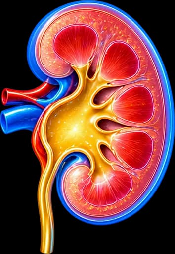
The ureteropelvic junction (UPJ) — also called the pelviureteric junction (PUJ) — is the point where the renal pelvis joins the ureter.
At this point urine passes from the renal pelvis into the ureter. Anatomical or functional disorders at this location can impede normal flow.
What Does Obstruction Mean
Obstruction means difficulty in draining urine from the kidney to the ureter. It does not necessarily mean:
Severity varies significantly from patient to patient — from mild, well-tolerated dilatation to significant functional impairment.
“I Have Hydronephrosis – Does That Mean I Have Obstruction?”
Not necessarily. Dilatation of the kidney on imaging does not, by itself, prove functional obstruction.
Hydronephrosis = dilatation of the kidney’s collecting system. It can be caused by UPJ obstruction, a ureteral stone, vesicoureteral reflux, distal ureteral obstruction, bladder outlet obstruction, physiological/non-obstructive dilatation, or other causes. See also our page on Hydronephrosis.
| Hydronephrosis | UPJ Obstruction | |
|---|---|---|
| What it is | An imaging finding of dilatation | A specific cause/mechanism of obstruction |
| Does it prove obstruction? | No | Must be documented |
| Diagnosis | Ultrasound/CT/MRI | Anatomy + functional assessment |
| Treatment | Depends on the cause | Surveillance or correction, case by case |
Congenital or Acquired?
UPJ obstruction is often a congenital anatomical/functional disorder, which can:
- Be diagnosed in childhood (or even antenatally).
- Or remain relatively asymptomatic and be diagnosed for the first time in an adult.
Acquired causes can also exist (e.g. scarring after a stone/infection/procedure). Not every case in an adult is a new, purely acquired condition — it is often a pre-existing congenital disorder that became clinically apparent later.
Intrinsic Causes
Intrinsic obstruction can be related to:
What Is a Crossing Vessel?
In some patients, a lower pole blood vessel of the kidney can pass close to the ureteropelvic junction (a crossing vessel) and contribute to:
- Kinking of the ureter at that point.
- External compression.
- Intermittent obstruction.
The presence of a crossing vessel does not, by itself, mean there is functionally significant obstruction.
It must be interpreted together with anatomy, symptoms, drainage and functional findings — not as an isolated finding that determines the diagnosis.
Intermittent Obstruction & Dietl’s Crisis
In some patients, obstruction can be intermittent, with symptoms appearing after a large fluid intake, or in some patients after alcohol consumption/increased diuresis or intense physical activity.
Dietl’s Crisis
The term describes episodic flank pain due to intermittent UPJ obstruction, often after increased diuresis. It is not a necessary diagnostic criterion, but can be a clinically useful term when describing the patient’s history.
“Why Does the Kidney Hurt After a Large Fluid Intake?”
In intermittent UPJ obstruction, a sudden increase in urine output can:
- Distend the renal pelvis further.
- Raise intrapelvic pressure.
- Cause transient flank pain.
This alone is not a diagnosis of UPJ obstruction — further evaluation is needed.
Symptoms of UPJ Obstruction
Possible symptoms include:
Several patients can be entirely asymptomatic.
Kidney Stones & UPJ Obstruction
Impaired drainage can be associated with an increased risk of stone formation in selected patients. Not every case of UPJ obstruction causes stones.
For the detailed diagnosis and treatment of stone disease, see our page on Kidney Stones.
Infections & Fever
Urinary stasis/impaired drainage can be associated with recurrent urinary infection and pyelonephritis in selected patients. See our page on Pyelonephritis.
Fever/rigors in a patient with significant obstruction and hydronephrosis can indicate an infected obstructed kidney and require immediate evaluation. Depending on the clinical context, urgent drainage (a JJ stent or nephrostomy) and antimicrobial treatment may be needed.
How Is the Diagnosis Made?
The correct diagnosis often requires a combination of anatomical and functional imaging.
A proper assessment must answer two different questions:
1. The anatomical question
Is there dilatation / narrowing / a crossing vessel / another cause?
2. The functional question
Is drainage genuinely obstructed and is kidney function affected?
Ultrasound
Ultrasound can show dilatation of the renal pelvis, calyceal dilatation, parenchymal thickness, kidney size and, where relevant, Doppler findings.
It cannot always prove whether the dilatation is functionally obstructive. Complementary anatomical or functional assessment is often needed.
CT Urography
CT urography can provide information on the anatomy of the renal pelvis and ureter, the presence of stones, any crossing vessels and surrounding structures, and other causes of obstruction. See also our page on CT Urography.
Contrast/excretory imaging is used when clinically appropriate — it is not mandatory for every patient.
MR Urography
MR urography can have a role in selected patients: anatomical assessment, avoiding ionising radiation, complex anatomy, or selected functional information, particularly in younger patients where relevant.
It is not a routine mandatory test for every patient with suspected UPJ obstruction.
Diuretic Renography (MAG3)
MAG3 helps assess both drainage and the relative function of each kidney.
The 99mTc-MAG3 diuretic renal scan is a widely used test for evaluating renal drainage, particularly when there is hydronephrosis and suspected obstruction. It assesses:
- Renal perfusion.
- Radiotracer uptake.
- Drainage.
- Relative (split) renal function.
and helps answer: is there genuine functional obstruction? As an alternative radiotracer in appropriate settings, 99mTc-DTPA may also be mentioned.
A single numeric parameter from the renal scan is not, by itself, a complete diagnosis.
Diuretic Administration
Diuretic administration during the renal scan is used to examine whether the dilated collecting system can empty when urine flow is increased. Protocol, dosing and timing are not identical across every nuclear medicine department — they are determined according to the local protocol and current standards.
The T½ (Drainage Half-Time) and Its Limitations
The T½ (drainage half-time) is a parameter estimated from the renal scan. Its interpretation, however, also depends on:
The T½ alone should not be used as the sole criterion of obstruction.
What Does “Right/Left Kidney Function” Mean?
Differential renal function / split renal function expresses the percentage of overall kidney function contributed by each kidney separately, as estimated from the renal scan.
An arbitrary percentage does not automatically mean surgery or nephrectomy is needed.
“My MAG3 Showed 35% Function – Do I Need Pyeloplasty?”
A single split renal function or T½ number should not decide, by itself, whether intervention is needed.
The split function percentage must be interpreted together with the drainage curve, symptoms, change over time and the overall clinical context.
Kidney Function & Creatinine
Overall creatinine/eGFR can remain normal even if one kidney has reduced function, because the other kidney can compensate.
Normal creatinine does not rule out unilateral functional damage. Split renal function from the renal scan provides the kidney-specific information that overall creatinine cannot.
When Is Surveillance Enough?
“Do I Need Surgery If I Don’t Have Pain?”
Not everyone.
Selected patients may be monitored when:
in line with current evidence. There is no universal surveillance protocol — frequency is determined individually.
What Do We Monitor?
This can include symptoms, ultrasound, degree of hydronephrosis, parenchymal thickness, kidney function, and repeat functional imaging when clinically indicated — MAG3 does not need to be repeated at a fixed interval for everyone.
When Is Intervention Needed?
Intervention may be considered when there is:
A single, rigid threshold is not used — the decision is always individualised.
Pyeloplasty
In symptomatic or functionally significant UPJ obstruction, pyeloplasty is the main reconstructive treatment.
Pyeloplasty aims to surgically correct the narrowed ureteropelvic junction and restore free drainage of the kidney. In the classic dismembered technique, the narrowed segment is excised or bypassed, the ureter is reconnected to the renal pelvis, and a wide anastomosis is created.
Anderson–Hynes Dismembered Pyeloplasty
This is a classic, widely used technique, with a particular advantage when a crossing vessel is present, as it allows appropriate transposition/reconstruction of the anastomosis in relation to the vessel. Not every case of UPJ obstruction has the same anatomy — the technique is adapted to the findings.
Laparoscopic & Robotic Pyeloplasty
3D Laparoscopic Pyeloplasty
- Minimally invasive access.
- Dismembered repair.
- Precise suturing.
- Restoration of drainage.
Robotic Pyeloplasty
- Robotic-assisted laparoscopic reconstruction.
- Magnified 3D view.
- Articulated instruments.
- Precision in anastomotic suturing.
Open Pyeloplasty
Open pyeloplasty remains an effective surgical option, but in adults minimally invasive approaches (laparoscopic/robotic) are frequently used where expertise and resources allow. Open surgery is not an obsolete method — it remains a relevant option in selected cases.
Laparoscopic vs Robotic Pyeloplasty
Both techniques are minimally invasive dismembered reconstructions, with differences in:
The experience of the surgical team and correct reconstruction matter more than the commercial name of the platform.
Endopyelotomy
Endopyelotomy is a less invasive endourological option in selected cases, incising the narrowed segment via an endoscopic route. However, it generally has a different success/recurrence profile and suitability compared with pyeloplasty.
Factors that can affect its suitability:
- A long stricture length.
- Severe hydronephrosis.
- The presence of a crossing vessel.
- Poor kidney function.
Simple balloon dilatation may be mentioned as a selected endourological approach, but it is not a definitive first-line standard treatment for primary adult UPJ obstruction.
EndopyelotomyPyeloplasty vs Endopyelotomy
| Pyeloplasty | Endopyelotomy | |
|---|---|---|
| Principle | Surgical reconstruction | Endoscopic incision |
| Crossing vessel | Can be addressed anatomically | A limitation in selected cases |
| Durability | High in suitable cases | Lower in some groups |
| Invasiveness | Minimally invasive surgery | Endoscopic |
| Patient selection | Broad | More selective |
JJ Stent & Nephrostomy
After pyeloplasty, a temporary ureteral (JJ) stent is often used for drainage and healing of the anastomosis. Protocol and duration vary by case and centre. See our page on JJ Stent Placement.
Possible symptoms with a stent in place include urinary frequency, urgency, haematuria, flank discomfort and bladder discomfort.
Nephrostomy
A nephrostomy may be used in selected circumstances: an infected obstruction, urgent drainage, or diagnostic/functional assessment in selected complex cases. It is not a routine treatment for uncomplicated UPJ obstruction. See our page on Nephrostomy Placement.
Elective pyeloplasty and emergency drainage are different situations. With fever, sepsis, or an infected obstructed kidney, priority may be drainage and treatment of the infection, with definitive reconstruction following at an appropriate time.
A Stone & UPJ Obstruction Together
In patients with coexisting UPJ obstruction and kidney stones, treatment may need combined planning. In selected minimally invasive surgery, both the stone burden and the anatomy of the junction can be addressed at the same time, depending on the surgeon’s expertise. Simultaneous treatment cannot be promised in every case — it depends on the findings.
What Happens If the Kidney Has Very Poor Function?
There is no simple threshold of the type “below X% the kidney is removed.”
The choice between reconstruction (pyeloplasty), temporary drainage/reassessment, and nephrectomy depends on:
Nephrectomy
It is not standard treatment for UPJ obstruction when the kidney has useful function and can be salvaged.
It may be considered only in selected cases with a severely damaged/poorly functioning kidney, recurrent infection, severe symptoms and no meaningful salvage potential, always depending on the clinical context.
What Does Pyeloplasty Success Mean?
Success is not defined only as “the hydronephrosis disappeared.” It can include:
- Symptom improvement.
- Unobstructed drainage.
- Stable or improved kidney function.
Persistence of some degree of hydronephrosis after successful pyeloplasty does not necessarily mean failure.
The renal pelvis does not need to return to a normal size for the pyeloplasty to be considered successful. Assessment is based on symptoms, functional drainage, kidney function, and the trend of imaging over time — not on the absolute diameter of the pelvis alone.
Can Function Return?
After relief of obstruction, symptoms and drainage can improve and kidney function can stabilise, while in selected cases some degree of improvement may occur. However, full recovery of kidney function should not be promised in every case.
Recurrence & Redo Pyeloplasty
Recurrence of UPJ obstruction after repair is possible, but is not expected in every patient. If recurrence is suspected, repeat ultrasound, renography, or CT/MRI may be needed, and depending on the findings, redo reconstruction or a selected endourological treatment.
Redo laparoscopic/robotic pyeloplasty is an option in selected recurrent cases. We do not quote recurrence percentages here without adequate sourcing.
UPJ Obstruction in Adults, Children & Pregnancy
In adults, presentation can include incidental hydronephrosis, intermittent flank pain, stones, infection, or a gradual decline in function.
UPJ obstruction is often diagnosed antenatally or in childhood. The paediatric management strategy is different and requires paediatric urology assessment — we do not give paediatric intervention thresholds here.
In pregnancy, a physiological dilatation of the collecting system can occur due to pregnancy itself and must be differentiated from genuine UPJ obstruction; imaging and treatment are adjusted accordingly.
Differential Diagnosis
Hydronephrosis/dilatation of the collecting system can be due to:
A parapelvic cyst can mimic hydronephrosis on ultrasound, but a parapelvic cyst ≠ dilatation of the collecting system. See our page on Simple Kidney Cyst.
This list is informational and not a self-diagnosis tool.
The Patient’s Journey
Hydronephrosis / intermittent pain / incidental finding
The starting point.
Ultrasound
Initial assessment of dilatation and kidney size.
Is there an obvious other cause (e.g. a stone)?
If so, treatment of that specific cause.
No / suspected UPJ ? CT or MR Urography
Anatomical assessment, detection of crossing vessels.
Diuretic Renography (MAG3)
Drainage + split renal function.
No significant functional obstruction / stable kidney
Surveillance.
Symptomatic / obstructed / declining function / recurrent complications
Consideration of pyeloplasty.
Follow-up after treatment
Symptoms, drainage, kidney function.
This is an educational overview, not a rigid diagnostic/treatment algorithm — every case is discussed individually with the treating urologist.
Frequently Asked Questions (FAQ)
What is ureteropelvic junction (UPJ) obstruction?
It is a functional or anatomical impediment to urine flow from the renal pelvis to the ureter, at the point called the ureteropelvic junction (also known as PUJ obstruction). It can cause hydronephrosis, pain, infections or stones, with substantial variability in severity.
Is it always congenital?
It is often a congenital anatomical/functional disorder, which can be diagnosed in childhood or remain relatively asymptomatic and be discovered for the first time in an adult. Acquired causes can also exist — not every case in an adult is a new, purely acquired condition.
Can it present for the first time in an adult?
Yes. Many adults are diagnosed incidentally (incidental hydronephrosis on imaging done for another reason) or due to intermittent flank pain, stones, infection, or a gradual decline in kidney function.
What are the symptoms?
Possible symptoms include flank pain (often intermittent), nausea during a severe episode, recurrent urinary infection, pyelonephritis, haematuria and stone disease. Many patients, however, are completely asymptomatic.
Why can the kidney hurt after drinking a lot of fluids?
In intermittent UPJ obstruction, a sudden increase in urine output can distend the renal pelvis further and raise pressure, causing transient pain. This alone, however, is not a diagnosis of UPJ obstruction.
Does hydronephrosis always mean obstruction?
No. Hydronephrosis is an imaging finding of dilatation of the kidney and can have many causes — a stone, reflux, another obstruction, or even non-obstructive dilatation. UPJ obstruction is one specific possible cause that needs to be documented separately.
How is the diagnosis made?
Two different questions need to be answered: the anatomical question (is there dilatation/narrowing/a crossing vessel/another cause?) via ultrasound, CT or MR urography, and the functional question (is drainage genuinely obstructed and is kidney function affected?) mainly through diuretic renography (MAG3).
What is the MAG3 renal scan?
It is a diuretic renal scintigraphy using a radiotracer (99mTc-MAG3), usually with a diuretic, that assesses perfusion, uptake and drainage of each kidney separately, helping determine whether there is genuine functional obstruction.
What does split renal function mean?
It is the percentage of overall kidney function contributed by each kidney separately, as estimated from the renal scan. A single number does not automatically determine the need for surgery — it is interpreted together with the drainage curve, symptoms and change over time.
What is a crossing vessel?
It is a blood vessel of the lower pole of the kidney that, in some patients, passes close to the ureteropelvic junction and can contribute to kinking or external compression. Its presence alone does not mean it is causing functionally significant obstruction — it is interpreted together with anatomy, symptoms and functional findings.
Does every case of UPJ obstruction need surgery?
No. Asymptomatic patients with stable function, adequate drainage and no recurrent complications may, in selected cases, be monitored without an immediate need for intervention.
When is pyeloplasty performed?
It is considered when there is significant or recurrent pain, documented functional obstruction, deterioration of split renal function, recurrent pyelonephritis or stone formation, or progressive worsening with relevant functional findings — always individualised.
What is laparoscopic pyeloplasty?
It is a minimally invasive surgical repair of the narrowed ureteropelvic junction, with excision/bypass of the narrowed segment and a wide ureteropelvic anastomosis, performed through small incisions.
What is the difference between pyeloplasty and endopyelotomy?
Pyeloplasty is a surgical reconstruction of the junction, while endopyelotomy is an endoscopic incision of the narrowed segment. They have a different suitability profile — endopyelotomy is more selective, particularly in the presence of a crossing vessel, a long stricture, or severe hydronephrosis.
Does hydronephrosis return after surgery?
The renal pelvis does not need to return to an entirely normal size for the pyeloplasty to be considered successful. Some degree of dilatation may persist; success is judged by symptoms, drainage and kidney function, not by imaging alone.
Can kidney function be lost?
In selected patients with prolonged or severe obstruction, there can be progressive functional decline. After relief of obstruction, function can stabilise or, in some cases, partly improve, but no one should promise full recovery in every case.
Related Topics
Book Your Appointment in Rhodes
The presence of hydronephrosis alone is not enough to decide on surgical treatment. Assessing the anatomy, drainage and relative kidney function is necessary to determine whether surveillance or repair of the ureteropelvic junction with pyeloplasty is needed.
References – Sources
- EAU Guidelines relevant to upper urinary tract obstruction — uroweb.org
- Taylor A Jr, et al. SNMMI Procedure Standard/EANM Practice Guideline for Diuretic Renography. J Nucl Med Technol 2018.
- O’Reilly PH, et al. Consensus on diuresis renography for investigating the dilated upper urinary tract. J Nucl Med 1996;37:1872–1876.
- Klingler HC, et al. Unpredictability of clinical outcome after pyeloplasty and endopyelotomy: comparative outcome review. Urology 2003.
- Autorino R, et al. Robot-assisted and laparoscopic pyeloplasty: a systematic review and meta-analysis. Eur Urol 2014;65:430–452.
Medical Editorial

Medically reviewed by Dr. Marinos Vasilas – Urologist – Andrologist
Dr. Marinos Vasilas runs a private urology practice in Rhodes, specialising in laparoscopic and robotic repair of ureteropelvic junction obstruction and the modern diagnostic approach to hydronephrosis.
View full profile


