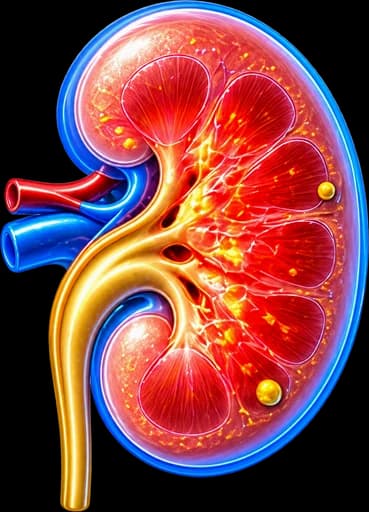
My Clinical Approach to Pyelonephritis
Pyelonephritis is one of the most common infections I manage in the urology clinic — and also one of the most underestimated by patients, who often mistake it for a “simple urinary infection”. In practice, the difference between an uncomplicated case and an obstructed or emphysematous one can mean the difference between simple outpatient antibiotics and a life-threatening urosepsis.
In daily practice I frequently see two very different scenarios: the young woman with recurrent pyelonephritis who has never been evaluated for an underlying anatomical abnormality, and the diabetic patient with an atypical presentation who already shows signs of urosepsis.
My approach follows the EAU Guidelines on Urological Infections:
- Urine culture before starting any antibiotic therapy.
- Assessment for an obstructive factor (stone, stricture, anatomical abnormality) in every atypical or recurrent case.
- Immediate mechanical decompression in obstructive pyelonephritis — within hours, not days.
- Full urological evaluation for every man with pyelonephritis, regardless of age.
- Rational antibiotic use, guided by culture and local resistance patterns.
What Is Pyelonephritis
Pyelonephritis is a bacterial infection of the renal parenchyma and renal pelvis — an infection of the upper urinary tract. It differs from simple cystitis, which is confined to the bladder, and can be more serious. Not every episode of flank pain or fever should be assumed to be pyelonephritis.
Acute pyelonephritis
A sudden bacterial infection with a characteristic clinical syndrome — the subject of this article.
Obstructive pyelonephritis
Coexistence of obstruction (most commonly from kidney stones) with infection — a vital urological emergency.
Complicated pyelonephritis
In patients with risk factors: diabetes mellitus, immunosuppression, chronic kidney disease, pregnancy, anatomical abnormalities, or a transplanted kidney.
Chronic pyelonephritis
A specific pathological/radiological entity resulting from repeated or undertreated infections that lead to renal scarring and, eventually, chronic kidney impairment — often linked to vesicoureteric reflux (VUR). It is not simply “pyelonephritis that lasts a long time” and should not be used loosely for recurrent flank pain/UTI.
Rare forms
- • Emphysematous pyelonephritis: rare, life-threatening, gas-forming infection. Typical in diabetics. Significant mortality without urgent management.
- • Xanthogranulomatous pyelonephritis: chronic destructive inflammation, often with coexisting stones. Frequently requires nephrectomy.
Prevalence & Risk Factors
Pyelonephritis is one of the most common infections leading to hospital admission. It is substantially more frequent in women than in men.
| Risk Factor | Mechanism |
|---|---|
| Female sex | Anatomically short urethra — easier ascending spread of pathogens. |
| Sexual activity | Increases bladder bacterial contamination. |
| Pregnancy | Physiological dilatation of the urinary tract + hormonal effects. |
| Diabetes mellitus | Immune suppression + glycosuria favour bacterial growth. |
| Urinary tract obstruction | Kidney stones, BPH, strictures. |
| Vesicoureteric reflux (VUR) | Reflux of infected urine from the bladder toward the kidney. |
| Bladder catheterisation | Mechanical introduction of pathogens + catheter biofilm. |
| Immunosuppression | Transplantation, chemotherapy, long-term steroid therapy. |
| Anatomical abnormalities | Horseshoe kidney, duplex collecting system, pyeloureteric junction stenosis. |
| Prior urinary infection | Clear association with recurrence. |
Pathogens & How Infection Reaches the Kidney
Routes of infection
- Ascending infection — the most common route: bacteria travel from the periurethral area → urethra → bladder → ureter → kidney.
- Haematogenous spread (rarer): from a distant focus, in specific clinical contexts (e.g. endocarditis from Staphylococcus aureus).
Common pathogens
| Pathogen | Frequency | Notes |
|---|---|---|
| Escherichia coli | >80% | The dominant pathogen in community-acquired infection. |
| Klebsiella pneumoniae | ~5–8% | More common in healthcare-associated infection, diabetics. |
| Proteus mirabilis | ~3–5% | Urease-producing — alkalinises urine, can favour stone formation. |
| Enterococcus faecalis | ~2–5% | Often in patients with prior antibiotic exposure. |
| Pseudomonas aeruginosa | <5% | Healthcare-associated, immunosuppression, chronic catheterisation. |
| Fungi (Candida) | <2% | Immunosuppressed, diabetic, prolonged broad-spectrum antibiotic use. |
Escherichia coli remains by far the most common cause of community-acquired urinary infections and pyelonephritis. Treatment choice must always take into account urine culture, susceptibility, local resistance patterns, and previous antibiotic exposure — there is no universal empirical regimen for every patient.
Resistant strains (ESBL+, carbapenem-resistant)
A growing concern, particularly with:
- Recent antibiotic use (last 6–12 months).
- Recent hospitalisation or travel to endemic areas.
- Care in chronic healthcare facilities.
- Recurrent urinary infections.
Symptoms & Clinical Presentation
The classic presentation includes a triad of symptoms, usually developing within 24–48 hours. Not every symptom needs to be present.
Systemic symptoms
- High fever (>38.5°C), often with chills — sudden onset.
- Marked malaise, myalgia, arthralgia.
- Nausea and vomiting in severe cases.
- Tachycardia and tachypnoea — warning signs of urosepsis.
Flank pain
- Unilateral or, more rarely, bilateral pain in the flank/loin.
- Constant rather than the classic wave-like pattern of renal colic.
- Positive costovertebral angle tenderness (Goldflam sign) on percussion.
Lower urinary tract symptoms (LUTS)
- Dysuria (burning/pain on urination).
- Frequency and urgency.
- Suprapubic discomfort.
Important: Lower urinary tract symptoms often precede the fever by 1–3 days, suggesting ascending spread from cystitis.
Atypical presentations
- Elderly patients: often only confusion, falls, general decline — without fever.
- Diabetic patients: may not develop fever due to blunted immune response.
- Immunosuppressed patients: minimal symptoms, rapid deterioration.
- Pregnant women: fever + back pain + nausea — often confused with gastroenteritis or pre-eclampsia.
Red Flags — Immediate Specialist Evaluation
- Fever >38.5°C with chills and flank pain in a pregnant woman — hospital admission immediately.
- No response to antibiotics within 48–72 hours — exclude abscess or obstruction.
- Signs of sepsis: hypotension, tachycardia, confusion, reduced urine output.
- Coexisting kidney stone with fever — urological emergency.
- Pyelonephritis in a transplanted kidney, a solitary kidney, or with immunosuppression.
- Every man with pyelonephritis.
Pyelonephritis vs Cystitis, Renal Colic & Prostatitis
Pyelonephritis vs Cystitis
Fever and flank pain change the clinical picture significantly in a urinary infection — this is the key distinguishing point.
Cystitis
- Infection primarily of the bladder.
- Dysuria, frequency, urgency.
- Usually no fever or systemic illness.
Pyelonephritis
- Infection of the upper urinary tract/kidney.
- Fever, chills, flank pain.
- Systemic illness more likely.
Pyelonephritis vs Renal Colic
Pyelonephritis
- More often fever, chills, malaise.
- Constant flank pain.
- Infection findings on urinalysis/culture.
Renal Colic
- More often very intense, wave-like pain.
- Radiation toward the groin.
- Nausea/vomiting, stone-related obstruction.
The two can coexist — and when they do, the situation can be a genuine emergency (see the Obstructive Pyelonephritis section).
Pyelonephritis vs Acute Prostatitis (in men)
Pyelonephritis
- Flank pain.
- Upper urinary tract infection.
Acute Prostatitis
- Perineal/pelvic pain, LUTS.
- Tender prostate on examination, systemic symptoms.
There is clinical overlap — which is one reason every man with pyelonephritis needs a full urological evaluation.
Diagnosis & Investigations
Laboratory investigations
Urinalysis
- Pyuria — white cells in the urine.
- Positive nitrites.
- Positive leucocyte esterase.
- Haematuria (in ~30%).
- White cell casts — suggestive of renal origin.
Negative nitrites do not exclude a urinary infection or pyelonephritis. Also, persistent haematuria after the infection resolves may need separate urological evaluation.
Urine culture
Mandatory before starting antibiotics where feasible. Positive in the large majority of cases. Susceptibility results determine the definitive treatment choice.
Blood tests
- Leucocytosis with left shift.
- Elevated CRP.
- Elevated procalcitonin (PCT) — marker of bacterial infection and sepsis prognosis.
- Creatinine and eGFR — assessment of kidney function.
- Electrolytes and general biochemistry.
Blood cultures
Recommended with severe illness, hospitalisation, suspected sepsis, immunosuppression or atypical presentation — not mandatory in every uncomplicated outpatient case.
Imaging
Renal ultrasound
First-line imaging. Detects hydronephrosis, renal cysts, stones, and abscesses >2 cm. Not sufficient alone to confirm pyelonephritis — a normal ultrasound does not exclude uncomplicated pyelonephritis.
Contrast-enhanced CT scan
Not needed in every patient. Investigation of choice with:
- No response to treatment within 72 hours.
- Suspected abscess or emphysematous pyelonephritis.
- Severe clinical presentation.
- Men with pyelonephritis.
- Immunosuppressed patients.
The exact protocol (with or without contrast, timing of phases) depends on the diagnostic question — suspected stone, abscess, renal parenchymal complication, anatomy and kidney function — and is decided case by case by the clinician.
MRI
Not routine first-line imaging in most acute pyelonephritis cases. May be used in selected situations — pregnancy where appropriate, contrast/radiation limitations, or a complex diagnostic question.
Recognising Urosepsis — qSOFA Score
Urosepsis is the most serious complication of pyelonephritis. The qSOFA score can help with rapid recognition at initial assessment.
| Parameter | Threshold |
|---|---|
| Systolic blood pressure | ≤100 mmHg |
| Respiratory rate | ≥22 breaths/min |
| Altered mental status | Glasgow Coma Scale <15 |
qSOFA \u22652 → high suspicion of sepsis → immediate hospital admission and close monitoring.
Septic shock
- Persistent hypotension despite adequate fluid resuscitation.
- Need for vasopressors to maintain mean arterial pressure ≥65 mmHg.
- Elevated serum lactate.
Treatment & Antibiotic Therapy
Treatment depends on severity, clinical stability, ability to tolerate oral medication, culture results, resistance, kidney function, pregnancy, obstruction, comorbidity and healthcare-associated risk — there is no one-size-fits-all regimen.
This page does not provide a self-prescribing guide. It deliberately avoids brand names, exact doses and a single “best” antibiotic for everyone. Antimicrobial choice depends on expected pathogens, resistance patterns, previous culture, kidney function, allergy, pregnancy, severity, tissue/urinary penetration and local antimicrobial policy — always determined by your treating physician.
Antimicrobial resistance
Increasing antimicrobial resistance — including fluoroquinolone resistance and ESBL-producing Enterobacterales — affects empirical treatment choices. This is one of the main reasons urine culture plays such a central role in guiding therapy.
How long does treatment last?
Duration depends on the antimicrobial class, severity, sex, clinical response, pathogen and complicating factors such as obstruction — there is no single fixed duration for everyone. Your clinician will determine the appropriate length of therapy based on current guidance and your specific case.
Outpatient Treatment vs Hospital Assessment
Outpatient management may be appropriate when:
- The patient is clinically stable.
- Oral medication can be tolerated.
- There is no sepsis.
- There is no significant obstruction.
- No other major complicating factor is present.
Hospital assessment/admission may be needed when there is:
- Sepsis/systemic instability.
- Persistent vomiting — inability to tolerate oral therapy.
- Obstruction.
- Pregnancy in the appropriate context.
- Severe pain.
- Kidney impairment.
- Significant comorbidity.
- Treatment failure.
IV Therapy & Step-down
Severe or systemic cases may initially require intravenous antimicrobial treatment, fluids/supportive care and monitoring, with a switch to oral therapy once the patient is clinically stable and afebrile for an appropriate period, guided by culture results.
Supportive Care
Adequate hydration where there is no contraindication (individualised in kidney/cardiac impairment), symptomatic control of fever and pain (accounting for kidney function, GI risk and comorbidities), and antiemetics where needed.
“I’m on Antibiotics and Still Have a Fever”
Persistent fever may indicate:
- Treatment not yet fully effective.
- A resistant pathogen.
- Unrecognised obstruction.
- A renal abscess.
- An alternative diagnosis.
This needs clinical reassessment, especially if the patient is worsening — not simply switching antibiotics without further evaluation.
Obstructive Pyelonephritis — Urological Emergency
Obstructive pyelonephritis — the coexistence of obstruction (most commonly from a kidney stone) with active infection — is one of the most dangerous situations in urology. Without prompt intervention, progression to urosepsis and multi-organ failure can be rapid.
Management Algorithm
- Immediate fluids + broad-spectrum antibiotics (intravenous).
- Blood cultures obtained before antibiotics.
- Immediate mechanical decompression within hours — two options, neither universally superior; the choice depends on anatomy, severity, availability and the treating team’s experience:
- Ureteral double-J (JJ) stent: placed cystoscopically. Less invasive — preferred where feasible.
- Percutaneous nephrostomy (PCN): a catheter placed through the skin into the renal pelvis under ultrasound guidance. Preferred in septic shock or when cystoscopic access is not feasible.
- ICU monitoring in septic shock.
- Definitive stone treatment (e.g. lithotripsy) is not the first priority — it is usually performed at a second stage, once sepsis has resolved and the infection is fully controlled.
Particular attention with a solitary kidney or bilateral obstruction: obstructive pyelonephritis in a solitary functioning kidney or with bilateral obstruction requires especially rapid assessment, due to the immediate risk of acute kidney injury.
Urease-producing organisms (e.g. Proteus mirabilis) can be associated with struvite (infection) stone formation — this does not mean every episode of pyelonephritis causes a struvite stone, but it is an additional reason for a full metabolic and stone work-up in selected patients.
Complications
Renal & Perinephric Abscess
A collection of pus within or around the kidney. Clinical suspicion: persistent fever despite appropriate antibiotics (>72 hours). Diagnosed by contrast-enhanced CT.
- • Small abscesses: prolonged IV antibiotic therapy.
- • Moderate-sized abscesses: image-guided (ultrasound or CT) percutaneous drainage.
- • Large or multiple abscesses: surgical drainage.
Emphysematous Pyelonephritis (EPN)
A rare, life-threatening form with gas production from anaerobic metabolism. Typical in diabetic patients. Substantial mortality without prompt management. More severe grades often require emergency nephrectomy.
Urosepsis & Septic Shock
Bacteraemia with a systemic inflammatory response. Requires ICU care, broad-spectrum IV antibiotics, immediate urological decompression if obstruction is present, and vasopressors in septic shock.
Acute Kidney Injury (AKI)
Temporary or permanent loss of kidney function due to sepsis or obstructive damage.
Chronic Kidney Disease
Repeated infections lead to focal renal scarring, hypertension and gradually CKD. Rare in healthy adults, more common in children with VUR.
Xanthogranulomatous Pyelonephritis (XGP)
Chronic destructive inflammation, often with a coexisting staghorn stone and chronic infection. Usually requires nephrectomy.
Pyelonephritis in Pregnancy
Pyelonephritis in pregnancy is one of the most serious infections and always requires hospital-based assessment.
Potential Effects
- Preterm labour.
- Low birth weight.
- Maternal septic shock.
- Kidney impairment or acute respiratory distress syndrome (ARDS).
Management
- Hospital admission required.
- Intravenous antibiotics — beta-lactams are typically first choice.
- Avoidance of fluoroquinolones, sulphonamides (in the third trimester), and tetracyclines.
- Close fetal monitoring.
- Exclusion of an obstructive cause with ultrasound or MRI (not CT, due to radiation).
All antibiotic choices in pregnancy must be individually checked for pregnancy safety against current guidance by the treating physician — self-treatment is not appropriate.
Differential Diagnosis
A similar presentation (fever, abdominal/flank pain) can occur in other conditions that must be considered during clinical evaluation:
This list is informational and not a self-diagnosis tool — differential diagnosis is always made by your treating physician.
The Patient’s Journey
Fever/chills + flank pain ± urinary symptoms
Initial clinical presentation.
Clinical assessment + vital signs
Temperature, blood pressure, pulse, respiratory rate.
Urinalysis & urine culture
Before antibiotic therapy, where feasible without dangerous delay.
Blood tests ± blood cultures
Depending on severity of clinical presentation.
Is there sepsis / obstruction / stone / severe illness?
The key branching question.
Yes ? Urgent imaging/hospital assessment
If an infected obstructed system is present ? urgent urological drainage + antimicrobial treatment. If severe/systemic infection without obstruction ? severe infection management.
No ? Clinically stable, uncomplicated pathway
Appropriate antimicrobial therapy ? adjustment guided by culture ? clinical follow-up.
This is an educational overview, not a self-treatment protocol — every case is assessed individually by your physician.
Follow-up, Recurrence & Prognosis
Follow-up Programme
- Reassessment within 48–72 hours — if there is no improvement, suspect abscess or obstruction ? imaging.
- Repeat urinalysis and culture 1–2 weeks after completing treatment.
- In pregnant women: repeat cultures monthly until delivery.
- In men or with anatomical abnormalities: renal/bladder ultrasound 4–6 weeks later.
- Kidney function monitoring (creatinine, eGFR) with recurrent infections.
Preventing Recurrence
In recurrent pyelonephritis (\u22652 episodes/year), the following are considered:
- Treating any underlying obstruction (stones, strictures, BPH).
- Assessment of diabetes and other immunosuppressive factors.
- Adequate hydration.
- Local oestrogen therapy in postmenopausal women.
- Non-antibiotic approaches: D-mannose, cranberry extract, immunostimulants (e.g. OM-89/Uro-Vaxom).
- Low-dose prophylactic antibiotics in selected cases.
- Methenamine as an antibiotic-sparing alternative in selected cases.
Prognosis
With correct and timely treatment, the large majority of uncomplicated cases recover fully, with low risk of permanent kidney damage in otherwise healthy adults. Prognosis is less favourable with recurrence, an untreated obstructive cause, immunosuppression, or the emphysematous form.
Frequently Asked Questions (FAQ)
How do I distinguish pyelonephritis from simple cystitis?
Cystitis is confined to the bladder — dysuria, frequency and burning, without fever or flank pain. Pyelonephritis involves the kidneys and presents with high fever (>38.5°C), chills, flank pain and marked malaise. Correct differentiation requires clinical evaluation and urine testing.
What are the symptoms of pyelonephritis?
Typically high fever with chills, flank/loin pain, nausea, vomiting and malaise, often together with lower urinary tract symptoms (frequency, dysuria, urgency). Not every symptom needs to be present for the diagnosis to be considered.
Can pyelonephritis occur without fever?
Yes, in some patients — particularly the elderly, immunocompromised patients, or those already taking antibiotics — pyelonephritis can occur without a high fever. Absence of fever does not exclude the diagnosis.
How does it differ from renal colic?
Renal colic (typically from a stone) causes very intense, wave-like pain radiating to the groin, with nausea/vomiting, usually without fever. Pyelonephritis more often presents with fever, chills, malaise and a more constant flank pain. The two can coexist, and when they do, the situation can be a genuine emergency.
How is the diagnosis made?
Through a combination of clinical history and examination, vital signs, urinalysis, urine culture, blood tests and, when indicated, imaging. No single test is an absolute gold standard for every case.
Is a urine culture always needed?
Urine culture is particularly important in pyelonephritis, to identify the pathogen and guide susceptibility-based treatment. Ideally it is obtained before starting antimicrobial therapy, when feasible without dangerous delay.
Is a CT scan always necessary?
No, not for every patient. Imaging has greater value when there is suspected obstruction, stone disease, severe illness, sepsis, recurrent infection, immunosuppression, poor treatment response, or suspicion of abscess.
Which antibiotics are used?
Antimicrobial choice depends on expected pathogens, local resistance patterns, prior culture results, kidney function, allergies, pregnancy, severity and tissue/urinary penetration — there is no single universal "best" antibiotic for every patient. Treatment is always guided by urine culture and sensitivity.
How long does antibiotic treatment last?
Duration depends on the antimicrobial class, severity, sex, clinical response, presence of obstruction/complication and the pathogen involved — there is no single fixed duration that applies to everyone. Your clinician will determine the appropriate duration based on your specific case and current guidance.
When is hospital admission needed?
Hospital assessment or admission may be needed with sepsis or systemic instability, vomiting preventing oral intake, obstruction, pregnancy in the appropriate context, severe pain, kidney impairment, significant comorbidity, or treatment failure.
What does it mean when a kidney stone is present together with fever?
A stone with fever is not treated like simple renal colic. It may require urgent drainage, antimicrobial treatment, hospital admission and sepsis management — it should not be treated with simple waiting at home.
Can pyelonephritis cause urosepsis?
Yes. In severe cases, upper urinary tract infection can progress to bacteraemia, sepsis and septic shock. Suspected sepsis is not something to manage with home treatment — it requires immediate medical evaluation.
What is a renal abscess?
A collection of pus within or around the kidney, which should be suspected when fever or pain persists, or when there is inadequate clinical response, significant comorbidity, or recurrent/severe infection. Management may involve imaging, antibiotics and, in selected cases, drainage.
Is pyelonephritis more dangerous in pregnancy?
Yes, it has greater clinical importance during pregnancy, with potential maternal complications and potential effects on the pregnancy. It requires prompt medical assessment; antibiotic choice must always account for pregnancy safety and current guidance.
Is follow-up needed after treatment?
Yes. Follow-up urinalysis and culture, and — in men, pregnant women or those with anatomical abnormalities — imaging follow-up, are generally recommended. Recurrent pyelonephritis warrants urological investigation for an underlying cause.
Related Topics
Book Your Appointment in Rhodes
After an episode of pyelonephritis — especially with recurrences, stones or hydronephrosis — urological investigation may be needed for factors that favour infection or obstruct urine drainage. If you have fever with severe flank pain, rigors, a stone with fever, urinary obstruction, confusion or hypotension, this requires immediate medical evaluation, not simply a scheduled appointment.
References – Sources
- EAU Guidelines on Urological Infections — uroweb.org
- Gupta K, et al. International Clinical Practice Guidelines for the Treatment of Acute Uncomplicated Cystitis and Pyelonephritis in Women. Clin Infect Dis 2011;52:e103.
- Wagenlehner FME, et al. Epidemiology, definition and treatment of complicated urinary tract infections. Nat Rev Urol 2020;17:586.
- IDSA Guideline on Complicated Urinary Tract Infections — Infectious Diseases Society of America.
- KDIGO Clinical Practice Guideline for Acute Kidney Injury.
- NIDDK — Kidney Infection (Pyelonephritis) — niddk.nih.gov
Medical Editorial

Medically reviewed by Dr. Marinos Vasilas – Urologist – Andrologist
Dr. Marinos Vasilas runs a private urology practice in Rhodes, with particular expertise in the management of urinary infections, the combined treatment of nephrolithiasis and infection (obstructive pyelonephritis), and the investigation of recurrent pyelonephritis for an underlying urological cause. He follows the EAU Guidelines on Urological Infections and the principles of antimicrobial stewardship.
View full profile


