My Clinical Approach
In a dilated renal pelvis, the most important question is not only “is there hydronephrosis?” but “is there really clinically significant obstruction that needs repair?”.
The decision for pyeloplasty is not based on a single imaging finding, but on a combination of symptoms, anatomy, functional drainage, and kidney function.
- I do not equate hydronephrosis with obstruction.
- I do not decide on surgery from a single T½ value or split renal function percentage.
- I clearly explain the difference between pyeloplasty and endopyelotomy to every patient.
- I do not promise zero recurrence risk or improved kidney function for everyone.
- I do not consider persistent hydronephrosis after surgery an automatic failure.
What Is Pyeloplasty
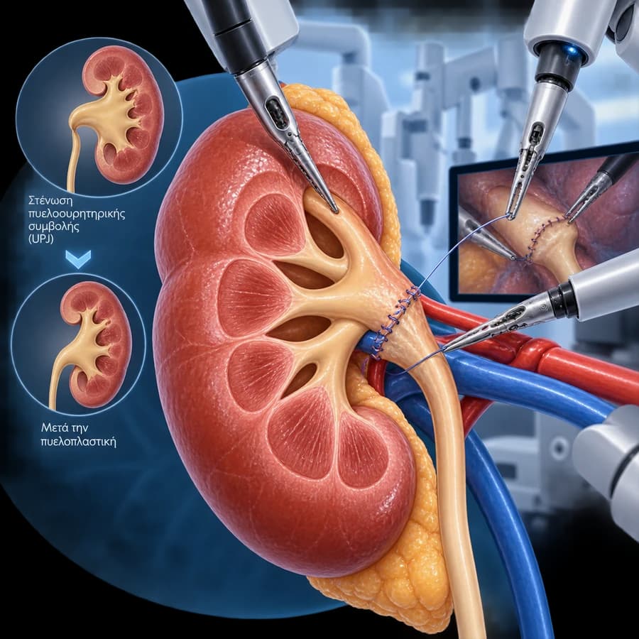
Robotic pyeloplasty is a reconstructive procedure for ureteropelvic junction obstruction. The narrowed segment of the junction is rebuilt to restore normal urine drainage from the renal pelvis to the ureter, while preserving the kidney.
The procedure does not remove the kidney. The goal is complete restoration of drainage and preservation of existing kidney function.
Pyeloplasty ≠ Dilation ≠ Endopyelotomy
Pyeloplasty is not simple dilation of the stricture, nor an endoscopic incision (endopyelotomy). It is a reconstruction of the junction — the narrowed segment is addressed and the ureter is reconnected to the renal pelvis.
The Ureteropelvic Junction (UPJ)
The ureteropelvic junction (UPJ/PUJ) is the transition point from the renal pelvis to the ureter:
Hydronephrosis ≠ Obstruction
Dilation of the renal pelvis on ultrasound or CT does not by itself prove clinically significant obstruction.
The decision must be based on a combination of:
- Symptoms.
- Anatomy.
- Functional drainage.
- Kidney function.
- Progression over time.
- Infections.
- Stones.
- Overall clinical context.
We do not present hydronephrosis as an automatic indication for surgery. See also ureteropelvic junction obstruction.
When Is Pyeloplasty Needed?
Potential indications may include:
- Symptomatic, clinically significant obstruction.
- Recurrent flank pain attributable to UPJ obstruction.
- Deterioration of renal function.
- Progressive functional obstruction.
- Recurrent infection in the relevant clinical context.
- Stone disease associated with impaired drainage.
- Selected significant obstruction in a solitary kidney.
- Other clinically meaningful complications.
There is no one-size-fits-all indication for every patient.
Pain & UPJ Obstruction
Classic symptoms may include intermittent flank pain, pain after high fluid intake in selected patients, and nausea in some episodes.
Symptoms are not specific. We do not diagnose UPJ obstruction from symptoms alone.
Congenital ≠ Always a Childhood Diagnosis
An anatomical UPJ obstruction may exist from a young age but be recognised in childhood, adolescence, or later in adult life, depending on severity and presentation. We do not present adult UPJ obstruction as always acquired.
Causes & Mechanisms
Because this page has procedure intent, causes are kept brief:
- Intrinsic narrowing of the wall.
- Abnormal muscular/peristaltic segment.
- High ureteral insertion.
- Crossing lower-pole vessel.
- Scarring/secondary stenosis in selected cases.
For more depth, see ureteropelvic junction obstruction.
What Is a Crossing Vessel?
A vessel to the lower pole of the kidney may cross near the UPJ and, in selected patients, may be associated with:
- Extrinsic compression.
- Anatomical kinking.
- Impaired drainage.
Not every crossing vessel is pathological. Its significance must be assessed together with anatomy, obstruction, symptoms, and operative findings.
Why Pyeloplasty Matters for a Crossing Vessel
Reconstructive pyeloplasty can allow excision/reconstruction of the narrowed segment and repositioning of the reconstructed UPJ relative to a relevant crossing vessel, where this is anatomically appropriate. This is an important difference from endopyelotomy.
We do not present every crossing vessel as an absolute indication for transposition.
What Dismembered Pyeloplasty Means (Anderson-Hynes)
In the classic reconstructive concept:
- The stenosed junction is disconnected.
- The non-functional/narrowed segment is addressed.
- The ureter is reconnected to the renal pelvis with a wide, tension-free passage.
This description is deliberately kept at a general level — it is not a surgical manual.
Anderson-Hynes ≠ the Only Technique
Other reconstructive variations exist for selected anatomy. We do not turn this page into a history of techniques, nor present one technique as universally necessary.
Robotic ≠ Autonomous
The robotic system does not perform the procedure on its own. Every movement is controlled by the surgeon through the console.
What Does the Robotic Approach Offer?
Potential technical features:
- Magnified 3D view.
- Articulated instruments.
- Fine tissue manipulation.
- Facilitated intracorporeal suturing.
- Precise reconstruction.
- Ergonomics for the surgeon.
Technological advantage ≠ automatic guarantee of a better functional outcome.
Why the Robot Makes Sense in Pyeloplasty
Pyeloplasty is a reconstructive operation that requires fine dissection, identification of anatomy, excision/reshaping where needed, tension-free anastomosis, and intracorporeal suturing.
Robotic articulation can facilitate these steps technically. We do not make a marketing claim.
Robotic vs Laparoscopic Pyeloplasty
| Robotic | Laparoscopic | |
|---|---|---|
| Minimally invasive | Yes | Yes |
| View | Magnified 3D | System-dependent |
| Instrument articulation | Robotic, wristed | Conventional laparoscopic |
| Intracorporeal suturing | Technically facilitated | Established in experienced hands |
| Reconstruction | Yes | Yes |
| Oncologic issue | Not an oncologic procedure | Not an oncologic procedure |
| Long-term goal | Restore drainage | Restore drainage |
| Best approach | Individualised | Individualised |
No approach is presented as the “winner”. See also laparoscopic pyeloplasty.
Do Not Underestimate Laparoscopic Pyeloplasty
Conventional laparoscopic pyeloplasty remains an established option, can provide excellent reconstructive results, and depends strongly on surgeon expertise. We do not present robotics as having made laparoscopy obsolete.
Robotic vs Open Pyeloplasty
Robotic/Minimally Invasive
Small incisions, reconstructive approach, typically less access-related surgical trauma.
Open
Established reconstructive operation, remains appropriate in selected situations, depending on anatomy, previous surgery, and surgeon/centre factors.
We do not characterise open surgery as outdated.
Pyeloplasty vs Endopyelotomy
| Pyeloplasty | Endopyelotomy | |
|---|---|---|
| Principle | Reconstruction | Endoscopic incision |
| Narrowed segment | Reconstructed/excised as appropriate | Incised |
| Crossing vessel | Can be addressed anatomically | Limited ability |
| Skin incisions | Small ports in robotic/laparoscopic approach | Usually none in retrograde approach |
| Invasiveness | Greater | Lower |
| Patient selection | Broad reconstructive role | More selective |
| Long-term durability | Generally stronger | Generally lower/more selective |
We do not use unsourced percentages. See also endopyelotomy.
“Smaller Surgery” ≠ “Better Surgery”
Endopyelotomy is a smaller procedure, but that does not mean it is the better choice for every anatomy.
The long-term choice should be based on:
- Anatomy.
- Stenosis characteristics.
- Crossing vessel.
- Previous surgery.
- Renal function.
- Durability expectations.
What Role Does MAG3 Play?
Diuretic renography can provide information about:
- Tracer uptake.
- Drainage pattern.
- Differential/split renal function.
- Response to diuretic challenge.
MAG3 is not just “a number”.
T½ Is Not Enough
T½ should not be used alone as absolute proof of obstruction or as the sole indication for surgery.
Interpretation depends on:
- Renal function.
- Hydration.
- Bladder status.
- Curve morphology.
- Gravity/post-void drainage.
- Protocol.
- Total clinical picture.
According to current nuclear-medicine guidance.
Split Renal Function
Differential renal function helps estimate how much each kidney contributes.
There is no single percentage that by itself decides “surgery or not”.
The treatment decision must consider function, symptoms, drainage, anatomy, trend, age, the opposite kidney, and infection/stones.
“MAG3 Showed 35% — Do I Need Surgery?”
A single percentage is not enough for a treatment decision.
It must be interpreted together with the rest of the clinical picture.
The Role of Ultrasound
Ultrasound is useful for hydronephrosis, renal parenchyma, and follow-up. However, it cannot alone reliably distinguish every dilated system from clinically significant functional obstruction.
CT/MR Urography
Can help define anatomy, crossing vessels, stones, level of narrowing, and alternative causes of obstruction.
Contrast CT is not required in every patient.
Should Hydronephrosis Disappear After Surgery?
Not necessarily.
The collecting system can remain dilated for a long period, or partially permanently, despite improved drainage.
Persistent dilation ≠ automatic surgical failure.
How We Judge Success
Successful pyeloplasty ≠ a perfectly normal ultrasound.
We do not define success only by imaging appearance. We use a combination of:
Preoperative Workup
- Symptom history.
- Previous imaging.
- Ultrasound.
- CT/MR anatomy where appropriate.
- MAG3/functional assessment.
- Creatinine/eGFR.
- Urinalysis.
- Urine culture according to context.
- Stones.
- Previous procedures.
- Prior pyeloplasty/endopyelotomy.
- Medications.
- Anticoagulants/antiplatelets.
- Anaesthetic assessment.
Infection & Fever Before Surgery
Clinically significant infection should be appropriately managed before elective upper-tract reconstruction.
Fever or rigors together with obstructive hydronephrosis may indicate an infected obstructed urinary tract and requires urgent evaluation.
In that setting, priority may be a JJ stent or nephrostomy, plus infection management. Definitive pyeloplasty is not normally the first goal during uncontrolled sepsis. See also pyelonephritis.
Anticoagulants
Do not stop anticoagulants or antiplatelets on your own.
The plan depends on the medication, indication, thrombotic risk, and bleeding risk.
How Robotic Pyeloplasty Is Performed
This description is deliberately kept at a general level — it is not a surgical manual with port placement, spatulation length, suture type/sequence, or vessel transposition technique.
Why a JJ Stent Is Placed After Pyeloplasty
A temporary ureteral stent may:
- Support drainage.
- Splint the reconstructed UPJ.
- Allow healing.
- Reduce temporary obstruction from oedema.
See also ureteral stent.
Is a JJ Stent Always Needed? How Long Does It Stay?
Many contemporary pyeloplasty protocols use temporary internal stenting, but the exact drainage strategy depends on surgeon technique, the patient, the reconstruction, age, any associated procedures, and local protocol.
We do not give a universal fixed number of weeks. Duration depends on the reconstruction, healing, protocol, and any complications.
Stent Symptoms & Surgical Drain
Possible stent symptoms: frequency, urgency, dysuria, haematuria, flank discomfort during voiding. See our ureteral stent page for more.
Fever or rigors are not normal stent symptoms.
A temporary surgical drain may be used in selected cases depending on the reconstruction, intraoperative findings, and surgeon protocol. It is not always a necessary step, and we do not give universal removal criteria.
Urine Leak
An anastomotic urine leak can occur, but it is not synonymous with complete failure. Management depends on volume, drainage, symptoms, infection, and stent function, and may involve observation or additional drainage in selected cases. We do not give self-management instructions.
Anaesthesia, Duration & Hospital Stay
Robotic pyeloplasty is performed under general anaesthesia, in an operating-room setting — it is not a day-case procedure under simple local anaesthesia.
There is no universal operating time. It depends on anatomy, crossing vessel, previous surgery, primary vs redo case, obesity, stones, adhesions, and reconstruction complexity.
Hospital stay depends on recovery, pain, oral intake, drainage/stent, any complications, and centre protocol. We do not promise next-day discharge or a fixed number of days.
Recovery & Timeline
Covers mobilisation, oral intake, wound care, pain control, fatigue, stent symptoms, return to work, driving, exercise, and heavy lifting. We prefer healing-based advice over rigid timetables.
Immediate postoperative period
Monitoring, pain control, mobilisation, urine output, renal function.
Early recovery
Wound healing, gradual increase in activity, temporary stent symptoms may occur.
Stent removal
According to the individualised surgical plan.
Functional follow-up
Symptoms, ultrasound, renal function, selected diuretic renography.
Mild haematuria, especially with the stent, can occur — heavy persistent bleeding or clots need assessment. Pain may come from the port incisions, the abdominal wall, the surgical area, the stent, or transient ureteric/spasm discomfort — we do not say the procedure is painless.
Fever After Surgery
Fever or rigors after pyeloplasty need prompt medical assessment.
Possible concerns include UTI, pyelonephritis, an infected collection, or a drainage problem.
Possible Complications
Bleeding
Uncommon but significant complication.
Infection
Requires assessment and targeted treatment.
UTI/pyelonephritis
May require infection management before or after surgery.
Urine leak
Not synonymous with complete failure.
Stent-related symptoms
Often transient, resolve with removal.
Injury to adjacent structures
Rare, depending on anatomy.
Vascular injury
Rare, particularly with difficult anatomy/redo.
Persistent/recurrent obstruction
Requires individualised reassessment.
Need for re-intervention
In selected cases.
Conversion to open surgery
A safety decision.
Anaesthetic complications
Rare, depending on the patient.
Wound/port-site complications
Rare with a minimally invasive approach.
We do not use unsourced incidence rates.
Can It Narrow Again?
Yes. No reconstructive procedure guarantees zero recurrence risk.
Evaluation of suspected recurrence should include symptoms, imaging, functional assessment, anatomy, and details of the previous operation. We do not define recurrence from persistent hydronephrosis alone.
Robotic Pyeloplasty After a Previous Failed Repair
Redo pyeloplasty can be considered in selected recurrent obstruction. Important issues:
- Scar tissue.
- Altered anatomy.
- Crossing vessels.
- Previous endopyelotomy.
- Previous stents.
- Renal function.
Redo surgery is more complex than primary repair. We do not present it as routine or simple.
Endopyelotomy After Failed Pyeloplasty
Options for recurrence can include redo pyeloplasty, selected endopyelotomy, or other reconstruction, depending on anatomy. We do not present one universally preferred option without current evidence.
Stones & UPJ Obstruction
Impaired drainage and renal stones can coexist. The treatment plan may sometimes combine reconstruction and stone management in selected cases, depending on stone burden, location, anatomy, and surgeon strategy — but may also require separate RIRS or PCNL.
Not every patient with UPJ obstruction forms stones.
Functional Success vs Anatomical Appearance
Success does not mean the renal collecting system must become perfectly normal on ultrasound.
Functional success focuses on improved drainage, stable/preserved renal function, symptom relief, and absence of clinically significant recurrent obstruction.
Follow-Up: Ultrasound & MAG3
Ultrasound is useful for the degree/trend of hydronephrosis and renal parenchyma in serial follow-up, but it cannot always prove success or failure by itself.
Selected patients may need repeat diuretic renography. We do not create one universal timing schedule — we use symptoms, ultrasound, renal function, preoperative severity, and clinical concern to guide follow-up.
Paediatric vs Adult UPJ Obstruction
This page is primarily adult urology. UPJ obstruction is also common in paediatric urology, but indications, follow-up, and surgical considerations may differ by age. We do not copy paediatric thresholds into adult guidance.
Kidney Function & Very Poor Function
The goal often includes preserving existing renal function, preventing deterioration, and improving drainage. Not every kidney will show dramatic improvement in split function — we do not promise renal-function recovery.
We do not use one arbitrary split-function percentage to declare a kidney “non-functional”, “must be removed”, or “cannot recover”. The decision may depend on symptoms, residual parenchyma, drainage, age, the contralateral kidney, reversibility, infection, and clinical context.
Nephrectomy is reserved for selected situations where preservation is not appropriate or meaningful. We do not frame a poor MAG3 number alone as a reason to remove the kidney.
The Patient Journey
Key Questions in Plain Language
I Have Hydronephrosis — Do I Need Pyeloplasty?
Not necessarily. Hydronephrosis must be interpreted together with symptoms, anatomy, drainage, and kidney function.
Robotic Pyeloplasty or Endopyelotomy?
Pyeloplasty is a reconstructive procedure with generally greater long-term durability, while endopyelotomy is less invasive but suitable for more selected cases.
Robotic or Laparoscopic Pyeloplasty?
Both are minimally invasive reconstructive techniques. The robotic platform can facilitate intracorporeal suturing and fine reconstruction, but laparoscopic pyeloplasty remains an established, effective option in experienced hands.
What Happens If There Is a Crossing Vessel?
A relevant crossing vessel can influence reconstruction. Pyeloplasty can allow the UPJ to be reconstructed in an anatomically favourable relationship to the vessel. It does not mean every vessel needs transposition.
What Is MAG3?
It assesses drainage, differential renal function, and response to a diuretic. One number alone is not the diagnosis.
Is a JJ Stent Needed?
Temporary internal stenting is commonly used in many pyeloplasty protocols, but exact practice and duration are individualised.
If Hydronephrosis Remains, Did the Surgery Fail?
No. Persistent dilation can remain despite restored drainage.
Can It Narrow Again?
Yes. Recurrence is possible and should be assessed functionally/anatomically, not only by ultrasound dilation.
Can Robotic Pyeloplasty Be Done Again?
In selected recurrent cases, redo robotic, laparoscopic, or open reconstruction can be considered depending on anatomy and expertise. Suitability is not guaranteed.
Will Kidney Function Improve?
The main goal is often to restore drainage and preserve existing kidney function. Function does not improve to the same degree in every patient.
Frequently Asked Questions (FAQ)
What is robotic pyeloplasty?
It is the surgical reconstruction of a stenosed ureteropelvic junction (UPJ), using a robotic system, to restore normal urine drainage from the renal pelvis to the ureter while preserving the kidney.
What is ureteropelvic junction obstruction?
It is a functional or anatomical impairment of urine flow at the point where the renal pelvis transitions into the ureter. See our page on ureteropelvic junction obstruction for more detail.
Does hydronephrosis always mean obstruction?
No. Dilation of the renal pelvis on ultrasound or CT is an imaging finding and does not by itself prove clinically significant obstruction. It needs to be interpreted together with symptoms, anatomy, functional drainage, and kidney function.
When is pyeloplasty needed?
It is considered for symptomatic, clinically significant obstruction, deterioration of renal function, recurrent pyelonephritis or stones related to impaired drainage, or selected significant obstruction in a solitary kidney — always individualised.
What is MAG3?
It is a dynamic renal scan using a radiotracer, usually with a diuretic, that assesses drainage and differential (split) kidney function of each kidney separately. A single percentage or T½ value is not sufficient by itself for diagnosis.
What is split renal function?
It is the percentage of overall kidney function contributed by each kidney, estimated from the renogram. There is no single percentage that automatically decides "surgery or not".
Is T½ alone enough to prove obstruction?
No. T½ must be interpreted together with renal function, hydration, bladder status, curve morphology, and the overall clinical picture, according to current nuclear-medicine guidance.
What is a crossing vessel?
It is a vessel to the lower pole of the kidney that, in some patients, crosses near the UPJ and may contribute to external compression or kinking. Its mere presence does not prove it is pathological — its significance is assessed together with anatomy, obstruction, and findings.
Does the vessel always need to be moved?
No. Pyeloplasty allows the reconstructed UPJ to be positioned in an anatomically appropriate relationship to a relevant crossing vessel where indicated, but not every vessel requires transposition.
What is Anderson-Hynes dismembered pyeloplasty?
It is the classic reconstructive technique in which the stenosed junction is disconnected, the narrowed segment is addressed, and the ureter is reconnected to the renal pelvis with a wide, tension-free passage.
What is the difference between pyeloplasty and endopyelotomy?
Pyeloplasty is a reconstructive procedure that rebuilds the junction, while endopyelotomy is an endoscopic incision of the narrowed segment without full reconstruction. Pyeloplasty generally has stronger long-term durability, but is a larger operation.
Robotic or laparoscopic pyeloplasty?
Both are minimally invasive reconstructive techniques. The robotic platform can facilitate intracorporeal suturing, but laparoscopic pyeloplasty remains an established, effective option in experienced hands.
Is a JJ stent needed?
Many contemporary pyeloplasty protocols use temporary internal drainage (JJ/DJ stent), but the exact practice and duration are individualised according to the patient, the reconstruction, and the surgeon’s protocol.
How long does the JJ stent stay in?
There is no universal duration. It depends on the reconstruction, healing, protocol, and any complications.
What symptoms can the JJ stent cause?
Frequency, urgency, dysuria, haematuria, or flank discomfort during voiding can occur with the stent. Fever or rigors, however, are not normal stent symptoms and need assessment.
How long is the hospital stay?
There is no universal duration. It depends on recovery, pain, oral intake, drainage/stent, any complications, and the centre’s protocol.
What are the possible complications?
These include bleeding, infection, urine leak from the anastomosis, stent-related symptoms, injury to adjacent structures, vascular injury, persistent or recurrent obstruction, need for re-intervention, conversion to open surgery, and anaesthetic complications.
Can there be a urine leak?
Yes, an anastomotic urine leak can occur, but it is not synonymous with complete failure. Management depends on volume, drainage, symptoms, infection, and stent function.
Can it narrow again?
Yes. No reconstructive procedure guarantees zero risk of recurrence. Evaluation of a suspected recurrence is based on symptoms, imaging, functional assessment, and anatomy — not persistent hydronephrosis alone.
What if a previous pyeloplasty has already failed?
In selected recurrent obstruction, a redo pyeloplasty — robotic, laparoscopic, or open — or selected endopyelotomy may be considered, depending on anatomy. Redo surgery is more complex than primary repair.
If hydronephrosis persists, does that mean the surgery failed?
Not necessarily. The collecting system can remain dilated for a long period, or even permanently, despite improved drainage. Persistent dilation does not automatically equal failure.
How is success assessed?
Success is assessed using a combination of symptom improvement, stable or improved drainage, preserved renal function, and absence of clinically significant recurrent obstruction — not ultrasound appearance alone.
Will kidney function improve?
The main goal is often to restore drainage and preserve existing kidney function. Function does not improve to the same degree in every patient.
Related Topics
Evaluation for Robotic Pyeloplasty in Rhodes
The decision for pyeloplasty is not based only on the image of a dilated renal pelvis. It requires a combination of symptoms, anatomical imaging, and functional assessment of drainage and kidney function. When there is truly significant obstruction, pyeloplasty reconstructs the ureteropelvic junction and can address anatomical factors, such as a relevant crossing vessel, that a simple endoscopic incision cannot correct in the same way.
Scientific References
- EAU Guidelines relevant to upper urinary tract obstruction / reconstruction. European Association of Urology. 2024–2025 update.
- Taylor E, et al. Contemporary Management of Ureteropelvic Junction Obstruction in Adults: A Systematic Review. Eur Urol Focus. 2023.
- Bagrodia A, et al. Impact of Crossing Vessels on Outcomes of Robotic Pyeloplasty. J Endourol. 2020.
- Society of Nuclear Medicine and Molecular Imaging (SNMMI) / EANM. Procedure Standard for Diuretic Renography.
Meet the Doctor

Dr. Marinos Vasilas, Urologist – Andrologist
Dr. Marinos Vasilas performs robotic and 3D laparoscopic reconstructive procedures of the upper urinary tract, with emphasis on clearly informing every patient about the functional significance of obstruction and the real treatment options available.
Full ProfileSurgical Team
Robotic pyeloplasty is performed in collaboration with a specialised surgical centre and urology team, ensuring access to modern robotic infrastructure.
Meet Our TeamThe content on this page is informational and does not replace individualised urological assessment. Hydronephrosis does not automatically mean obstruction. Not every UPJ obstruction requires surgery. MAG3 should not be interpreted from a single number. Persistent hydronephrosis after repair does not automatically mean failure. The surgical approach is individualised. The final treatment choice requires specialist review of anatomy and kidney function.


