My Clinical Approach
Renal colic from ureteral stones is among the most intense pains a patient can experience. Modern urology offers targeted options — from observation and medical assistance to endoscopic lithotripsy with holmium laser achieving stone-free rates >95%.
Per EAU Urolithiasis 2026 and AUA Surgical Management of Stones 2016 (Assimos):
- Immediate assessment: NCCT or U/S, urinalysis, urine culture, biochemistry, CRP.
- Exclude pyonephrosis/urosepsis — mandates emergency decompression.
- Determine size and location — drives spontaneous passage probability and treatment choice.
- α-blockers (tamsulosin 0.4mg/day) for 5-10mm distal ureteral stones (EAU 2026 Strong recommendation; off-label use).
- Endoscopic modality selection: ESWL, URS+laser, PCNL based on size, location, anatomy.
- Comprehensive metabolic workup after first episode for recurrence prevention.
What are ureteral stones
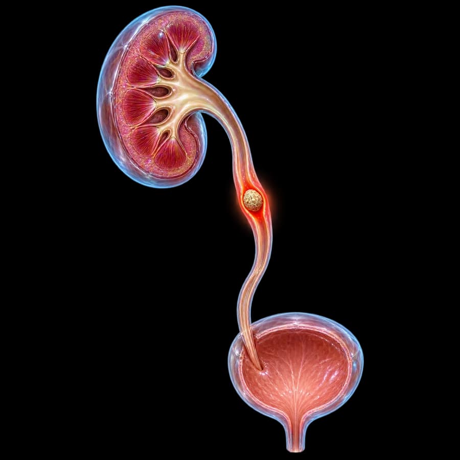
Calculi located within the ureter — the 25-30cm muscular tube transporting urine from kidney to bladder. They usually originate as kidney stones that migrated and lodged at one of three physiologic narrowings: ureteropelvic junction (UPJ), crossing over the iliac vessels, ureterovesical junction (UVJ).
Lifetime prevalence in Western populations is 10-15%. Male:female ratio 2:1. Peak age 30-50 years. Mediterranean climate increases incidence via dehydration and urinary supersaturation.
Important: Stone location (proximal, mid, distal ureter) and size determine both spontaneous passage probability and treatment selection.
Stone types (pathophysiology)
Calcium Oxalate (70-80%)
Most common type. Drivers: hypercalciuria, hyperoxaluria, hypocitraturia, low fluid intake. Radiopaque.
Uric Acid (5-10%)
Acidic urine (pH<5.5), hyperuricemia/hyperuricosuria. Radiolucent (invisible on KUB). Dissolvable with urinary alkalinization (potassium citrate, target pH 6.5-7).
Struvite (10-15%)
Infection-related Mg-NH₄-PO₄ stones. Associated with urease-producing bacteria (Proteus, Klebsiella, Pseudomonas). Form large staghorn calculi. Require complete removal + antibiotics.
Cystine (1-2%)
Hereditary cystinuria (autosomal recessive). Presents in childhood, recurs frequently. Treatment: alkalinization + d-penicillamine/tiopronin.
Calcium Phosphate (5-10%)
Alkaline urine (pH>6.5), distal renal tubular acidosis (RTA-1), primary hyperparathyroidism.
Rare types
Xanthine, dihydroxyadenine, drug-induced (indinavir, sulfadiazine, triamterene, ephedrine).
Symptoms & renal colic
Pain (colic)
Severe, sudden, wave-like (writhing). Radiates from flank to groin/testicle/labia majora. Patient cannot find a relieving position (unlike peritonitis).
Urinary signs
Hematuria (micro- or macroscopic in 80-90%), frequency, urinary urgency (when stone sits in distal ureter near bladder), dysuria.
Systemic
Nausea, vomiting (reflex via celiac plexus), diaphoresis, tachycardia, pallor.
Atypical
Some stones remain asymptomatic or cause only mild dull flank pain or microscopic hematuria.
Red flags — seek immediate care
- • Fever >38.5°C + chills (pyonephrosis/urosepsis)
- • Anuria or significant decreased urine output (bilateral obstruction or solitary kidney)
- • Uncontrolled pain despite analgesia
- • Confusion, hypotension, tachycardia (sepsis)
- • Persistent vomiting precluding hydration
Diagnosis (NCCT, U/S, KUB)
History & physical examination
Pain characteristics, hematuria, nausea/vomiting, fever. CVA tenderness (Murphy renal punch). Rule out peritonitis (appendicitis, diverticulitis, ectopic pregnancy).
Laboratory workup
Urinalysis (RBCs, WBCs, nitrites, pH, crystals), urine culture, CBC, CRP, BUN/creatinine, electrolytes, calcium, uric acid, PTH if indicated.
NCCT — gold standard
Sensitivity >97%, specificity >95%. Identifies size, location, density (HU), obstructive signs (hydronephrosis, perinephric stranding). Low-dose CT (<3 mSv) for follow-up.
Ultrasound
First choice in pregnancy and children. Shows hydronephrosis, kidney stones, UPJ/UVJ stones. Limited sensitivity for mid ureter.
KUB X-ray
Useful for radiopaque stones (Ca-Ox, Ca-PO₄, struvite, cystine). Misses uric acid. Mainly used for follow-up of known stone.
Urography (IVU/CT urography)
For suspected anatomic anomaly (duplex system, kinking, stricture), preoperative planning.
Watch for false negatives: Indinavir stones don't show on CT. Stones <3mm may be missed on US. With high clinical suspicion and negative US, NCCT is mandatory.
Treatment approach
Treatment depends on stone size, location, composition, and presence of obstruction or infection.
1. Watchful waiting (conservative)
Stones <5mm in distal ureter — observation up to 4 weeks. NSAIDs (diclofenac/ketoprofen) + antiemetics + hydration + urine straining for stone capture.
2. MET (Medical Expulsive Therapy)
Tamsulosin 0.4mg/day for 4 weeks. Increases spontaneous passage by 30% in 5-10mm distal ureteral stones (Hollingsworth meta-analysis BMJ 2016). Alternative: nifedipine.
3. ESWL (Extracorporeal Shock Wave Lithotripsy)
Non-invasive, no anesthesia. Effective for <1cm proximal ureteral stones (stone-free 70-80%). Lower yield in hard stones (cystine, monohydrate Ca-Ox, density >1000 HU). Contraindications: pregnancy, coagulopathy, anomalous anatomy.
4. URS with Holmium Laser — gold standard
Ureteroscopy + holmium:YAG laser lithotripsy. Stone-free rate >90% at one year. Effective for all stone types and locations. Typically followed by JJ stent placement for 1-2 weeks. New thulium fiber laser technology offers even higher speeds.
5. PCNL (Percutaneous Nephrolithotomy)
For large proximal stones or concurrent significant kidney stone burden (>2cm). Stone-free rate 80-90% in single session. Mini-PCNL and micro-PCNL reduce morbidity.
6. Emergency decompression (pyonephrosis/sepsis)
JJ stent or percutaneous nephrostomy within 24 hours + IV broad-spectrum antibiotics (3rd-gen cephalosporin + aminoglycoside or piperacillin/tazobactam). Definitive stone treatment after complete infection resolution (2-4 weeks).
7. Open/laparoscopic/robotic surgery
Rare indication — only after endoscopic failure or very large impacted stones precluding advancement.
Recurrence prevention
Recurrence rate is 50% at 10 years. Metabolic workup recommended after first episode in selected patients.
- Hydration: target urine output >2.5 L/day — dilutes lithogenic substances.
- Adequate dietary calcium (1000-1200 mg/day) — not restriction, reduces oxalate absorption.
- Low sodium intake (<2.3 g/day) — reduces hypercalciuria.
- Moderate animal protein (<1 g/kg/day) — lowers uric acid and acid load.
- Increase citrate (lemons, oranges) — crystallization inhibitor.
- Thiazide (hydrochlorothiazide 25mg) for hypercalciuria.
- Allopurinol 100-300mg for hyperuricosuria.
- Potassium citrate for hypocitraturia and cystine.
Follow-up & prognosis
- KUB or U/S at 4-6 weeks post-procedure to confirm stone-free status.
- Stone analysis (if captured) for targeted prevention.
- 24-hour urine (Ca, uric acid, oxalate, citrate, Na, volume) in recurrent stone formers.
- Annual U/S or low-dose CT in patients with stone history.
Summary: Proper treatment of ureteral stones has excellent prognosis with stone-free rates >90% using modern modalities. Recurrence prevention via individualized metabolic workup is the cornerstone of long-term management.
Frequently Asked Questions
What are ureteral stones?
Ureteral stones are calculi within the ureter — the muscular tube connecting kidney to bladder. They typically migrate from the kidney and cause acute renal colic, hematuria, nausea, and obstruction.
What is the most common stone composition?
Calcium oxalate (70-80%), followed by uric acid (5-10%), struvite (10-15%, infection-related Mg-NH₄-PO₄), cystine (1-2%, hereditary cystinuria), and calcium phosphate (5-10%).
What is the imaging modality of choice?
Non-contrast CT (NCCT) is the gold standard with sensitivity >97% and specificity >95%. Detects most stones except indinavir. Alternatives: ultrasound (first choice in pregnancy and children), KUB X-ray.
What is the chance of spontaneous passage?
Depends on size and location: stones <5mm in the distal ureter pass spontaneously in 80-90% of cases within 4 weeks. 5-10mm: 50%. >10mm: rarely — intervention usually required.
What is Medical Expulsive Therapy (MET)?
Administration of α-blockers (tamsulosin 0.4mg/day) to relax ureteral wall and facilitate stone passage. Effective for 5-10mm distal ureteral stones. EAU 2026: Strong recommendation for 5–10mm distal stones — use is off-label.
When is urgent intervention required?
In obstruction + infection (pyonephrosis/urosepsis): urgent decompression within 24 hours via JJ stent or percutaneous nephrostomy. Also: solitary kidney with obstruction, bilateral obstruction, uncontrolled pain.
What are the surgical options?
Three core modalities: ESWL (extracorporeal shock wave lithotripsy, non-invasive, effective for <1cm proximal ureteral stones), URS with holmium laser (gold standard for distal ureter), PCNL (percutaneous nephrolithotomy for large proximal stones or concurrent kidney stones).
How is recurrence prevented?
Through metabolic workup (24-hour urine, serum Ca/PO₄/uric acid), individualized diet (adequate calcium, low sodium, moderate animal protein, increased water >2.5L/day), thiazides for hypercalciuria, allopurinol for uric acid, potassium citrate for cystinuria.
Is ureteral stone disease hereditary?
There is genetic predisposition. Family history increases risk 2.5-fold. Several metabolic conditions are clearly hereditary: cystinuria (autosomal recessive), primary hyperoxaluria, renal tubular acidosis.
Related Topics
Have ureteral stones or renal colic?
Contact us for prompt evaluation: NCCT, pain management, MET, ESWL, URS with holmium laser or PCNL — with individualized recurrence prevention plan.
Scientific References
- EAU Guidelines on Urolithiasis (2026) — uroweb.org
- Assimos D, Krambeck A, Miller NL, et al. Surgical Management of Stones: AUA/Endourological Society Guideline. J Urol 2016;196(4):1153-69 — auanet.org
- Hollingsworth JM, Canales BK, Rogers MA, et al. Alpha blockers for treatment of ureteric stones: systematic review and meta-analysis. BMJ 2016;355:i6112 — pubmed.ncbi.nlm.nih.gov
- Pearle MS, Goldfarb DS, Assimos DG, et al. Medical management of kidney stones: AUA guideline. J Urol 2014;192(2):316-24 — pubmed.ncbi.nlm.nih.gov
- Türk C, Petřík A, Sarica K, et al. EAU Guidelines on Diagnosis and Conservative Management of Urolithiasis. Eur Urol 2016;69(3):468-74 — pubmed.ncbi.nlm.nih.gov
Meet the Doctor

Dr. Marinos Vasilas, Urologist – Andrologist
Dr. Marinos Vasilas treats ureteral stones using modern techniques: emergency decompression in pyonephrosis/sepsis, ureteroscopy with holmium laser, ESWL and percutaneous nephrolithotomy (PCNL), with comprehensive metabolic workup for recurrence prevention per EAU 2026 and AUA 2016 (Assimos).
View full profile


