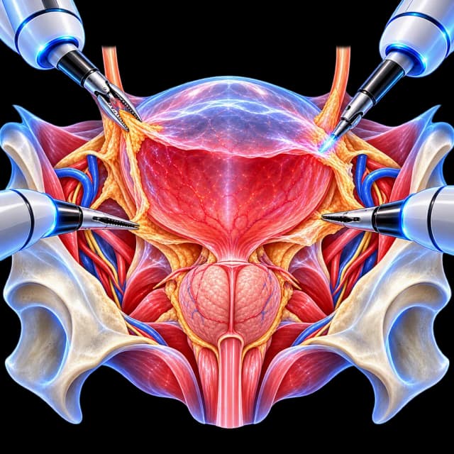
My Clinical Approach
A bladder diverticulum often causes concern for patients, but its anatomical presence alone does not automatically mean surgery is needed. My approach always starts with two questions: does it actually cause a clinical problem, and why did it form?
Particularly in adult men, the investigation always includes assessment of possible bladder outlet obstruction, because removing the diverticulum without addressing the cause can leave the problem unresolved.
- A diverticulum by itself is not an indication for surgery.
- Size alone does not decide the treatment.
- Not every patient needs simultaneous prostate surgery.
- A tumour inside a diverticulum requires particular oncological attention.
What Is a Bladder Diverticulum
A bladder diverticulum is a sac-like outpouching of the bladder wall. Its presence alone is not automatically an indication for surgery.
The decision for diverticulectomy depends on symptoms, bladder emptying, complications, the underlying cause, and the possibility of neoplasia.

Diverticulum ≠ Automatic Indication for Excision
Finding a diverticulum on imaging or cystoscopy does not automatically mean it needs to be removed. The need for surgery is judged clinically, not only radiologically.
Acquired vs Congenital Diverticulum
Acquired Diverticulum
More common in adults, often related to increased intravesical pressures/bladder outlet obstruction.
- • BPH/BPO
- • Urethral stricture
- • Neurogenic dysfunction
- • Chronic voiding dysfunction
Congenital Diverticulum
Different mechanism and anatomy, not necessarily linked to obstruction.
Not every diverticulum should be assumed to be caused by BPH.
“Why Did the Diverticulum Form?”
Possible mechanisms:
Removing the diverticulum without assessing the cause may leave the problem that created it unresolved.
Does Every Diverticulum Need to Be Removed?
No.
An asymptomatic diverticulum without relevant complications may be monitored in selected patients. The decision depends on:
Possible Indications for Surgery
Possible indications in selected patients:
This is not a rigid checklist, each case is individualised.
Diverticulum Size Alone ≠ Indication
Rules such as “>X cm always operate” or arbitrary thresholds are avoided. Clinical relevance matters more than a single measurement.
Symptoms
Many diverticula may be asymptomatic. Symptoms are not specific to a diverticulum by themselves.
Double Voiding
Some patients may void once and then pass additional urine as retained fluid empties from the diverticulum back into the bladder. This is described as double voiding, but is not diagnostic by itself.
Diagnosis
Evaluation may include, depending on context:
Not every test is needed for every patient.
Ultrasound
Useful for:
Ultrasound alone may not fully characterise the mucosa or the relationship to the ureter.
Flexible Cystoscopy
Cystoscopy can assess:
Flexible cystoscopy ≠ diverticulectomy.
See the page on Flexible Cystoscopy.
Uroflowmetry & Post-Void Residual (PVR)
Uroflowmetry
Assesses the flow pattern.
Post-Void Residual (PVR)
Assesses residual urine after voiding.
Neither test alone proves the cause of the obstruction.
Urodynamic Testing
It is not presented as mandatory. It may help in selected complex patients to distinguish between:
A large post-void residual does not automatically equal BPH-related obstruction.
Bladder Diverticulum & the Prostate
In adult men, acquired diverticula are frequently associated with chronic bladder outlet obstruction. Assessment of possible BPO may include:
It is not implied that BPH is always the cause. See the page on Benign Prostatic Hyperplasia.
“If BPH Is Present, Does the Prostate Also Need Surgery?”
Not automatically.
If clinically significant obstruction contributes to:
then treating the BPO may be important. Management may be outlet procedure alone, diverticulectomy plus outlet treatment, or staged treatment, depending on anatomy and functional findings.
Same Session or Staged Surgery
Selected patients may undergo diverticulectomy + BPO treatment as part of the same overall treatment strategy. Others may benefit from staged procedures.
Simultaneous treatment is not implied to always be preferable.
Prostate Procedures in Relevant Context
Depending on the patient and available techniques, possible bladder outlet obstruction procedures may include:
It is not implied that every technique is suitable to combine with diverticulectomy. This page is not a comparative guide to BPH treatments.
Is Treating the Obstruction Alone Enough?
In selected acquired diverticula, treating bladder outlet obstruction alone may improve emptying, symptoms, and stasis, and may avoid diverticulectomy in some patients.
It is not promised that the anatomical diverticulum will disappear.
Stone Within the Diverticulum
Urinary stasis may promote stone formation. Management may require:
See the page on Laser Bladder Stone Lithotripsy. Not every stone requires diverticulectomy.
Recurrent Urinary Tract Infections
Urinary stasis within a poorly emptying diverticulum may contribute to recurrent infection.
Bacteriuria ≠ symptomatic UTI. Recurrent infection has multiple possible causes, the diverticulum should not automatically be blamed.
Haematuria
Haematuria requires appropriate evaluation. It should not be assumed to originate from the diverticulum.
Tumour Within a Bladder Diverticulum
Urothelial carcinoma can rarely develop within a diverticulum.
Important anatomical concept: acquired bladder diverticula may have absent or very attenuated muscularis propria. This can complicate pathological staging and can allow a tumour to reach perivesical tissues with less anatomical depth than in a normal bladder wall.
Not every lesion within a diverticulum is aggressive, and unsupported prognosis percentages are not used here.
Tumour in a Diverticulum ≠ Routine Diverticulectomy
If tumour is suspected or confirmed, management becomes an oncological decision, depending on:
Management may selectively include:
See the pages on TURBT and Bladder Cancer. No automatic algorithm is given here.
Why Staging Is More Difficult
Therefore, conventional bladder tumour staging may be more difficult within a diverticulum and requires careful pathological and oncological assessment.
Preoperative Cystoscopy
Direct inspection before surgery may help to:
It is not presented as mandatory in every single case without justification.
Relationship to the Ureteric Orifice
Some diverticula may lie near the ureteric orifice or distal ureter. This can affect surgical planning. Dissection technique is not described here.
Is Ureteric Reimplantation Always Needed?
No.
It may be required in selected anatomy when:
Ureteric (JJ) Stent
A ureteric stent may be used selectively depending on ureteric anatomy, proximity to the diverticulum, any reimplantation, and surgical strategy. It is not used routinely in every procedure. See the page on Ureteral Stent Placement.
What Is Laparoscopic Diverticulectomy
The diverticulum is identified laparoscopically, separated from surrounding structures, excised, and the bladder wall is reconstructed.
Transperitoneal vs Extraperitoneal Approach
Both approaches may be used, depending on the location of the diverticulum, surgeon experience, anatomy, and any prior surgery. Neither is presented here as universally superior.
Laparoscopic vs Open Surgery
Laparoscopic
Minimally invasive access, small incisions, magnified view, intracorporeal bladder reconstruction.
Open
Established approach, may remain appropriate for selected complex anatomy/combined surgery.
Open surgery is not characterised here as obsolete.
Laparoscopic vs Robotic
Laparoscopic
Conventional laparoscopic instruments, intracorporeal suturing, minimally invasive approach.
Robotic
Articulated instruments, potential ergonomic/suturing advantages in selected cases.
Robot ≠ autonomous surgery. Neither method is presented here as superior oncologically or functionally without evidence.
3D Laparoscopy
3D laparoscopic visualisation can improve depth perception during minimally invasive reconstruction. See the page on 3D/Robotic Laparoscopic Surgery.
3D ≠ robotic. No specific equipment model is stated here.
The Procedure Step by Step (Patient Level)
Anaesthesia
General anaesthesia is administered.
Laparoscopic access
Small incisions.
Identification of the diverticulum
Visualisation of the anatomy.
Assessment/protection of adjacent structures
Particularly the ureter.
Excision of the diverticular sac
Removal of the diverticulum.
Bladder-wall reconstruction
Repair of the defect.
Leak assessment per surgical protocol
Confirmation of watertightness.
Catheter ± drain depending on the case
To allow healing.
Pathological examination where required
Histological assessment.
No trocar positions, port distances, insufflation pressures, dissection planes, ureteric mobilisation steps, suture materials, suture pattern, cystotomy closure layers, leak-test volume/pressure, or drain-placement technique are given here. These are exclusively for the surgical team.
Histological Examination of the Diverticulum
Excised tissue is normally submitted for pathological assessment, which may identify:
It is not implied that occult cancer is a common finding.
Do I Need a Catheter?
Following bladder-wall reconstruction, urinary catheter drainage is commonly used to allow healing. No single universal duration is given here. It depends on the repair, anatomy, concern for leak, any simultaneous procedures, and the clinical course.
Cystography Before Catheter Removal
It is not claimed to be mandatory for every patient. Some surgeons/centres may perform cystography before catheter removal depending on the complexity of the repair, the postoperative course, and institutional protocol.
Drain
A surgical drain may be used selectively. Not every patient needs one. Drain-management instructions are not given here.
Hospital Stay
Fixed universal numbers are avoided. Hospital stay depends on surgical complexity, any simultaneous prostate operation, the catheter/drain, bleeding, bowel recovery, comorbidity, and any complications. A “48 hours for everyone” claim is not made.
Recovery
Return to work, exercise, driving, and lifting should be individualised. There is no rigid universal timeline.
Possible Complications
No unsupported percentages are used here.
Urinary Leak
Because the bladder wall is surgically reconstructed, urinary leakage is a recognised potential complication.
Management depends on severity, drain output, imaging, catheter drainage, and clinical condition. A management algorithm is not given here.
Ureteric Injury
Particularly relevant when the diverticulum is close to the ureteric orifice/distal ureter. This remains at a patient-facing, high-level description.
Recurrent Diverticulum
It is not promised that recurrence is impossible. Persistent high-pressure voiding or untreated dysfunction may contribute to continued bladder structural problems. Unsupported recurrence statistics are avoided.
Will Bladder Function Return to Normal?
Not always.
It depends on:
Removing the diverticulum does not automatically restore impaired detrusor contractility.
Detrusor Underactivity
A patient with a large bladder, high residual, and a diverticulum does not necessarily have purely mechanical obstruction.
If detrusor underactivity is significant, surgery may not normalise voiding. Urodynamic assessment is used selectively to distinguish this.
“Big Diverticulum = Big Problem?”
Not necessarily.
Large size may matter technically/functionally, but symptoms, emptying, and complications remain central. There is no size-only logic.
“Can the Diverticulum Shrink After Prostate Surgery?”
Treating the obstruction can improve bladder emptying and symptoms. The anatomical diverticulum itself may persist. Spontaneous disappearance is not promised.
“Can the Stone and Diverticulum Be Treated Together?”
In selected cases, yes. The strategy depends on:
A universal same-session promise is not made.
Diverticulectomy ≠ Partial Cystectomy
Diverticulectomy
Removes a bladder diverticulum.
Partial Cystectomy
Removes a full-thickness segment of the bladder, often for different indications, including selected tumours.
They may overlap anatomically in selected oncological cases but are not synonymous.
The Patient Journey
Incidental Diverticulum / LUTS / Recurrent UTI / Retention / Haematuria / Stone
Initial clinical indication.
Ultrasound + Post-Void Residual ± Uroflowmetry
Initial assessment.
Cystoscopy / Imaging Where Indicated
Further investigation.
Why Did the Diverticulum Form?
BPO/BPH · urethral obstruction · bladder dysfunction · congenital anatomy · other cause.
Does It Cause a Clinically Relevant Problem?
The central question.
NO → Observation/Treat the Cause | YES → Define the Complication
Stasis, infection, stone, retention, symptoms, tumour concern.
Is Diverticulectomy Required?
The treatment decision.
Laparoscopic Excision + Bladder Reconstruction
In selected patients.
Simultaneous or Staged Treatment of Obstruction Where Indicated
Depending on the case.
Pathology + Functional Follow-Up
Final step.
This is an informational diagram, not a strict diagnostic/treatment algorithm. Each case is discussed individually with the treating urologist.
Frequently Asked Questions (FAQ)
What is a bladder diverticulum?
It is a sac-like outpouching of the bladder wall in which the mucosa protrudes through a weakness in the muscular layer.
Why does it form?
In acquired diverticula in adults, it is often related to increased intravesical pressures from bladder outlet obstruction (e.g. BPH), urethral stricture, or dysfunctional voiding. Congenital diverticula have a different mechanism.
Is it always the result of an enlarged prostate?
No. BPH is common but not the only cause. Neurogenic dysfunction, urethral stricture, and congenital factors can also contribute.
Does every diverticulum need surgery?
No. An asymptomatic diverticulum without relevant complications may be monitored in selected patients.
Does size matter?
Size alone does not decide the need for surgery. Clinical relevance (symptoms, emptying, complications) matters more than a single measurement.
When is diverticulectomy needed?
In selected patients with persistent bothersome symptoms, significant urinary stasis, recurrent infections, stone formation, haematuria requiring evaluation, recurrent retention, or suspected tumour.
Can it cause urinary tract infections?
Stasis of urine within a poorly emptying diverticulum can contribute to recurrent infection. However, bacteriuria does not necessarily mean symptomatic infection, and not every infection should automatically be attributed to the diverticulum.
Can stones form inside the diverticulum?
Yes, urinary stasis favours stone formation. Management may require stone removal, assessment of the diverticulum, and assessment of any underlying obstruction.
Can cancer develop inside the diverticulum?
Rarely, urothelial carcinoma can develop within a diverticulum. This requires particular oncological attention.
Why is staging more difficult for a tumour inside a diverticulum?
Because acquired diverticula may have a deficient or absent muscularis propria, which can complicate conventional pathological staging of invasion depth.
How is it diagnosed?
Depending on context, this may include ultrasound, post-void residual, uroflowmetry, flexible cystoscopy, CT/CT urography, and in selected circumstances voiding cystography or urodynamics.
Do I need a cystoscopy?
It can be useful for assessing the mucosa, the diverticular neck, a stone, suspicion of tumour, and the bladder outlet/prostatic area. It is not necessarily mandatory in every case without justification.
Do I need urodynamic testing?
It can help in selected complex patients to distinguish between obstruction and impaired detrusor contractility. It is not mandatory for everyone.
If I have BPH, does the prostate also need surgery?
Not automatically. If clinically significant obstruction is documented that contributes to high-pressure voiding, residual urine, or retention, treating it may be important, but the decision is individualised.
Can prostate and diverticulum surgery be done together?
In selected patients, yes, as part of the overall treatment strategy. Other patients may benefit from staged treatment.
Can treating the prostate alone be enough?
In selected cases, yes, treating the obstruction can improve emptying and symptoms. However, the anatomical diverticulum itself may persist.
How is laparoscopic diverticulectomy performed?
The diverticulum is identified laparoscopically, separated from surrounding structures, excised, and the bladder wall is reconstructed.
What is the difference between laparoscopic and robotic surgery?
Both are minimally invasive. Robotic surgery offers articulated instruments with potential ergonomic advantages for suturing in selected cases. Neither method is presented here as universally superior oncologically or functionally.
Is ureteric reimplantation always needed?
Not for every procedure. It may be needed when the ureteric orifice is very close to or involved by the diverticulum, or when safe excision requires ureteric reconstruction.
Do I need a catheter?
After bladder-wall reconstruction, a catheter is usually used to allow healing. The duration is not universal, it depends on the repair, the course, and any simultaneous procedure.
How long is recovery?
There is no rigid universal timeline. It depends on the complexity of the procedure, any simultaneous prostate surgery, the catheter, and general health.
What are the possible complications?
Bleeding, infection, urinary leak, ureteric injury, ureteric obstruction, need for ureteric reconstruction, urinary retention, urinary tract infection, and rarely bowel/vascular injury.
Will the bladder empty normally afterwards?
Not always. It depends on the duration of the obstruction, detrusor function, residual urine, bladder compliance, and any neurological disease. Removing the diverticulum does not automatically restore impaired detrusor contractility.
When should I contact a doctor urgently?
With fever, chills, inability to urinate, heavy or persistent bleeding, or significant urinary leakage/abdominal pain.
Related Topics
Book an Appointment in Rhodes
The correct management of a bladder diverticulum begins with answering two questions: does it actually cause a clinical problem, and why did it form? Treatment may address the diverticulum itself, the underlying obstruction, or both, depending on the anatomy and function of the bladder.
References & Sources
- EAU Guidelines on Management of Non-Neurogenic Male LUTS/BPO — uroweb.org
- EAU Guidelines on Non-Muscle-Invasive Bladder Cancer — uroweb.org
- EAU Guidelines on Urological Infections — uroweb.org
- There is no formal high-level guideline specifically for laparoscopic/robotic bladder diverticulectomy. Current practice is based mainly on retrospective series and specialised surgical experience, not on strong guideline-level evidence.
Medical Review

Medically reviewed by Dr. Marinos Vasilas – Urologist – Andrologist
Dr. Marinos Vasilas performs laparoscopic bladder diverticulectomy in Rhodes, with emphasis on investigating and treating the underlying cause of the diverticulum.
View full profileThis page is for informational purposes only and does not replace individualised medical examination. A bladder diverticulum is not by itself an indication for surgery. Size alone does not determine treatment. Not every patient needs prostate surgery. Impaired detrusor contractility may limit the functional outcome even after successful excision. A tumour within a diverticulum requires specialised oncological assessment. Anticoagulants should not be stopped without medical advice. Fever, inability to urinate, or significant urinary leakage after the procedure require prompt medical assessment.