My Clinical Approach
A bladder diverticulum is almost always a sign of an underlying problem, not a stand-alone disease. My approach always starts with “why did it form” before any treatment decision is made.
I follow the EAU Guidelines on Non-Neurogenic Male LUTS/BPO 2026 and the EAU Guidelines on Non-Muscle-Invasive Bladder Cancer 2026 where relevant to a tumour within a diverticulum:
- Distinguishing BPH from BPO — benign prostatic hyperplasia is a histological diagnosis, while bladder outlet obstruction (BPO) is a urodynamic diagnosis. It is the obstruction that is treated, not the prostate size itself.
- Actively considering detrusor underactivity as an alternative or coexisting cause, especially when the clinical picture does not fit “typical” obstruction.
- Cystoscopy in every patient with haematuria, recurrent infection, or suspected tumour within a diverticulum.
- Careful interpretation of pathology reports on a tumour within a diverticulum, taking into account that the muscularis propria may be thin or absent in the diverticulum itself.
- Individualised decision-making: observation, treatment of the underlying cause only, or diverticulectomy — not automatic surgical removal based on size.
- Laparoscopic/robotic diverticulectomy as a modern option in appropriately selected patients, with or without simultaneous treatment of the obstruction.
What is a bladder diverticulum
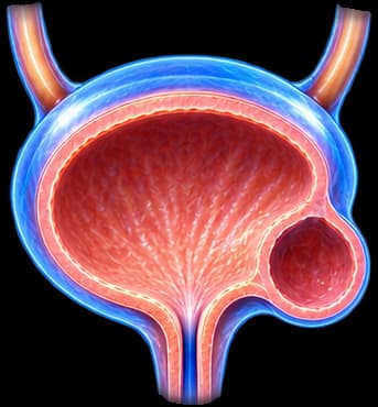
A bladder diverticulum is an outpouching (sac) of the bladder wall, communicating with the bladder through an opening or neck. In most adults it is a pseudodiverticulum (acquired) — the sac consists mainly of mucosa, without a complete muscular wall, which is why it does not have the ability to contract and actively empty.
Important: A true (congenital) diverticulum includes all layers of the wall, whereas an acquired pseudodiverticulum does not. This anatomical difference has practical consequences, especially when a possible tumour within it is being assessed.
Acquired vs congenital diverticulum
Acquired diverticulum
Usually related to chronic high intravesical pressure or outlet resistance. More common in adults, particularly older men, but not exclusively.
Congenital diverticulum
May arise without acquired outlet obstruction and is often related to the ureteric insertion point and anatomical features of the wall.
Adult acquired diverticulum and paediatric congenital diverticulum are not the same clinical problem — they have different mechanisms, different assessment, and different management.
Why it forms
Possible underlying mechanisms include:
- Bladder outlet obstruction (BPO) from prostate enlargement.
- Bladder neck obstruction.
- Urethral stricture.
- Detrusor-sphincter dysfunction or neurogenic causes.
- Long-standing high-pressure voiding over an extended period, regardless of the cause.
- Congenital wall weakness/anatomy, without acquired obstruction.
A diverticulum does not automatically mean a “prostate problem.” Although BPO is common in adult men, other causes must always be considered before deciding on treatment.
Prostate: BPH ≠ BPO
Chronic bladder outlet obstruction from prostate enlargement can lead to increased detrusor workload, trabeculation, and, over time, diverticulum formation.
BPH ≠ BPO. Benign prostatic hyperplasia (BPH) is a histological diagnosis — it describes enlargement of the gland. Bladder outlet obstruction (BPO) is a urodynamic diagnosis — it describes whether there is genuine resistance to urine flow. A large prostate does not automatically prove there is obstruction.
Likewise, diverticulum plus a large prostate does not automatically indicate prostate surgery. There must be evidence that obstruction is genuinely the primary cause of the patient’s problems before any outlet treatment is decided. See the full page on benign prostatic hyperplasia.
Urethral stricture & bladder neck
Obstruction of the urethra — for instance from a urethral stricture — or bladder neck dysfunction can also increase voiding pressures and contribute to diverticulum formation, independent of the prostate. The prostate should not be assumed to be the only possible obstructive cause, especially when the history is atypical.
Detrusor underactivity
A patient can have a large diverticulum, high PVR, and weak flow without true outlet obstruction. Underactivity of the detrusor itself may be a significant contributing factor, either alone or combined with some degree of obstruction.
High PVR ≠ proof of BPO. Outlet surgery (e.g. of the prostate) should not be recommended solely because PVR is elevated — it must first be clarified whether the cause is true obstruction, detrusor underactivity, or both.
Bladder wall trabeculation
Chronic high-pressure voiding can cause trabeculation (thickening of the muscle bundles of the wall), sacculation, and eventually diverticula. However, the imaging appearance of trabeculation alone does not quantify the degree of functional obstruction — it always requires clinical, and where indicated urodynamic, correlation.
Congenital diverticulum & the ureter
In younger patients, a diverticulum should not automatically be attributed to prostatic enlargement. Congenital diverticulum, urethral stricture, bladder neck dysfunction, or functional/neurological causes must be considered. Some congenital diverticula are located near the ureteric orifice (paraureteral) and may be related to particular features of the ureterovesical junction — assessed individually, without automatically applying the same protocol as for an adult acquired diverticulum.
Symptoms
Many diverticula are asymptomatic and are discovered incidentally. When symptomatic, possible symptoms include:
Urinary
Weak stream, incomplete emptying sensation, frequency, double voiding, discomfort.
Complications
Recurrent UTI, haematuria, retention, rarely stone formation.
Important: Symptoms often originate from the underlying bladder/outlet disorder rather than the diverticulum itself — which is why assessment always focuses on the cause, not just the anatomical picture.
Double voiding
Some patients notice that they urinate, and shortly afterward feel the need to void again — urine retained in the diverticulum gradually returns to the bladder. This can explain post-void dribbling, a “second void,” and a sensation of incomplete emptying. Not every patient with a diverticulum experiences this.
Asymptomatic diverticulum
An asymptomatic, uncomplicated diverticulum does not automatically need surgical removal. Observation can be an appropriate option, without applying a treatment criterion based solely on size.
There is no single diverticulum size that alone means diverticulectomy must be performed. The decision depends on symptoms, emptying, infections, stones, tumour, obstruction, effect on the kidneys/ureters, and bladder function — not a number of centimetres.
Residual urine & flow
Post-void residual (PVR) may accumulate in the bladder proper, within the diverticulum, or both. An elevated PVR suggests incomplete emptying, but it does not identify the mechanism by itself.
Uroflowmetry may suggest slowed flow or outlet resistance, but a low maximum flow rate (Qmax) does not by itself prove obstruction — it is always interpreted together with voided volume, PVR, symptoms, and, if needed, a pressure-flow study as part of urodynamic testing. A universal PVR cutoff is not used as an indication for surgery.
Recurrent urinary infections
Urinary stasis within the diverticulum can contribute to recurrent infection. However, recurrent UTI must not automatically be attributed to the diverticulum.
Residual urine, catheter use, stones, obstruction, or other causes must be assessed. Treating the underlying cause is the priority — antibiotics alone do not substitute for a structural assessment.
Stones within the diverticulum
Urinary stasis within a diverticulum may predispose to stone formation — either within the diverticulum or in the bladder itself — with possible symptoms of haematuria, recurrent infection, irritative symptoms, or pain.
Stone removal (e.g. via laser bladder stone lithotripsy) addresses the immediate problem, but does not necessarily correct the reason it formed — the underlying stasis/obstruction must be assessed separately.
Haematuria
Blood in the urine in a patient with a diverticulum can result from infection, stone, mucosal irritation, tumour, or other urinary tract pathology.
Haematuria in a patient with a diverticulum must not automatically be attributed to the diverticulum. It always requires full investigation (cystoscopy, imaging) to rule out a more serious cause, particularly tumour.
Tumour within a Bladder Diverticulum
Urothelial carcinoma can arise within a diverticulum. This is one of the most important points of attention on this page.
Anatomical issue: Acquired diverticula may have a thin, attenuated, or completely absent muscularis propria in parts of their wall. This can complicate:
- • pathological staging,
- • assessment of depth of invasion,
- • interpretation of the TURBT specimen,
- • surgical planning.
A tumour within a diverticulum does not automatically mean advanced cancer. But staging may be more challenging because the normal muscle layer may be absent or attenuated — which is why assessment requires specialist expertise.
Diagnostic Approach
History & LUTS assessment
Storage/voiding symptoms, bladder diary, infection history, prior surgery, neurological history.
Bladder/kidney ultrasound + PVR
First-line imaging: demonstrates the diverticulum, residual urine, stones, and bladder morphology. A normal ultrasound does not rule out a mucosal tumour inside the diverticulum.
Uroflowmetry, where indicated
May suggest slowed flow, but always interpreted together with voided volume and PVR — never in isolation.
Cystoscopy
Assesses the diverticular neck, the mucosa where visible, looks for tumour, stones, or trabeculation. Not every diverticulum can be fully inspected endoscopically.
CT urography, selectively
Useful for anatomy, relationship to the ureter, and when tumour is suspected. Not needed for every asymptomatic diverticulum.
VCUG / cystography, in selected cases
More useful in congenital or complex cases — not a mandatory test for everyone.
Urodynamic testing, when there is uncertainty
Useful when it is unclear whether the problem is obstruction, detrusor underactivity, or a combination — especially before major surgery.
Before removing the diverticulum, it may be more important to understand whether the bladder itself can empty effectively. If the underlying detrusor contractility is poor, removing the diverticulum or treating the prostate may not fully restore normal voiding — anatomical correction does not guarantee functional recovery.
Ureteric orifice & reflux
Some diverticula lie near the ureteric orifice. This can influence the likelihood of reflux, obstruction, and surgical planning. Ureteric reimplantation is not needed in every case — only when genuine anatomical involvement of the ureter is documented.
Vesicoureteral reflux can coexist in selected diverticula, particularly congenital/paraureteral cases. Assessment here remains at the bladder-disease level — a full paediatric reflux algorithm is not applied on this page.
Effects on the kidneys
In selected cases, underlying high-pressure voiding, obstruction, or ureteric involvement may contribute to hydronephrosis, upper tract deterioration, or renal impairment. The diverticulum itself does not always damage the kidneys — the effect depends on the underlying cause and the relationship to the ureter.
When treatment is needed
Treatment is considered when there is:
- Significant symptoms affecting quality of life.
- Recurrent infection genuinely attributable to stasis.
- Stones within the diverticulum.
- Retention or problematic emptying.
- Tumour within the diverticulum.
- Obstruction or ureteric complications.
- Progressive complications noted during follow-up.
Presence of the diverticulum alone is not an indication for treatment.
Observation
Observation can be a reasonable option for selected asymptomatic, uncomplicated, stable diverticula. Follow-up frequency depends on age, symptoms, haematuria, underlying cause, malignancy risk, and bladder function — there is no universal surveillance schedule for every patient.
Treating the cause first
If the diverticulum developed because of outlet obstruction, treating the underlying obstruction may sometimes improve emptying, symptoms, and diverticular stasis. In selected patients, outlet treatment alone may be sufficient.
Not every patient needs diverticulectomy. In selected patients, where there is no persistent problem attributable to the diverticulum itself (infection, stone, tumour, poor emptying), treating the underlying cause may be enough.
Outlet procedure vs diverticulectomy
Outlet procedure alone
May be reasonable when obstruction is the dominant problem, the diverticulum is uncomplicated, and bladder function is expected to improve. Outlet options (where clinically appropriate): HoLEP, TURiS, Aquablation, or Rezūm, depending on the case.
Diverticulectomy
Considered when significant problems related to the diverticulum itself persist — recurrent infection/stasis, stone, tumour, poor emptying related to the diverticulum, or anatomical complications.
Simultaneous vs staged surgery
In selected patients, outlet surgery and diverticulectomy can be performed simultaneously or in two stages. The decision depends on age, prostate/outlet pathology, bladder function, diverticulum anatomy, surgical complexity, and comorbidity. Neither approach is universally superior — the choice is individualised.
Diverticulectomy
Diverticulectomy removes the diverticular sac and reconstructs the bladder wall. It can be performed via an open, laparoscopic, or robotic approach, depending on anatomy, centre expertise, available equipment, and any associated pathology.
No approach is universally superior — robotic is not always better, laparoscopic is not always better, and open surgery is not obsolete. The choice depends on complexity, anatomy, relationship to the ureter, any simultaneous procedure, and surgeon experience.
For a full discussion of indications, operative planning, relationship to the ureteric orifice, postoperative care, and complications, see the dedicated page: Laparoscopic Bladder Diverticulectomy.
Treating a tumour within the diverticulum
If a tumour is diagnosed, treatment may include, depending on the case: TURBT, re-evaluation, diverticulectomy/partial cystectomy, or radical cystectomy — depending on grade, stage, diverticular anatomy, presence/absence of muscle, any associated CIS, multifocality, and overall bladder status. There is no single treatment that fits every case.
Endoscopic resection (TURBT) within a diverticulum can be technically and pathologically more challenging, because the wall may be thin, the muscle layer may be absent, perforation risk can differ, and staging may be limited. A pathology report noting absence of muscle in a diverticular specimen must be interpreted with knowledge of the diverticular anatomy — it does not always indicate an inadequate resection.
For a full discussion of staging, grade, BCG, cystectomy, and systemic therapy, see the dedicated page: Bladder Cancer.
Age, sex & special populations
Younger patients
In younger men, congenital diverticulum, urethral stricture, bladder neck dysfunction, or neurological/functional causes should be considered — not automatically prostate enlargement.
Women
Bladder diverticula can occur in women too, but the causes of outlet obstruction differ from those in men — this is not an exclusively male condition.
Children
Paediatric diverticula may have different congenital mechanisms, reflux associations, and treatment pathways — adult BPH logic does not apply here.
When to seek help immediately
Seek prompt assessment if you notice:
- • Gross haematuria
- • Catheter blockage from clots
- • Recurrent febrile UTI
- • Urinary retention
- • Worsening hydronephrosis
- • Decline in renal function
- • Persistent unexplained pain
- • A suspicious lesion within the diverticulum on imaging/cystoscopy
Your Patient Journey
Incidental finding or symptoms
A diverticulum is discovered incidentally, or the patient presents with LUTS, recurrent infection, or haematuria.
Clinical assessment
History, examination, ultrasound with PVR, uroflowmetry where indicated.
Cystoscopy / CT / urodynamics where indicated
Looking for tumour, stones; assessing anatomy and function.
Why did the diverticulum form?
Outlet obstruction (prostate/urethra/bladder neck) — or detrusor dysfunction — or congenital/anatomical cause, assessed individually.
Does it cause real complications?
Infection, stone, retention, haematuria, tumour, effects on the ureter.
Observation or treatment of the cause
Without a major complication: observation ± treatment of the underlying cause.
Outlet procedure alone, in selected patients
If obstruction is the primary driver and the diverticulum is uncomplicated.
Diverticulectomy when needed
When the diverticulum itself remains clinically significant despite treating the cause.
Long-term follow-up
Based on bladder function and pathology findings, individualised.
Frequently Asked Questions (FAQ)
What is a bladder diverticulum?
It is an outpouching (sac) of the bladder wall, connecting to the bladder through an opening called the neck. In most adults it is a pseudodiverticulum (without a complete muscular wall), while congenital forms may include all wall layers.
Why does it form?
It is usually related to chronically increased voiding pressure — most often from bladder outlet obstruction (e.g. prostate, urethral stricture) — but it can also result from detrusor dysfunction, or it can be congenital, without obstruction.
Is it always caused by the prostate?
No. Although benign prostatic enlargement is a common cause in adult men, other causes must always be considered: urethral stricture, bladder neck dysfunction, detrusor dysfunction or underactivity, neurogenic causes, or congenital anatomy — especially in younger patients and in women.
What is the difference between congenital and acquired diverticulum?
An acquired diverticulum (the most common in adults) is usually related to chronically increased voiding pressure/outlet resistance. A congenital diverticulum can occur without obstruction and is often related to the ureteric insertion point and anatomical features. These are not the same clinical problem, and they are not managed in the same way.
Does every diverticulum need treatment?
No. An asymptomatic, uncomplicated diverticulum can be observed without surgical intervention. The need for treatment depends on symptoms, emptying, infections, stones, tumour, and effect on the kidneys/ureters — not simply on the presence of the diverticulum.
Does size matter?
Size is one of several factors assessed, but there is no single size that alone constitutes an indication for diverticulectomy. The decision is based on the overall clinical picture.
Can it cause residual urine?
Yes — urine can accumulate in the bladder itself, within the diverticulum, or both. Elevated post-void residual (PVR) indicates incomplete emptying, but it does not by itself identify the mechanism (obstruction or detrusor weakness).
Why might I urinate a second time shortly after voiding?
This is called double voiding. Urine that was retained in the diverticulum during the first void gradually returns to the bladder, creating the sensation of needing to void again shortly afterward. Not every patient with a diverticulum experiences this.
Can it cause urinary tract infections?
Urinary stasis within the diverticulum can contribute to recurrent infection, but recurrent UTI must not automatically be attributed to the diverticulum — stones, obstruction, catheter use, or other causes must be ruled out.
Can stones form inside the diverticulum?
Yes, urinary stasis can predispose to stone formation within the diverticulum, potentially causing haematuria, recurrent infection, or discomfort. Removing the stone alone does not necessarily correct the reason it formed.
Can it cause haematuria?
Yes, but haematuria in a patient with a diverticulum must not automatically be attributed to it — it may result from infection, stone, mucosal irritation, tumour, or other urinary tract pathology, and always requires full evaluation.
Can a tumour develop inside a diverticulum?
Yes, urothelial carcinoma can arise within a diverticulum. This is one of the most important points of attention on this page, due to the particular challenges in staging.
Is a tumour in a diverticulum always more aggressive?
Not automatically. A tumour within a diverticulum does not automatically mean advanced or invasive disease. However, its staging can be more challenging, as explained below.
Why is staging a tumour inside a diverticulum difficult?
An acquired diverticulum may have a thin, attenuated, or absent muscularis propria in parts of its wall. This can complicate pathological staging, assessment of depth of invasion, and interpretation of the TURBT specimen — absence of muscle in the specimen does not always indicate an inadequate resection, but may reflect the diverticular anatomy itself.
Is cystoscopy needed?
Yes, it is an essential tool for assessing the neck, the bladder, and looking for tumour or stones. However, not every diverticulum can be completely inspected endoscopically, depending on its size and the position of the neck.
What does ultrasound show?
It can demonstrate the diverticular sac, residual urine, stones, and bladder morphology. However, a normal ultrasound does not rule out a mucosal tumour inside the diverticulum.
Is a CT scan needed?
CT urography can help assess the anatomy, the relationship to the ureter, and cases with suspected tumour. It is not needed for every asymptomatic diverticulum.
What does uroflowmetry show?
It may suggest slowed flow or outlet resistance, but a low maximum flow rate (Qmax) does not by itself prove obstruction — it is always interpreted together with voided volume, PVR, symptoms, and, if needed, a pressure-flow study as part of urodynamic testing.
When is urodynamic testing needed?
When there is uncertainty about whether the problem is obstruction, detrusor weakness, or a combination of both — especially before major surgery, when PVR is large, flow is poor, or symptoms do not match the anatomical picture.
Does a high PVR mean prostate obstruction?
No. A large diverticulum with high PVR and weak flow does not automatically prove prostate outlet obstruction. True obstruction must be distinguished from detrusor underactivity or a mixed picture — which often requires urodynamic testing.
Can prostate treatment alone be enough?
In selected patients, yes — if the main cause is true outlet obstruction, treating it may improve emptying and symptoms without needing removal of the diverticulum itself.
When is diverticulectomy needed?
When the diverticulum itself remains clinically significant despite treatment of the underlying cause — for example, persistent infection, stone formation, poor emptying related to the diverticulum, tumour, or anatomical complications.
Can it be done laparoscopically or robotically?
Yes, laparoscopic or robotic diverticulectomy are modern options in appropriately selected patients, as alternatives to the open approach. The choice depends on anatomy, the relationship to the ureter, and the experience of the surgical centre.
Does prostate surgery always need to be done at the same time?
Not always. In selected patients, outlet surgery (e.g. of the prostate) and diverticulectomy can be performed simultaneously or in two stages, depending on age, pathology, bladder function, and complexity.
What if the diverticulum is close to the ureter?
Proximity to the ureteric orifice can influence surgical planning and, rarely, is associated with reflux. Ureteric reimplantation is not needed in every case — only when there is genuine anatomical involvement.
Is follow-up always needed?
Yes, especially for patients managed conservatively. Frequency depends on age, symptoms, haematuria, underlying cause, and risk of progression — there is no universal surveillance schedule for everyone.
When does haematuria need urgent evaluation?
Any new, unexplained haematuria in a patient with a diverticulum needs prompt evaluation (cystoscopy, imaging) to rule out tumour or another serious cause — it should never be assumed to be expected or benign.
Related Topics
Bladder diverticulum: understand the cause first
Managing a bladder diverticulum should not be based on its size alone. It requires assessing why it formed, whether there is outlet obstruction or detrusor underactivity, whether significant residual urine remains, and whether infections, stones, haematuria, or an intravesical lesion have occurred. In selected patients, treating the underlying cause is enough, while others may need diverticulectomy.
Scientific References
- EAU Guidelines on Non-Neurogenic Male LUTS/BPO 2026 — uroweb.org
- EAU Guidelines on Non-Muscle-Invasive Bladder Cancer 2026 — uroweb.org
- Golijanin D, Yossepowitch O, Beck SD, et al. Carcinoma in a bladder diverticulum: presentation and treatment outcome. J Urol. 2003;170(5):1761-1764
- Idrees MT, Alexander RE, Kum JB, Cheng L. The spectrum of histopathologic findings in vesical diverticulum: implications for staging and follow-up. Hum Pathol. 2013;44(7):1223-1232
- Tareen BU, Mufarrij PW, Godoy G, Stifelman MD. Robot-assisted laparoscopic transperitoneal bladder diverticulectomy. Urology. 2008;71(2):299-302
- Macejko AM, Viprakasit DP, Nadler RB. Robot-assisted bladder diverticulectomy: technique and outcomes. Urology. 2010;75(6):1344-1347
Meet the Doctor

Dr. Marinos Vasilas, Urologist & Andrologist
Individualised assessment of bladder diverticulum following EAU 2026 guidance: distinguishing BPH from BPO, urodynamic confirmation, and modern surgical treatment (laparoscopic/robotic diverticulectomy) when needed.
View Full Profile


