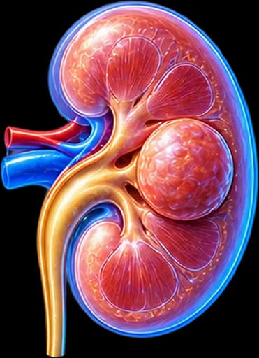
My Clinical Approach to Renal Oncocytoma
Oncocytoma is one of the most common benign kidney tumours, but also one of the most demanding to manage. The problem is usually not the tumour itself. It is the inability to reliably distinguish a benign renal mass from a malignant one using imaging alone.
Renal mass biopsy has significantly improved management, allowing selected patients to avoid unnecessary surgery. However, even with biopsy, differentiation from chromophobe renal cell carcinoma is not always completely clear-cut.
My approach is based on:
- Individualised assessment of diagnostic certainty before every treatment decision.
- Renal mass biopsy in appropriate patients, to avoid unnecessary surgery.
- A nephron-sparing philosophy in every interventional decision — partial rather than radical nephrectomy where feasible.
- Close collaboration with pathology for accurate differentiation from chromophobe RCC.
What Is Renal Oncocytoma
Renal oncocytoma is a benign epithelial tumour that, according to current pathology classification, arises from cells of the renal tubular system.
What “oncocytic” means
The term describes a characteristic cell phenotype: oncocytes have abundant eosinophilic cytoplasm and a large number of mitochondria. The word “oncocytic” describes this cell type — it does not by itself mean “cancer”.
It is the second most common benign solid kidney tumour after angiomyolipoma, and is usually discovered incidentally on imaging performed for another reason.
Is Renal Oncocytoma Cancer?
Classic renal oncocytoma is considered a benign kidney tumour.
The issue is often not the behaviour of a histologically confirmed oncocytoma. The problem is that, before a reliable histological diagnosis exists, a solid renal mass may not always be distinguished with certainty from RCC by imaging alone.
This distinction — between “a benign histological entity” and “diagnostic certainty before histology” — is the central theme running through the entire clinical management of oncocytoma.
Renal Oncocytoma vs Renal Cell Carcinoma (RCC)
Renal Oncocytoma
- Usually benign — epithelial oncocytic tumour.
- Does not have the typical malignant behaviour of RCC.
- May be observed with surveillance in selected cases when the diagnosis is sufficiently supported.
Renal Cell Carcinoma (RCC)
- Malignant renal epithelial tumour.
- Can have invasive/metastatic potential.
- Treatment depends on subtype/stage — see kidney cancer and renal cell carcinoma.
CT/MRI findings can increase or decrease suspicion, but do not always prove oncocytoma with certainty.
Oncocytoma vs Chromophobe RCC
Renal oncocytoma and chromophobe renal cell carcinoma can have overlapping:
- Morphology on microscopy.
- Imaging characteristics.
- Oncocytic appearance.
In some cases, distinguishing them requires expert pathological assessment, immunohistochemical markers and, where necessary, molecular findings.
CT should not be assumed to always reliably separate oncocytoma from chromophobe RCC.
Oncocytic Renal Tumours & Current Classification
Current pathology classification of kidney tumours now includes several oncocytic/eosinophilic renal entities beyond classic oncocytoma and chromophobe RCC. The precise categorisation of such a lesion is made by the pathologist, based on morphological and immunohistochemical criteria.
Hybrid oncocytic/chromophobe tumours
In certain clinical contexts — particularly specific hereditary syndromes — tumours with mixed oncocytoma/chromophobe RCC features have been described. Their clinical significance and management are assessed individually by the medical team, based on the full histopathological picture.
Birt-Hogg-Dubé Syndrome (BHD)
Bilateral or multiple oncocytic/chromophobe renal neoplasms can, in selected circumstances, be associated with hereditary syndromes such as Birt-Hogg-Dubé syndrome (mutations in the FLCN/folliculin gene).
Genetic evaluation may be considered in the appropriate clinical context, such as:
- Young age at presentation.
- Bilateral disease.
- Multiple tumours.
- Family history.
- A characteristic spectrum of associated findings (e.g. skin lesions, history of pneumothorax).
Genetic testing is not recommended for every solitary, sporadic oncocytoma.
Symptoms of Renal Oncocytoma
Most oncocytomas are discovered incidentally and cause no symptoms. When symptoms do occur, they may include:
Flank/loin discomfort
A dull ache or sense of heaviness with larger lesions.
Haematuria
Blood in the urine — a rarer presentation, which always warrants evaluation.
Abdominal discomfort
Generalised discomfort with larger tumours.
Palpable mass (rare)
Rarely, a mass may be felt in the abdomen with large lesions.
These symptoms are not specific to oncocytoma — they can occur with many types of kidney tumours.
“I Was Told I Have a Kidney Mass — Could It Be Oncocytoma?”
Yes, but it cannot be assumed to be oncocytoma just because it looks benign on imaging.
The differential diagnosis of a solid renal mass can include:
Imaging Evaluation
Ultrasound
Ultrasound can detect a renal mass, but it usually cannot reliably confirm renal oncocytoma. It is used for initial detection and, in selected clinical contexts, for surveillance.
CT Scan
Multiphasic contrast-enhanced CT assesses size, enhancement, morphology, vascularity, relationship to vessels, the collecting system, and surrounding structures. However, there is no single imaging finding that confirms oncocytoma with absolute certainty in every case.
The “central scar” is not diagnostic
Some larger oncocytomas may show a central stellate scar (“spoke-wheel” pattern). However, the central scar is not a specific finding and does not prove oncocytoma — it can also appear in other kidney tumours.
Segmental Enhancement Inversion
This has been proposed as a possible distinguishing imaging feature in selected studies, but its diagnostic performance does not support treating it as a pathognomonic finding — it does not replace clinical/histological evaluation.
MRI
MRI can help characterise a renal mass, particularly in selected cases. However, MRI also cannot always reliably distinguish oncocytoma from RCC — it is not a histological diagnosis.
99mTc-Sestamibi SPECT/CT
There is growing research interest in molecular imaging (99mTc-sestamibi SPECT/CT) for distinguishing selected oncocytic renal lesions from RCC. However, it is not a universally established standard test in current EAU/AUA guidelines, it does not replace biopsy, and it does not have 100% diagnostic accuracy. Availability and its role in clinical practice vary.
Is a Biopsy Needed for Suspected Oncocytoma?
Renal mass biopsy can be useful when:
- Imaging cannot provide a safe diagnosis.
- Histology would change the treatment strategy.
- Active surveillance is being considered.
- Avoiding unnecessary surgery is an important goal.
- Patient comorbidity influences the choice of treatment.
Routine biopsy of every renal mass is not required. The decision should be individualised and guideline-based.
What a biopsy can show
Biopsy can show an oncocytic neoplasm, oncocytoma, an RCC subtype, or other pathology, depending on sample quality and histopathological findings.
A result of “oncocytic renal neoplasm” is not always equivalent to a definitive benign oncocytoma diagnosis — further discussion with your urologist is needed.
Limitations of biopsy
Renal mass biopsy remains a useful tool, but has known limitations: non-diagnostic sampling, sampling error, tumour heterogeneity, and difficulty differentiating between certain oncocytic entities.
“If Oncocytoma Is Benign, Why Am I Being Offered Surgery?”
Because in many cases the real issue before surgery is not an already-proven oncocytoma, but a renal mass that cannot be confidently differentiated from a malignant tumour using CT or MRI alone.
Renal mass biopsy can help in selected cases, but it is not diagnostic in every situation — and the treatment decision always depends on the overall clinical context, not just the imaging appearance.
Growth, Metastasis & Coexistence with RCC
Can it grow large without being cancer?
Yes. Size alone does not turn a benign oncocytoma into RCC. However, a larger tumour can cause symptoms, create diagnostic/surgical challenges, or lead to treatment being considered.
Can it metastasise?
A histologically confirmed classic oncocytoma is considered a benign tumour without the metastatic behaviour of RCC. Older reports should be interpreted with caution, as they likely reflected cases of initial diagnostic misclassification.
Can it turn into RCC?
Classic oncocytoma and RCC are considered different tumour entities. This is not a transformation of a confirmed oncocytoma into cancer — the real clinical issue is mainly the certainty of the initial diagnosis.
Coexistence with RCC
Rarely, synchronous different kidney tumours can occur — multifocal lesions, or an oncocytoma together with RCC in the same or the opposite kidney. This is why evaluation should not assume every lesion shares the same histology.
How Is Treatment Chosen?
The choice depends on:
When Is Surveillance Alone Enough?
In selected patients with:
- A biopsy-supported benign oncocytoma.
- A small renal mass.
- A low-risk clinical profile.
- Significant comorbidity.
- A suitable ability to undergo imaging follow-up.
Active Surveillance may be chosen.
Active Surveillance ≠ ignoring it
It involves an organised, planned reassessment programme — not simply ignoring the finding.
Follow-up
Follow-up may include ultrasound, CT or MRI, depending on diagnostic confidence, size, growth, kidney function and patient characteristics. There is no single universal imaging schedule that applies identically to every patient — frequency is determined in consultation with your treating physician.
When Is Surgery Needed?
Surgery may be considered when:
A single size cutoff is not used as an absolute indication for surgery.
Partial Nephrectomy
When surgical treatment of a localised renal mass that may be an oncocytoma is required, the nephron-sparing approach allows removal of the mass while preserving the rest of the kidney, when this is technically and clinically appropriate.
Robotic Partial Nephrectomy
A robotic-assisted approach with a magnified 3D view and articulated instruments, facilitating tumour excision and kidney reconstruction. As with any surgery, it carries a possible risk of bleeding or other complications, and the lesion’s histology is not known with certainty before removal unless a biopsy has already been performed.
3D Laparoscopic Partial Nephrectomy
A minimally invasive alternative with 3D imaging, suitable in selected cases depending on anatomy and the surgical team’s experience.
Radical Nephrectomy & Thermal Ablation
Radical nephrectomy
Radical nephrectomy is not the desired routine treatment for a confirmed small benign oncocytoma. It may, however, be required in selected circumstances when:
- The tumour is very large or complex.
- Nephron-sparing surgery is not technically feasible.
- The diagnosis raises strong concern for malignancy.
- The involved kidney has poor function.
- Other patient-specific factors apply.
Thermal ablation
In appropriate, selected cases, cryoablation or radiofrequency ablation may be discussed. Patient selection is critical, histological confirmation carries particular importance, and follow-up remains necessary — this is not a universal treatment for every oncocytoma.
Active Surveillance vs Partial Nephrectomy
| Active Surveillance | Partial Nephrectomy | |
|---|---|---|
| Goal | Avoid unnecessary surgery | Definitive removal of the mass |
| Histology | Usually biopsy-supported when needed | Definitive pathology after excision |
| Kidney tissue | Preserved | Largely preserved |
| Surgical risk | No immediate surgical risk | Surgical risks are present |
| Follow-up | Required | Oncological/renal follow-up per pathology |
| Suitable for | Selected patients | Selected renal masses |
Oncocytoma & Kidney Function
Treatment decisions must take into account creatinine, eGFR, the function of the contralateral kidney, chronic kidney disease (CKD), and a solitary kidney. Avoiding unnecessary loss of functional kidney tissue is particularly important when a benign tumour is likely.
Solitary kidney
In a patient with a solitary functional kidney, nephron preservation takes on even greater importance — management must be highly individualised.
Bilateral/multifocal oncocytomas & renal oncocytosis
Multiple or bilateral oncocytic findings require careful imaging evaluation, consideration of a possible hereditary context, and a strong emphasis on nephron preservation — without automatically meaning a genetic syndrome. The related literature also describes “renal oncocytosis”, a rare condition involving multiple oncocytic lesions.
What Should It Be Distinguished From?
The differential diagnosis mainly includes:
- Clear-cell RCC.
- Chromophobe RCC — with particular emphasis due to genuine histopathological overlap.
- Papillary/eosinophilic renal neoplasms.
- Angiomyolipoma.
- Other oncocytic renal neoplasms.
Oncocytoma vs Angiomyolipoma
Oncocytoma
- Epithelial tumour.
- Usually not characterised by macroscopic fat.
- Can mimic RCC on imaging.
Classic Angiomyolipoma
- Mesenchymal tumour (PEComa).
- Often contains macroscopic fat.
- Bleeding risk is an important part of management — see renal angiomyolipoma.
The Patient’s Journey
Incidental finding of a solid renal mass
Discovered on ultrasound or CT performed for another reason.
Contrast-enhanced CT ± MRI
Further imaging to characterise the lesion.
Can it be characterised as a clearly benign lesion?
A key question — if not, there is oncocytoma vs RCC uncertainty.
Assessment of size, anatomy, kidney function and patient risk
Would a biopsy change management?
Yes → Renal mass biopsy
If a benign oncocytoma or low-risk oncocytic neoplasm is supported → surveillance or treatment depending on clinical context.
Malignancy / persistent diagnostic uncertainty
Nephron-sparing treatment where appropriate.
This is an educational overview, not a rigid management algorithm — every case is discussed individually with your urology team.
Is It Urgent?
An incidentally discovered oncocytoma/renal mass is usually not an emergency.
When immediate evaluation is needed
- Significant haematuria with clots.
- Severe acute bleeding.
- Haemodynamic instability.
- Acute, severe symptoms.
Follow-up & Prognosis
A histologically confirmed oncocytoma generally has a favourable prognosis. The main clinical risk relates to diagnostic certainty — not the biological behaviour of a genuine oncocytoma.
In summary
The need for treatment of an oncocytoma is not determined by size alone, but mainly by diagnostic certainty and the overall clinical context. When surgery is needed, the nephron-sparing approach remains a priority wherever technically and clinically feasible.
Frequently Asked Questions (FAQ)
What is a renal oncocytoma?
It is a benign epithelial kidney tumour made up of oncocytes — cells with abundant eosinophilic cytoplasm due to a large number of mitochondria. It is the second most common benign solid kidney tumour after angiomyolipoma and is usually discovered incidentally on imaging.
Is renal oncocytoma cancer?
A classic, histologically confirmed oncocytoma is considered a benign kidney tumour. The real challenge is that, before a reliable histological diagnosis exists, a solid renal mass may not always be distinguished with certainty from renal cell carcinoma by imaging alone.
Can oncocytoma turn into cancer?
A classic, histologically confirmed oncocytoma and renal cell carcinoma are considered different tumour entities — this is not a "transformation". The real clinical issue is mainly the certainty of the initial diagnosis, not a genuine oncocytoma converting into cancer.
How is oncocytoma distinguished from RCC?
CT and MRI can increase or decrease suspicion, but do not always prove oncocytoma with certainty. Distinguishing it from chromophobe renal cell carcinoma is particularly difficult, and the definitive differentiation often requires histopathological and immunohistochemical assessment.
Can a CT scan reliably confirm oncocytoma?
Not with absolute certainty. CT assesses size, contrast enhancement, morphology and vascularity, but there is no single imaging finding that confirms oncocytoma in every case.
What does a "central scar" on CT mean?
Some larger oncocytomas may show a central stellate scar. However, the central scar is not a specific finding and does not prove oncocytoma on its own — it can also appear in other kidney tumours, including RCC.
Is a biopsy always needed?
Not in every case. Renal mass biopsy can be particularly useful when imaging cannot give a safe diagnosis, when histology would change management, or when active surveillance is being considered. Routine biopsy of every renal mass is not required.
What does "oncocytic renal neoplasm" mean on a biopsy?
This result describes a tumour with oncocytic features, but it is not always equivalent to a definitive benign oncocytoma diagnosis — in some cases it may correspond to chromophobe RCC or another related entity. Further discussion with your urologist is needed to understand what it means in your specific case.
Can an oncocytoma grow?
Yes, a benign oncocytoma can increase in size over time. Growth alone does not mean malignant transformation, but it may influence the treatment decision due to diagnostic uncertainty, technical feasibility, or symptoms.
Can oncocytoma metastasise?
A histologically confirmed classic renal oncocytoma is considered a benign neoplasm without the metastatic behaviour of RCC. Older reports of "metastatic oncocytoma" likely reflected cases of initial diagnostic misclassification.
When is surveillance alone enough?
In selected patients with a biopsy-supported benign oncocytoma, a small renal mass, a low-risk clinical profile, or significant comorbidity, active surveillance with an organised reassessment programme may be an appropriate strategy.
When is surgery needed?
Surgery may be needed when the diagnosis remains uncertain, when RCC cannot be sufficiently excluded, with significant growth, with symptoms, or based on other individualised clinical considerations. The decision is never based on a single size cutoff alone.
What is partial nephrectomy?
It is nephron-sparing surgery that removes the renal mass while preserving as much functional kidney tissue as possible — the preferred approach when surgical treatment of a possible oncocytoma is required.
Why would a benign tumour need surgery?
Because in many cases the real issue before surgery is not an already-proven oncocytoma, but a renal mass that cannot be confidently differentiated from a malignant tumour using CT or MRI alone.
What is the difference between oncocytoma and angiomyolipoma?
Oncocytoma is an epithelial tumour that usually does not contain macroscopic fat and can mimic RCC on imaging. Classic angiomyolipoma is a mesenchymal tumour (PEComa) that often contains macroscopic fat, with bleeding risk being an important part of its management.
Related Topics
Book Your Appointment in Rhodes
Oncocytoma is usually a benign tumour, but safely differentiating it from other solid renal masses is not always possible with imaging alone. The decision between surveillance, biopsy and nephron-sparing surgery should be based on the mass’s characteristics, kidney function and the overall clinical context.
References – Sources
- EAU Guidelines on Renal Cell Carcinoma 2024 (benign renal tumour section) — uroweb.org
- WHO Classification of Tumours Editorial Board. Urinary and Male Genital Tumours (5th edition). IARC, Lyon 2022.
- Kutikov A, et al. Renal Mass Biopsy — AUA Renal Mass Guidance.
- Schmidt LS, Linehan WM. Genetic predisposition to kidney cancer. Semin Oncol 2016;43:566–574.
- Siegel RL, et al. Cancer statistics 2024. CA Cancer J Clin 2024.
Medical Editorial

Medically reviewed by Dr. Marinos Vasilas – Urologist – Andrologist
Dr. Marinos Vasilas runs a private urology practice in Rhodes, specialising in the management of renal masses, benign kidney tumours, and nephron-sparing surgical treatment.
View full profile


