My Clinical Approach
Kidney cancer is one of the diagnoses I encounter daily in the urology clinic. The evolution from open radical nephrectomy to laparoscopic or robotic partial nephrectomy represents one of the greatest advances in modern urology. The goal is not only to cure the tumour but to preserve renal function.
I place particular emphasis on precise staging before every treatment decision: triphasic CT, assessment of renal function, biopsy in selected cases and multidisciplinary discussion for possible immunotherapy in high-risk or metastatic patients.
My approach follows the EAU 2024 guidelines on renal cell carcinoma:
- Partial nephrectomy as first choice for T1 (≤7 cm) when technically feasible.
- Active surveillance for small renal masses (SRM) in elderly or comorbid patients.
- Renal mass biopsy before ablation or in equivocal cases.
- Adjuvant immunotherapy (pembrolizumab) for high-risk pT2G4/pT3+ or N+ after nephrectomy.
What Is Kidney Cancer
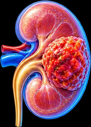
Kidney cancer refers mainly to renal cell carcinoma (RCC), which arises from the epithelium of the proximal tubules of the kidney. It accounts for approximately 90% of malignant kidney neoplasms in adults.
Other rarer malignancies of the kidney include urothelial carcinoma of the renal pelvis, nephroblastoma (Wilms tumour, mainly in children) and, rarely, lymphomas or metastases from other primary neoplasms.
An important distinction: not every renal mass is cancer. Benign tumours such as oncocytoma (>3–7%) and angiomyolipoma (~1–3%) can mimic RCC on CT, and their differentiation directly influences the treatment decision.
Key Statistics
RCC is the 3rd most common urological cancer in men and the most lethal per case among urological cancers. The 5-year survival for localised disease (T1) exceeds 90%, while for metastatic disease the outlook is variable but continuously improving.
Incidence & Risk Factors
Kidney cancer accounts for approximately 3% of all adult malignancies. It most commonly presents in the 6th–7th decade of life, with a male-to-female ratio of roughly 2:1.
Smoking
The most important modifiable risk factor — increases risk by ~50% and is associated with more aggressive disease.
Obesity
A clear dose-dependent relationship — every 5 kg/m² increase in BMI raises risk by ~25%, possibly through elevated insulin and oestrogen levels.
Hypertension
An independent risk factor — both hypertension per se and certain antihypertensive medications (diuretics) have been associated with increased risk.
Chronic kidney disease & dialysis
Dialysis patients have a 10–20× increased risk due to acquired cystic kidney disease.
Hereditary syndromes
Von Hippel-Lindau (clear-cell), Birt-Hogg-Dubé (chromophobe/oncocytoma), HLRCC (papillary type 2), HPRC, tuberous sclerosis.
Occupational exposure
Trichloroethylene, cadmium, asbestos and other chemicals are associated with increased RCC risk.
Symptoms & Clinical Presentation
Kidney cancer has been called a “silent” tumour because in early stages it is almost always asymptomatic. Today >50–60% is discovered incidentally on imaging performed for another reason.
Haematuria (blood in urine)
Gross, painless, visible haematuria is the classic symptom — but occurs in only ~40% of symptomatic patients. Any unexplained visible haematuria requires urological investigation.
Loin / flank pain
A dull, persistent ache in the lumbar or flank region. Acute, colicky pain may occur if a blood clot obstructs the ureter or if there is intrarenal haemorrhage.
Palpable abdominal mass
Rarely palpated as a non-tender mass in the upper quadrant — usually in lean patients or large tumours. Now a rare finding due to earlier detection.
Paraneoplastic syndromes
Hypertension, hypercalcaemia, polycythaemia, unexplained fever, weight loss, Stauffer syndrome (hepatic dysfunction without metastases). Occur in ~20% and may be the first manifestation.
When immediate urological evaluation is needed
- Sudden, painless, visible haematuria at any age — urgent urological assessment.
- Incidental renal mass finding on any ultrasound — referral to a urologist.
- Unexplained weight loss + fatigue + flank pain in a smoker or obese patient.
- Ankle oedema + varicocele (particularly right-sided) appearing suddenly — possible renal vein thrombosis.
Diagnosis & Staging
Diagnosis relies mainly on imaging. Unlike other cancers, biopsy is not always required before surgical removal — but it is indicated in specific situations.
Imaging investigations
Renal ultrasound
First-line — often the first incidental finding. Not sufficient alone for full characterisation of a renal mass.
Triphasic CT scan
Investigation of choice. Characterises the mass, assesses renal vein / inferior vena cava, lymph nodes and metastases. Bosniak classification system for cystic masses.
MRI kidneys
Alternative for patients with contrast allergy or to differentiate pseudoenhancement. Superior for evaluating the extent of venous thrombus extension.
Renal mass biopsy
Indicated before thermal ablation, in suspected metastatic disease or lymphoma, and in equivocal masses with atypical characteristics.
TNM 2017 Staging
Tumour ≤4 cm — confined to the kidney
Tumour 4–7 cm — confined to the kidney
Tumour >7 cm — confined to the kidney
Extension into renal vein / perinephric fat / inferior vena cava
Invasion beyond Gerota’s fascia
Histological Types of RCC
The histological type determines prognosis, treatment sensitivity and the need for genetic testing:
Clear-cell RCC (ccRCC) — ~75%
The most common subtype. Associated with VHL gene mutations. Responds best to immunotherapy and TKI/mTOR inhibitors. Tumour cells characteristically contain clear cytoplasm due to glycogen and lipid accumulation.
Papillary RCC (pRCC) — ~15%
Type 1: indolent, often multifocal, associated with MET mutations. Type 2: more aggressive, associated with HLRCC. Generally lower response rates to immunotherapy than ccRCC.
Chromophobe RCC (chRCC) — ~5%
Best prognosis among the major subtypes. Associated with Birt-Hogg-Dubé syndrome. Challenging to manage in the metastatic phase — resistant to immunotherapy.
Rare subtypes
Collecting duct carcinoma (extremely aggressive), translocation carcinoma (young adults, Xp11 translocations), medullary carcinoma (in sickle cell disease). Each requires an individualised approach.
Treatment Options
Treatment choice depends on stage, histological type, patient fitness and kidney function:
Localised disease (T1–T2, N0M0)
Partial nephrectomy (laparoscopic / robotic / open) for T1 where feasible — gold standard. Radical nephrectomy for larger, centrally located or technically complex tumours. Thermal ablation (radiofrequency / cryoablation) for SRM in surgically unfit patients — requires prior biopsy.
Locally advanced disease (T3–T4 / N+)
Radical nephrectomy with thrombectomy from renal vein or inferior vena cava if venous extension is present. Extended lymph node dissection for N+ disease. Adjuvant pembrolizumab (KEYNOTE-564) for high risk of recurrence.
Metastatic RCC (mRCC)
First line: Immunotherapy + TKI (pembrolizumab/axitinib, pembrolizumab/lenvatinib, nivolumab/cabozantinib) or IO+IO (nivolumab/ipilimumab) for favourable/intermediate/poor risk. Cytoreductive nephrectomy in selected patients. VEGF/TKI monotherapy (sunitinib, pazopanib) as alternatives.
Important: RCC is resistant to conventional chemotherapy and standard-dose radiotherapy. Personalised systemic therapy based on histological type and genomic profile is the modern approach.
Surgical Management
Surgical resection remains the only potentially curative treatment for localised RCC. The evolution from open radical nephrectomy to laparoscopic/robotic partial nephrectomy is one of the greatest advances in modern urology:
Partial nephrectomy (laparoscopic / robotic)
Removal of the tumour only with a surgical margin. Standard of care for T1 (≤7 cm) where feasible. Preserves renal mass and reduces long-term cardiovascular risk. Warm ischaemia time < 25 minutes.
Radical nephrectomy (laparoscopic / robotic)
Removal of the entire kidney — preferred for T2, centrally located or technically complex T1 tumours, or when the contralateral kidney function is sufficient.
Radical nephrectomy with venous thrombectomy
For T3b/T3c tumours with thrombus extending into the renal vein or inferior vena cava. Requires a multidisciplinary team — vascular surgeon ± cardiac surgeon if the thrombus reaches the right atrium.
Thermal ablation (RFA / Cryoablation)
Alternative for small renal masses (≤3 cm) in comorbid or elderly patients. Requires pre-procedural biopsy. Lower complete ablation rate — close surveillance required.
Active surveillance without immediate intervention is an option for SRM (≤4 cm) in selected patients — provided the growth rate is <5 mm/year and no concerning features are present.
Bosniak Classification (Cystic Masses)
Not every renal mass is solid — some are cystic. The Bosniak classification (2019 version) stratifies cystic renal masses by CT/MRI appearance into categories that estimate the probability of malignancy:
Simple, benign cyst. Essentially 0% malignancy risk. No follow-up required.
Minimally complex, benign-appearing cyst (thin septa, fine calcification). No follow-up required.
"F" for follow-up. More septa or minimal wall thickening. ~5% malignancy risk — periodic imaging surveillance.
Indeterminate cystic mass with thickened irregular walls/septa. ~50% malignancy risk — surgery generally recommended.
Clearly malignant-appearing cystic mass with enhancing soft-tissue components. >90% malignancy risk — surgery indicated.
Important: the Bosniak category is a probability estimate, not a certain diagnosis. Final classification and management should be discussed individually with your urologist. See also Kidney Cysts.
Partial vs Radical Nephrectomy
Both operations aim to remove the tumour completely — the choice depends on tumour size, location and the patient’s overall kidney function:
| Partial Nephrectomy | Radical Nephrectomy | |
|---|---|---|
| What is removed | Tumour only, with a margin | The entire kidney |
| Nephron preservation | Yes — preserves renal mass | No — full loss of that kidney |
| Main goal | Oncological control + function preservation | Oncological control |
| Technical complexity | Higher — requires reconstruction | Lower — no reconstruction needed |
| Typically chosen for | T1 tumours (≤7 cm), favourable location | T2+, central/complex tumours, poor PN candidates |
Partial nephrectomy is not automatically “better” in every case — the final decision balances tumour anatomy, surgical complexity, and the patient’s baseline kidney function.
Robotic vs Laparoscopic Approach
Both are minimally invasive, but differ in instrumentation and control:
| Robotic | Laparoscopic (3D) | |
|---|---|---|
| Visualisation | 3D high-definition, magnified | 3D high-definition |
| Instruments | Wristed, 7 degrees of freedom | Rigid, straight instruments |
| Reconstruction complexity | Easier suturing for complex renorrhaphy | More demanding for complex reconstruction |
| Recovery | Comparable, minimally invasive | Comparable, minimally invasive |
| Availability | Requires robotic platform & team experience | Widely available, lower cost |
No single technology is universally superior — outcomes depend heavily on surgeon and team experience with the chosen approach.
The Patient’s Journey
Incidental finding
A renal mass is discovered on ultrasound or CT performed for another reason.
Dedicated imaging
Triphasic CT or MRI to characterise the mass — solid or cystic.
Solid vs cystic assessment
Solid masses are staged for probable RCC; cystic masses are classified by the Bosniak system.
Staging
Assessment of local extent, lymph nodes and distant metastases (TNM).
Biopsy (if indicated)
Considered before ablation, in equivocal masses, or suspected metastatic/lymphomatous disease.
Localised disease pathway
Partial or radical nephrectomy (robotic/laparoscopic/open), thermal ablation, or active surveillance.
Advanced/metastatic pathway
Systemic immunotherapy ± TKI, cytoreductive nephrectomy, metastasectomy in selected cases.
This is an educational overview, not a rigid algorithm — every case is discussed individually with your urology team.
Follow-up & Prognosis
After surgical removal, follow-up is vital for early detection of recurrence. The schedule depends on the risk of recurrence (low/intermediate/high):
- Chest-abdomen CT: every 6 months for the first 3 years, then annually.
- Kidney function (creatinine, eGFR, urinalysis): 4–6 weeks after surgery — regularly thereafter annually.
- Blood pressure: systematic monitoring — increased risk of hypertension after nephrectomy.
- On adjuvant immunotherapy: clinical assessment at each treatment cycle.
- Genetic testing when hereditary disease is suspected (young age, bilateral disease, family history).
Prognosis by stage
T1a (≤4 cm): 5-year survival >95% — excellent prognosis.
T1b–T2: 5-year survival 80–90%.
T3: 5-year survival 50–70% — depends on extent of venous extension.
T4 / N+ / M+: Variable — improving significantly with modern immunotherapy.
Frequently Asked Questions (FAQ)
Is every kidney tumour cancerous?
No. More than 20% of solid renal masses are benign — mainly oncocytoma or angiomyolipoma. The distinction is made with contrast-enhanced CT or MRI and, in equivocal cases, with renal mass biopsy.
Does kidney cancer cause symptoms early on?
Usually not. Over 50–60% of new diagnoses are incidental findings on ultrasound or CT done for another reason. The classic triad of pain, haematuria and a palpable mass now appears in fewer than 10% of cases and typically indicates advanced disease.
Is surgery mandatory for a small kidney tumour?
For tumours under 4 cm (T1a) partial nephrectomy is the preferred approach where technically feasible. In elderly or comorbid patients, active surveillance or thermal ablation are discussed, in accordance with EAU 2024 guidelines.
Can I live normally with one kidney after nephrectomy?
Yes. The remaining healthy kidney compensates and takes over the function of both. Regular monitoring of blood pressure, proteinuria and kidney function (eGFR) is needed, along with avoidance of nephrotoxic drugs and control of cardiovascular risk factors.
Is kidney cancer hereditary?
In most cases it is sporadic. In 5–8% it is linked to inherited syndromes (Von Hippel-Lindau, Birt-Hogg-Dubé, HLRCC, tuberous sclerosis). Genetic testing is recommended for early-onset disease, bilateral or multifocal tumours, and positive family history.
Does immunotherapy replace surgery?
Not for localised disease. Surgery remains the treatment of choice for localised RCC. Immunotherapy combinations (ipilimumab/nivolumab, pembrolizumab + TKI) have fundamentally changed the prognosis of metastatic disease, and are increasingly used as adjuvant therapy after surgery for high-risk patients.
What histological types of renal cell carcinoma exist?
The most common is clear-cell RCC (ccRCC, ~75%), followed by papillary RCC (~15%) and chromophobe RCC (~5%). Rarer types include collecting duct carcinoma, translocation carcinoma and others. The histological type determines prognosis and the choice of systemic therapy.
When is active surveillance recommended instead of surgery?
Active surveillance is recommended mainly for small renal masses (SRM, ≤4 cm) in elderly or comorbid patients who carry a high surgical risk. It involves periodic imaging every 3–6 months to assess the growth rate.
Related Topics
Book Your Appointment in Rhodes
Incidental renal mass, haematuria or concern about a hereditary syndrome? Early urological evaluation is critical.
References – Sources
- EAU Guidelines on Renal Cell Carcinoma 2024 — uroweb.org
- Motzer RJ, et al. Kidney Cancer, Version 3.2022. NCCN Clinical Practice Guidelines in Oncology.
- Choueiri TK, Motzer RJ. Systemic Therapy for Metastatic Renal-Cell Carcinoma. N Engl J Med 2017;376:354.
- Powles T, et al. Pembrolizumab after surgery for RCC (KEYNOTE-564). N Engl J Med 2021;385:683.
- Siegel RL, et al. Cancer statistics 2024. CA Cancer J Clin 2024.
Medical Editorial

Dr. Marinos Vasilas, Urologist – Andrologist
Dr. Marinos Vasilas runs a private urology practice in Rhodes with expertise in the surgical management of kidney tumours, kidney-sparing surgery and the management of metastatic RCC with modern systemic therapies. He follows the EAU 2024 guidelines on renal cell carcinoma.
Read full bio


