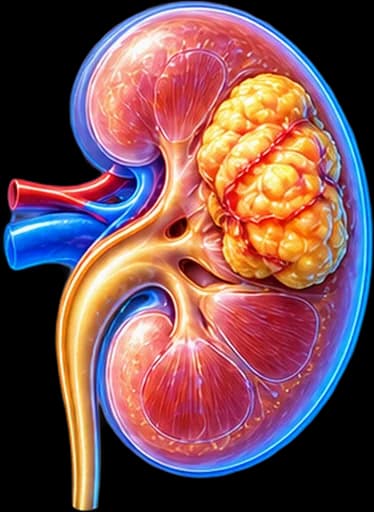
My Clinical Approach to Angiomyolipoma
Angiomyolipoma (AML) is one of the most common incidental kidney findings I see in clinic. The real challenge is not the diagnosis — it is making the right, individualised decision: when surveillance is enough and when intervention is genuinely needed. Over-treating a small, asymptomatic AML is just as problematic as delaying treatment of a tumour with a real bleeding risk.
I place particular emphasis on correctly distinguishing classic AML, fat-poor AML and the rare epithelioid AML, as well as recognising when a finding warrants specialist evaluation for a syndromic (TSC) context.
My approach is based on:
- Individualised bleeding-risk assessment — not a simple application of a single size threshold.
- Active surveillance as the first choice for typical, asymptomatic, sporadic AML.
- A nephron-sparing philosophy in every interventional decision — embolization before surgery where feasible.
- Close collaboration with radiology and, when needed, TSC/genetic counselling specialists.
What Is Renal Angiomyolipoma
Angiomyolipoma (AML) is a benign mesenchymal kidney tumour belonging to the PEComa family (Perivascular Epithelioid Cell tumours). It is classically made up of three histological components, in varying proportions from one tumour to another:
- Blood vessels — often abnormal, thick-walled.
- Smooth muscle fibres.
- Fat tissue.
The differing proportion of these three components matters, because it directly affects how the tumour appears on imaging — a fat-rich AML has a typical, easily recognisable appearance, while a minimal-fat AML can create genuine diagnostic difficulty.
AML is the most common benign kidney tumour
Many AMLs remain entirely asymptomatic throughout a patient’s life and are discovered incidentally on imaging performed for another reason.
Is Angiomyolipoma Cancer?
Classic angiomyolipoma is, as a rule, a benign tumour and is not renal cell carcinoma (RCC).
However, the full picture is more nuanced:
- Some lesions without visible fat (fat-poor AML) can be difficult to distinguish from RCC on imaging.
- Epithelioid AML is a rare, distinct category with potential malignant behaviour — it is not equivalent to classic AML.
Angiomyolipoma vs Renal Cell Carcinoma (RCC)
Angiomyolipoma (AML)
- Usually benign — mesenchymal tumour.
- Often contains macroscopic fat on imaging.
- Treatment is driven mainly by bleeding risk/symptoms.
Renal Cell Carcinoma (RCC)
- Malignant epithelial tumour.
- Different biology and natural history.
- Different oncological management — see our kidney cancer page.
The presence of fat can be particularly useful in diagnosing classic AML, but not every AML contains easily visible fat. For the oncological biology and treatment of renal cell carcinoma, see the dedicated page.
Sporadic Angiomyolipoma, Tuberous Sclerosis (TSC) & LAM
Most angiomyolipomas are sporadic and usually present as a solitary, unilateral lesion — unlike syndromic forms, which can be multiple, bilateral, generally larger, and more prone to growth/bleeding.
Tuberous Sclerosis Complex (TSC)
AML-TSC is associated with the genetic syndrome Tuberous Sclerosis Complex. In this context, AMLs:
- May appear at a younger age.
- Are more often multiple.
- Are more often bilateral.
- May grow larger.
- Carry increased clinical importance and require closer surveillance.
A single AML alone is not sufficient to establish a diagnosis of TSC — full clinical evaluation is required.
Lymphangioleiomyomatosis (LAM)
In a specific clinical context, mainly in women of reproductive age, renal AML can coexist with lymphangioleiomyomatosis (LAM), a rare lung disease. This association can occur both in the setting of TSC and sporadically.
When to Consider a Syndromic/Genetic Context
Greater suspicion of a syndromic association may exist with:
- Young age at presentation.
- Bilateral AML.
- Multiple AML.
- A large overall tumour burden.
- Other associated clinical features.
In these cases, appropriate specialist evaluation is warranted — genetic testing is not recommended for every small, sporadic AML.
Symptoms of Angiomyolipoma
Most small AMLs are asymptomatic and are discovered incidentally. Possible symptoms in larger or more complicated tumours include:
Flank pain / abdominal discomfort
A dull, persistent ache due to pressure or stretching of the renal capsule from the growing tumour.
Haematuria
Blood in the urine due to bleeding within the collecting system.
Palpable mass (rarer)
In very large AMLs or multiple bilateral AMLs, a mass may rarely be felt in the abdomen.
Spontaneous bleeding
In selected cases, sudden internal bleeding — see the next section.
Anaemia
In cases of significant bleeding, a drop in haemoglobin may occur.
These symptoms should not be considered expected in every angiomyolipoma — most small AMLs remain silent.
Bleeding Risk
AMLs contain abnormal blood vessels that, in selected cases, can cause spontaneous retroperitoneal haemorrhage. This risk relates to more factors than tumour size alone — including vascular morphology, the presence of intralesional aneurysms, a prior bleeding episode, and growth rate.
Spontaneous Bleeding — Wunderlich Syndrome
Wunderlich syndrome is the spontaneous rupture of a tumour vessel with bleeding into the renal/perirenal space. It can cause:
- Sudden, severe flank/abdominal pain.
- Weakness.
- Dizziness / syncope.
- A drop in blood pressure (hypotension).
- A fall in haemoglobin.
- Shock in severe cases.
Requires immediate medical evaluation.
Red Flags — When Urgent Evaluation Is Needed
Sudden severe pain in the kidney area together with weakness, dizziness or a drop in blood pressure may indicate bleeding and requires urgent evaluation. If you have a known angiomyolipoma and develop any of these signs, seek medical attention immediately.
Angiomyolipoma Over 4 cm: Does It Always Need Surgery?
Not necessarily. The 4-cm threshold should not be used on its own as an automatic indication for intervention.
The old, simplified rule of “AML >4 cm = surgery” no longer reflects modern clinical practice as a standalone rule. The treatment decision must weigh multiple factors together:
Size and Risk
In general, a larger AML may be associated with an increased bleeding risk, but size alone does not fully predict the risk. Before applying any specific threshold to an individual patient, the decision should be individualised in consultation with your urologist, taking into account the whole clinical and imaging picture.
Intralesional Aneurysms & Growth Rate
Intralesional aneurysms
The presence and size of intralesional aneurysms (dilations of the abnormal vessels within the AML) have been associated with increased bleeding risk in the literature. However, there is no single, universally accepted millimetre cutoff used automatically as the sole criterion for intervention — the assessment is made in the context of the overall imaging by an experienced radiologist/urologist.
Aneurysms are one of the factors used to assess risk — not the sole criterion for a decision to intervene.
Growth rate
Significant growth on follow-up imaging can influence management. There is no universal growth-rate threshold that applies identically to every patient — many sporadic AMLs show little or very slow growth over time, which is one reason surveillance remains a safe option for most typical findings.
Imaging Evaluation
Ultrasound
Classic AML can often appear hyperechoic on ultrasound because of its fat content. However, hyperechogenicity alone does not prove AML — some renal cell carcinomas can have an overlapping appearance, so ultrasound is not a 100% diagnostic test on its own.
CT Scan
Detecting macroscopic fat within an appropriate renal lesion is characteristic of classic AML, always considered alongside the differential diagnosis. CT also helps assess size, vascularity, any bleeding, and the anatomical relationship with kidney structures. Interpretation is always performed by an experienced radiologist, without arbitrary density cutoffs outside specialised radiological evaluation.
MRI
MRI can help with characterisation, fat detection in fat-poor cases, and in patients where CT findings remain indeterminate or where specific follow-up scenarios call for it. It is not a mandatory test for every angiomyolipoma.
Fat-poor / Minimal-fat Angiomyolipoma
Some AMLs contain so little macroscopic fat that they can resemble RCC or another solid kidney tumour on imaging. This creates genuine diagnostic uncertainty.
It cannot be assumed that fat-poor AML can always be reliably distinguished from RCC using CT or MRI alone — these cases require careful, individualised evaluation, and potentially a biopsy (see the next section).
Is a Biopsy Needed for Angiomyolipoma?
No, when imaging is typical for classic AML.
Renal mass biopsy may be considered when:
- The lesion is fat-poor.
- Imaging is indeterminate.
- There is a genuine differential diagnosis with renal cell carcinoma.
- The biopsy result would meaningfully change the treatment strategy.
Biopsy is not a routine investigation for every angiomyolipoma — it is performed according to the principles of current renal mass guidelines.
Epithelioid Angiomyolipoma
Epithelioid AML is a rare variant that differs significantly from classic angiomyolipoma and can have malignant potential, including the possibility of local aggressiveness or, more rarely, metastatic disease.
Depending on histopathological features, it may require:
- Detailed histopathological assessment by an experienced pathologist.
- A different, closer surveillance schedule compared with classic AML.
- Oncological management, depending on the lesion’s characteristics.
Epithelioid AML should not be presented as a simple classic AML — but nor does every angiomyolipoma have malignant potential. It is a distinct, rare category.
When Is Surveillance Alone Enough?
In many patients with an AML that is asymptomatic, small, stable and typical sporadic, the appropriate strategy is Active Surveillance.
What “active surveillance” means
It means a structured programme of planned imaging follow-up, with regular reassessment of symptoms and growth rate — not "ignoring" the finding.
Follow-up frequency
There is no universal schedule (e.g. "ultrasound every 6 months" or "CT every year") that applies identically to every patient. Frequency is individualised based on size, prior growth rate, symptoms, imaging characteristics, whether the AML is sporadic or TSC-associated, kidney function, and the patient’s overall risk profile — always in consultation with your treating physician.
When Is Treatment Needed?
Intervention may be considered when there is:
Size is one factor — not the only one. The treatment decision is based on the overall picture, not a single criterion.
Angiomyolipoma and Pregnancy
The hormonal and haemodynamic changes of pregnancy may be associated with tumour growth and an increased bleeding risk in selected angiomyolipomas.
Counselling should be individualised before pregnancy when a significant AML is present, so that the realistic risk and any need for surveillance or intervention can be discussed.
This does not mean every woman with an angiomyolipoma needs surgery before pregnancy — the decision is individualised.
Reproductive plans can more broadly influence decision-making, especially with larger or higher-risk lesions — without female sex alone being an indication for treatment.
Selective Arterial Embolization
Selective arterial embolization (SAE) can be used in three main scenarios:
- For active bleeding.
- Prophylactically, in selected high-risk AMLs.
- As a kidney-preserving treatment, maintaining as much functional kidney tissue as possible.
The goal of the technique is to occlude the vessels feeding the tumour, control bleeding, and reduce the lesion’s vascularity/volume. It does not always eliminate the tumour completely — it usually shrinks it and reduces its vascularity.
Embolization in acute bleeding
In haemodynamically stable patients with active bleeding, embolization can offer renal-preserving haemorrhage control and often represents an important first treatment option. In unstable/severe cases, the strategy depends on the emergency clinical circumstances and is decided individually by the treating team.
Post-embolization syndrome
After embolization, some patients may temporarily experience:
- Pain.
- Fever.
- Nausea.
- General inflammatory symptoms.
This is known as post-embolization syndrome and usually resolves gradually. The procedure is not without possible, expected effects in the immediate post-procedural period.
Retreatment after embolization
Tumour shrinkage after embolization varies between patients. Recurrence or revascularisation can occur, and in selected patients repeat embolization may be needed. It is not a guaranteed, one-time definitive cure in every case.
Partial Nephrectomy
Nephron-sparing surgery can be used in selected AMLs when:
- Intervention is genuinely needed.
- Embolization is not suitable or has failed.
- There is diagnostic uncertainty regarding benignity.
- Anatomy or other clinical factors favour a surgical approach.
Main goal: removal of the lesion while preserving the maximum possible functional kidney tissue.
Radical Nephrectomy & Thermal Ablation
Radical nephrectomy
Radical nephrectomy is not a routine treatment for a typical angiomyolipoma. It may only be required in selected, difficult situations, such as:
- A massive tumour occupying most of the kidney.
- Severe destruction of the kidney parenchyma.
- Uncontrolled bleeding.
- Inability to preserve a functional kidney.
- Significant diagnostic/oncological concern.
Thermal ablation
In appropriate, small lesions, thermal ablation (cryoablation, radiofrequency ablation) may be discussed for selected patients. It is not a first-line treatment for every angiomyolipoma — suitability is assessed individually.
Surveillance vs Embolization vs Surgery
An indicative comparison of the main strategies — without rigid size criteria:
| Strategy | When considered |
|---|---|
| Active Surveillance | Asymptomatic, low-risk AML |
| Selective Embolization | Bleeding, or a selected high-risk lesion |
| Partial Nephrectomy | Selected cases / diagnostic uncertainty / embolization not suitable |
| Thermal Ablation | Selected lesions / selected patients |
| Radical Nephrectomy | Rare, when kidney preservation is not feasible |
TSC-associated AML & mTOR Inhibitors
AMLs associated with tuberous sclerosis (TSC) have a distinct biology related to the mTOR (mechanistic Target Of Rapamycin) pathway — its overactivity drives uncontrolled cell growth in the tissues affected by the syndrome.
In selected patients with TSC-associated AML, systemic treatment with an mTOR inhibitor may be used, according to approved indications and current guidelines.
Everolimus
Everolimus is the main mTOR inhibitor used in the appropriate TSC-associated AML context — always under specialised medical monitoring, due to possible systemic adverse effects that require regular follow-up with the treating physician. It is not a self-directed therapy and does not apply to every sporadic angiomyolipoma.
mTOR-inhibitor therapy is used in a specific, appropriate TSC-associated context — it is not standard treatment for sporadic AML.
Preserving Kidney Function
The treatment strategy, wherever possible, should protect functional kidney parenchyma and long-term kidney function.
This is particularly important in:
- Bilateral AML.
- Patients with TSC.
- Multiple lesions.
- A solitary kidney.
- Chronic kidney disease (CKD).
Bilateral/multifocal AML requires particular attention to nephron preservation, and in these cases a syndromic (TSC) context is considered where relevant.
Common Clarifications
AML and haematuria
AML can cause haematuria, but not every episode of haematuria in a patient with a known AML should be automatically attributed to the tumour. Haematuria may need a full urological evaluation — see our page on blood in urine.
AML and pain
Flank discomfort can occur with larger lesions, but chronic back pain has many other possible causes — pain alone does not prove an AML-related complication.
AML and hormones
There is evidence of possible hormonal sensitivity in some AMLs, but it is not established that oral contraceptives "cause" AML or that pregnancy always increases every AML — the relationship is assessed individually based on currently available evidence.
AML in men
Angiomyolipoma is not an exclusively female condition — it also occurs in men. In these cases, the possibility of a syndromic context is equally evaluated, where relevant.
Differential diagnosis
The differential diagnosis can include renal cell carcinoma, oncocytoma, retroperitoneal lipomatous lesions, and other fat-containing renal or retroperitoneal masses, depending on the imaging appearance.
The Patient’s Journey
Incidental finding of a renal mass
Discovered on ultrasound or CT performed for another reason.
Ultrasound / CT / MRI
Further imaging to characterise the lesion.
Is there characteristic macroscopic fat?
A key question in distinguishing typical AML from indeterminate findings.
Typical classic AML
Assessment of symptoms, size, growth rate, vascular risk and patient characteristics → low risk: active surveillance; higher risk: embolization or nephron-sparing treatment.
Indeterminate / fat-poor lesion
Further imaging ± biopsy, when the result would change management, to distinguish AML from RCC.
This is an educational overview, not a rigid management algorithm — every case is discussed individually with your urology team.
Follow-up & Prognosis
Classic angiomyolipoma generally has a favourable prognosis — it is not a malignancy and does not metastasise. The main clinical risk relates to the possibility of bleeding, not the presence of the tumour itself.
In summary
Appropriate follow-up is individualised per patient. After successful treatment (embolization or surgery, where needed), quality of life remains good in most cases. Epithelioid AML is an exception and requires its own, closer surveillance programme.
Frequently Asked Questions (FAQ)
What is a renal angiomyolipoma?
It is a benign mesenchymal kidney tumour (part of the PEComa family) made up of blood vessels, smooth muscle and fat tissue in varying proportions. It is the most common benign solid kidney tumour and is usually found incidentally on ultrasound or CT.
Is angiomyolipoma cancer?
Classic angiomyolipoma is, as a rule, a benign tumour and is not renal cell carcinoma. Some lesions without visible fat (fat-poor AML) can be difficult to distinguish from RCC on imaging, and the rare epithelioid AML variant has malignant potential — so evaluation should always be individualised.
Can angiomyolipoma turn into cancer?
Classic angiomyolipoma does not transform into renal cell carcinoma. The exception is the rare epithelioid AML, a distinct histological entity with its own malignant potential — it is not a "transformation" of a classic AML.
Does an angiomyolipoma over 4 cm always need surgery?
Not necessarily. The historical 4-cm threshold should no longer be used as an automatic rule for intervention. The decision takes into account symptoms, growth, vascular characteristics, prior bleeding, tuberous sclerosis complex (TSC), pregnancy/reproductive plans and patient preference.
When is there a real risk of bleeding?
Bleeding risk is multifactorial — related to size, but also to vascular morphology, the presence of intralesional aneurysms, prior bleeding episodes and growth rate. Size alone does not fully predict the risk.
Can an angiomyolipoma "burst"?
In selected cases, one of the tumour’s abnormal vessels can rupture, causing internal (retroperitoneal) bleeding — not a "burst kidney". This is known as Wunderlich syndrome and is linked to red flags that require immediate evaluation.
How is angiomyolipoma diagnosed?
Mainly through imaging — ultrasound, CT and, where needed, MRI. Detecting macroscopic fat within an appropriate renal lesion is characteristic of classic AML, always considered alongside the differential diagnosis.
Is a CT or MRI always required?
CT is usually the investigation of choice. MRI is particularly helpful in indeterminate cases, fat-poor lesions, or when more detailed characterisation is needed — it is not mandatory for every angiomyolipoma.
What is fat-poor angiomyolipoma?
It is an AML variant with minimal or no detectable macroscopic fat on imaging, which can make it resemble RCC or another solid kidney tumour. This creates genuine diagnostic uncertainty and often requires further assessment.
When is a biopsy needed for angiomyolipoma?
Not when imaging is typical for classic AML. Biopsy may be considered when the lesion is fat-poor, when imaging is indeterminate, when there is a genuine differential diagnosis with RCC, or when the result would meaningfully change management.
When is surveillance alone enough?
In many patients with an asymptomatic, small, stable, typical sporadic AML, active surveillance (a structured imaging follow-up programme with reassessment of symptoms and growth) is the appropriate strategy — not "ignoring" the finding.
What is selective arterial embolization?
It is an interventional radiology technique that selectively occludes the vessels feeding the tumour, aiming to control or prevent bleeding and reduce the tumour’s vascularity/volume, while preserving as much functional kidney tissue as possible.
When is partial nephrectomy needed?
When intervention is genuinely needed but embolization is not suitable or has failed, when there is a real differential diagnosis with malignancy, or when anatomy/clinical factors favour a surgical approach — with the goal of removing the lesion while preserving the maximum functional kidney tissue.
What is the relationship between angiomyolipoma and tuberous sclerosis (TSC)?
In the context of Tuberous Sclerosis Complex, AMLs tend to appear at a younger age, are more often multiple and bilateral, and may grow larger — with increased clinical significance. A single AML alone does not establish a diagnosis of TSC.
Does pregnancy affect angiomyolipoma?
The hormonal and haemodynamic changes of pregnancy may be associated with growth and an increased bleeding risk in selected AMLs. Counselling should be individualised before pregnancy when a significant AML is present — this does not mean every woman with an AML needs surgery before having children.
Related Topics
Book Your Appointment in Rhodes
The need for angiomyolipoma treatment is not determined by diameter alone. Symptoms, growth, vascular characteristics, bleeding risk and individual patient factors determine whether surveillance, embolization or a kidney-preserving surgical approach is preferable.
References – Sources
- EAU Guidelines on Renal Cell Carcinoma 2024 (angiomyolipoma section) — uroweb.org
- Fernández-Pello S, et al. European Association of Urology Guidelines on Diagnosis and Conservative Management of Renal Angiomyolipoma. Eur Urol 2017;71:233–234.
- Bissler JJ, et al. Everolimus for angiomyolipoma associated with tuberous sclerosis complex or sporadic lymphangioleiomyomatosis (EXIST-2). N Engl J Med 2013;368:291–301.
- Krueger DA, Northrup H, on behalf of the International Tuberous Sclerosis Complex Consensus Group. Tuberous Sclerosis Complex Surveillance and Management Recommendations. Pediatr Neurol 2013;49:255–265.
- Siegel RL, et al. Cancer statistics 2024. CA Cancer J Clin 2024.
Medical Editorial

Medically reviewed by Dr. Marinos Vasilas – Urologist – Andrologist
Dr. Marinos Vasilas runs a private urology practice in Rhodes, specialising in the management of renal masses, benign kidney tumours, and nephron-sparing surgical treatment of angiomyolipomas.
View full profile


