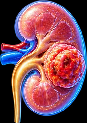
My Clinical Approach to Renal Cell Carcinoma
RCC is the pathology that has changed most in urology over the past decade. Laparoscopic and robotic partial nephrectomy has enabled preservation of renal function without compromise on oncological outcomes. Simultaneously, the introduction of immunotherapy in metastatic disease has created realistic prospects for long-term survival.
I do not manage all RCCs the same way. The histological subtype, WHO/ISUP grade, presence of sarcomatoid differentiation, and molecular profile in metastatic disease now determine both the surgical strategy and the choice of systemic therapy.
My approach follows EAU Guidelines RCC 2024:
- Partial nephrectomy as first choice for T1 (≤7 cm) where technically feasible.
- Renal mass biopsy before ablation or in ambiguous findings.
- IMDC classification before every systemic therapy decision.
- Adjuvant immunotherapy (pembrolizumab) in high-risk patients after nephrectomy.
What is Renal Cell Carcinoma
Renal cell carcinoma (RCC) is a malignant tumour arising from the epithelial cells of the renal tubule. It accounts for ~90% of malignant kidney tumours in adults.
The WHO 2022 classification recognises >20 histological subtypes, each with distinct molecular characteristics. This heterogeneity explains why two patients with "kidney cancer" may require completely different treatment approaches.
Important distinction: not every kidney tumour is RCC. Benign tumours such as oncocytoma (~3–7%) and angiomyolipoma (~1–3%) may resemble RCC on imaging. In equivocal cases, renal mass biopsy is key for differential diagnosis.
Key Statistics
3rd most common urological cancer in men. Peak incidence in the 6th–7th decade, male-to-female ratio ~2:1. 5-year survival >95% at T1a; <20% in untreated metastatic disease — substantially improved with modern immunotherapy.
Epidemiology & Risk Factors
RCC accounts for ~3% of all malignancies in adults. Although most cases are sporadic, 5–8% are associated with hereditary syndromes — with significant therapeutic and preventive implications for the family:
Smoking
The most important modifiable risk factor — increases risk by ~50% and is associated with more aggressive ccRCC.
Obesity
A clear dose-dependent relationship — each 5 kg/m² BMI increase raises risk by ~25%, possibly through hyperinsulinaemia and elevated oestrogen levels.
Arterial Hypertension
An independent risk factor — both hypertension itself and certain antihypertensives (diuretics) have been associated with increased risk.
Chronic Kidney Disease / Dialysis
Dialysis patients have 10–20-fold increased risk due to acquired cystic kidney disease.
Hereditary Syndromes
Von Hippel-Lindau (ccRCC), Birt-Hogg-Dubé (chRCC/oncocytoma), HLRCC (aggressive papillary type 2), HPRC (papillary type 1), tuberous sclerosis.
Occupational Exposure
Trichloroethylene, cadmium, asbestos — among substances associated with increased RCC risk.
Symptoms & Clinical Presentation
RCC follows a prolonged silent course — >50–60% are diagnosed incidentally on ultrasound or CT performed for another reason. The classic triad (haematuria–pain–palpable mass) today occurs in <10% and usually indicates advanced disease.
Haematuria
Sudden, painless, macroscopic haematuria is the classic finding — present in ~40% of symptomatic patients. Any unexplained macroscopic haematuria requires urological investigation.
Loin / Flank Pain
Dull, persistent pain in the loin or flank area. Acute colicky pain may occur with an obstructing blood clot in the ureter or intrarenal haemorrhage.
Palpable Abdominal Mass
A rare finding today — palpable as a painless resistance in the epigastrium/flank, mainly in thin patients or with large tumours. Imaging evaluation is mandatory.
Paraneoplastic Syndromes
Present in ~20% — hypertension, hypercalcaemia, polycythaemia, unexplained fever, Stauffer syndrome (hepatic dysfunction without metastases). May be the first manifestation of disease.
When to Seek Immediate Urological Evaluation
- Sudden, painless, macroscopic haematuria at any age — urgent urological assessment.
- Incidental finding of a renal mass on ultrasound — referral to a urologist.
- Unexplained weight loss + fatigue + flank pain in a smoker or obese patient.
- Lower limb oedema + varicocele (especially right-sided) appearing suddenly — possible renal vein thrombosis.
Diagnosis & Staging
Diagnosis relies primarily on imaging. Unlike many other cancers, pre-operative biopsy is not always required — but is critical in specific situations.
Imaging Investigations
Renal Ultrasound
Usually the first examination to detect the tumour — often incidentally. Insufficient alone for complete characterisation of a renal mass.
Triphasic CT of the Kidneys
Investigation of choice. Characterises the mass, assesses renal vein/inferior vena cava, lymph nodes and metastases. Bosniak classification for cystic masses.
Renal MRI
Alternative for contrast allergy or venous thrombus extent assessment — superior in mapping the extent of venous involvement.
Renal Mass Biopsy
Indicated before ablation, active surveillance, in a history of another cancer, atypical masses, and before initiating systemic therapy in metastatic disease.
TNM Staging 2017 & WHO/ISUP Grade
Tumour ≤4 cm — confined to kidney
Tumour 4–7 cm — confined to kidney
Tumour >7 cm — confined to kidney
Extension into renal vein / perinephric fat / inferior vena cava
Invasion beyond Gerota's fascia
WHO/ISUP Grade 1–4: assesses nuclear aggressiveness. Presence of sarcomatoid or rhabdoid differentiation confers adverse prognosis regardless of stage.
Histological Subtypes & Molecular Biology
Understanding the histological subtype is critical for treatment selection:
Clear Cell RCC (ccRCC) — ~75%
The most common subtype. Characterised by loss-of-function mutation of VHL gene (chromosome 3p25), activating the HIF/VEGF axis. Additional mutations in PBRM1, BAP1, SETD2 further influence prognosis. Best response to immunotherapy and VEGF/TKI agents.
Papillary RCC (pRCC) — ~15%
Type 1: indolent, often multifocal, MET mutations — hereditary in HPRC. Type 2: more aggressive, FH mutations — hereditary in HLRCC, lower immunotherapy response rates. Treatment algorithm differs from ccRCC.
Chromophobe RCC (chRCC) — ~5%
The best prognosis among major subtypes. Often associated with Birt-Hogg-Dubé syndrome. In metastatic disease, resistance to immunotherapy is notable — mTOR inhibitors may be a preferred option.
Rare Subtypes
Collecting duct (Bellini): extremely aggressive, poor prognosis. Translocation (Xp11): young adults, TKI resistance. Medullary carcinoma: exclusively in sickle cell disease, highly aggressive. Each subtype requires individualised management.
Treatment Options
Treatment choice depends on stage, histological subtype, grade, patient performance status and — in metastatic disease — on IMDC risk classification:
Localised Disease (T1–T2, N0M0)
Partial nephrectomy (laparoscopic/robotic/open) for T1 where feasible — gold standard. Radical nephrectomy for larger or centrally located tumours. Thermal ablation (RFA/cryoablation) for SRM ≤3 cm in non-surgical candidates — after prior biopsy.
Locally Advanced Disease (T3–T4/N+)
Radical nephrectomy with thrombectomy of renal vein or inferior vena cava. Extended lymph node dissection in N+ disease. Adjuvant therapy with pembrolizumab (KEYNOTE-564) in high-risk patients (pT2G4/pT3+/N+ or M1 after nephrectomy).
Metastatic RCC (mRCC) — First-line Therapy
IMDC favourable risk: Pembrolizumab + lenvatinib (CLEAR), pembrolizumab + axitinib (KEYNOTE-426) or sunitinib as alternative. IMDC intermediate/poor risk: Nivolumab + ipilimumab (CheckMate 214), nivolumab + cabozantinib (CheckMate 9ER), pembrolizumab + axitinib/lenvatinib. Cytoreductive nephrectomy in selected IMDC favourable/intermediate patients.
Key principle: RCC is resistant to conventional chemotherapy. Individualised systemic therapy based on histology, molecular profile and IMDC risk is the modern standard of care.
Surgical Management
Surgical resection remains the only potentially curative treatment in localised disease:
Partial Nephrectomy (laparoscopic/robotic)
Removal of the tumour only with a surgical margin. Standard of care for T1 (≤7 cm) where feasible. Preserves renal mass and reduces long-term cardiovascular risk. Warm ischaemia time <25 minutes.
Radical Nephrectomy (laparoscopic/robotic)
Removal of the entire kidney — preferred for T2, centrally located T1 tumours that are technically complex, or when contralateral renal function is sufficient.
Radical Nephrectomy with Thrombectomy
For T3b/T3c tumours with thrombus extending into the renal vein or inferior vena cava. Requires a multidisciplinary team — vascular ± cardiac surgeon if thrombus reaches the right atrium.
Cytoreductive Nephrectomy
Removal of the primary tumour in mRCC. In carefully selected IMDC favourable/intermediate patients may improve response to systemic therapy. Not routinely recommended in poor IMDC risk patients.
Active surveillance without immediate intervention is an option for SRM (≤4 cm) in selected patients — provided growth rate is <5 mm/year and no concerning features are present.
Follow-up & Prognosis
Surveillance intensity after nephrectomy is guided by the Leibovich score (low/intermediate/high recurrence risk) and UISS prognostic score:
- Chest-abdominal CT: every 6 months for the first 3 years, then annually (high risk).
- Renal function (creatinine, eGFR, urinalysis): 4–6 weeks post-operatively — then lifelong annual monitoring.
- Blood pressure: systematic monitoring — increased hypertension risk after nephrectomy.
- On adjuvant immunotherapy: clinical assessment at each treatment cycle.
- Genetic testing where hereditary syndrome is suspected (young age, bilateral/multifocal disease, positive family history).
Prognosis by Stage
T1a (≤4 cm): 5-year survival >95% — excellent prognosis.
T1b–T2: 5-year survival 80–90%.
T3: 5-year survival 50–70% — depending on extent of venous involvement.
T4/N+/M+: Variable — continuously improving with modern immunotherapy.
Frequently Asked Questions (FAQ)
How does renal cell carcinoma differ from general "kidney cancer"?
Renal cell carcinoma (RCC) is the predominant subtype — accounting for approximately 90% of malignant kidney tumours in adults. The term "kidney cancer" also includes urothelial carcinoma of the renal pelvis and, rarely, lymphoma or metastases. RCC has its own distinct biology, staging system, and treatment algorithm.
What are the main histological subtypes of RCC?
Clear cell RCC (ccRCC, ~75%), papillary types 1 and 2 (pRCC, ~15%), and chromophobe RCC (chRCC, ~5%) are the main subtypes. Rare subtypes include collecting duct (Bellini), translocation (Xp11), and medullary carcinoma (in sickle cell disease). Histological classification directly guides immunotherapy and TKI treatment selection.
How important is the nuclear grade in RCC?
Critical. The WHO/ISUP grading system (grades 1–4, replacing the Fuhrman system) combined with sarcomatoid/rhabdoid differentiation, necrosis, and vascular invasion determines surveillance intensity and the indication for adjuvant therapy after nephrectomy.
What is IMDC classification in metastatic RCC?
The IMDC (International Metastatic RCC Database Consortium) uses 6 criteria (time from diagnosis to treatment <1 year, haemoglobin level, corrected calcium, neutrophil count, platelet count, Karnofsky PS) to classify patients into favourable, intermediate, or poor risk — guiding selection of first-line systemic therapy.
When is a renal mass biopsy indicated?
Biopsy is not routine when imaging features are typical and the decision is upfront surgery. It is indicated before ablation, active surveillance, in patients with a history of another cancer, atypical masses, and before initiating systemic therapy in metastatic disease.
I have metastatic ccRCC — are there effective treatments?
Yes. Modern immunotherapy combinations (pembrolizumab + axitinib, pembrolizumab + lenvatinib, nivolumab + ipilimumab, nivolumab + cabozantinib) have significantly extended overall survival. In oligometastatic disease, local therapy (metastasectomy, SBRT) is also considered. Treatment is individualized based on IMDC risk and histology.
What is cytoreductive nephrectomy?
Removal of the primary kidney tumour in a patient with distant metastases. In carefully selected patients (IMDC favourable-intermediate risk, PS 0-1) it may improve response to systemic therapy. It is not routinely recommended in poor-risk IMDC patients.
How long does follow-up last after nephrectomy?
At least 5 years, with intensity guided by Leibovich score (low/intermediate/high recurrence risk). High risk: chest-abdominal CT every 6 months for the first 3 years, then annually. Renal function monitoring (creatinine, eGFR, proteinuria, blood pressure) is lifelong.
Related Topics
Book Your Appointment in Rhodes
Incidental renal mass finding, haematuria, or concern about a hereditary syndrome? Early urological assessment is decisive.
References – Sources
- EAU Guidelines on Renal Cell Carcinoma 2024 — uroweb.org
- WHO Classification of Urinary and Male Genital Tumours, 5th edition, 2022.
- Motzer RJ, et al. Nivolumab plus Ipilimumab versus Sunitinib (CheckMate 214). N Engl J Med 2018;378:1277.
- Powles T, et al. Pembrolizumab after surgery for RCC (KEYNOTE-564). N Engl J Med 2021;385:683.
- Motzer R, et al. Lenvatinib plus Pembrolizumab or Everolimus for Advanced RCC (CLEAR). N Engl J Med 2021;384:1289.
- Choueiri TK, et al. Cabozantinib plus Nivolumab in mRCC (CheckMate 9ER). N Engl J Med 2021;384:829.
Medical Review

Dr. Marinos Vasilas, Urologist – Andrologist
Dr. Marinos Vasilas runs a private urology practice in Rhodes with expertise in kidney tumour surgery, nephron-sparing surgery, and metastatic RCC management with modern systemic therapies. He follows the EAU Guidelines 2024 on renal cell carcinoma.
View full profile


