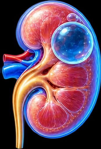
My Clinical Approach to Kidney Cysts
Kidney cysts are among the most common incidental findings I see in clinic — and they often cause disproportionate anxiety compared with the actual clinical risk. The challenge is not the cyst itself, but making the correct distinction: is this genuinely a simple cyst, or a complex cystic mass that needs further evaluation?
I place particular emphasis on accurate Bosniak classification, avoiding unnecessary surveillance for typical simple cysts, and promptly identifying the few cases that genuinely need closer follow-up or intervention.
My approach is based on:
- A clear distinction between simple cysts, complex cystic lesions and parapelvic cysts.
- Bosniak classification based on imaging features — not size alone.
- An individualised surveillance or treatment strategy depending on category.
- A nephron-sparing philosophy whenever intervention is genuinely needed.
What Is a Kidney Cyst
A kidney cyst is a fluid-filled cavity within or on the kidney parenchyma. It is a very common incidental imaging finding, typically seen on ultrasound or CT performed for another reason.
It is important not to confuse different entities:
Simple renal cyst
A benign cavity with simple fluid, without suspicious features.
Complex cystic renal mass
A cystic lesion with additional features (septa, wall thickening, enhancement) requiring Bosniak classification.
Parapelvic/peripelvic cyst
A cyst near the renal sinus/pelvis — different from obstruction/hydronephrosis.
Cystic renal tumour
A neoplasm with a cystic appearance — must be distinguished from a simple/complex cyst.
“I Was Told I Have a Kidney Cyst” — Should I Worry?
In most cases, when it is a typical simple cyst, no.
What matters is clarifying:
Is It Cancer? Can a Simple Cyst Turn Into Cancer?
Simple kidney cyst ≠ kidney cancer.
A typical simple renal cyst is not considered a lesion that “turns into” renal cell carcinoma. The real clinical issue is whether the lesion was genuinely a simple cyst from the start, or a complex cystic renal mass that required different characterisation from the outset.
A complex cyst does not automatically mean cancer, but it requires proper imaging classification (see the Bosniak section below) before deciding on surveillance or treatment. For the oncological biology and treatment of renal cell carcinoma and kidney cancer more broadly, see the dedicated pages.
Simple Renal Cyst
A typical simple cyst usually has the following characteristics:
- A thin, smooth wall.
- Simple, homogeneous fluid content.
- No solid component.
- No suspicious enhancement after contrast.
- No nodules.
A typical simple cyst is not a precancerous condition.
What Does a Complex Kidney Cyst Mean?
A cystic lesion is classified as complex when, depending on the category, it may show:
Complex cyst ≠ cancer. The degree of complexity determines the Bosniak category, which in turn guides the decision between surveillance and treatment.
What Is the Bosniak Classification?
The Bosniak classification is used to characterise cystic renal masses and estimate malignancy risk. It is based on imaging features — particularly contrast enhancement, wall/septal thickness and regularity, and the presence of solid/nodular components — not on size alone.
Bosniak I
A simple, benign cyst without suspicious features. Usually does not need oncological follow-up.
Bosniak II
A minimally complex, benign category — may have a few thin septa or other benign features per current criteria. Usually no further follow-up.
Bosniak IIF
The “F” stands for follow-up. It does not mean cancer. It requires structured imaging surveillance — progression to more complex features matters more than a simple increase in diameter.
Bosniak III
An indeterminate, higher-risk cystic lesion. A significant proportion may be malignant, but Bosniak III ≠ a certain cancer diagnosis. Modern management may include active surveillance, surgery, or other individualised strategies depending on patient/tumour factors.
Bosniak IV
A highly suspicious cystic lesion, usually with enhancing nodular/solid components. It does not equal 100% certainty of cancer in every case. Strategy is based on size, patient age, comorbidity, kidney function, surgical feasibility and the overall oncological context.
Bosniak Summary Table
| Bosniak | General meaning | Typical approach |
|---|---|---|
| I | Simple cyst | No oncological follow-up needed |
| II | Benign / minimally complex | Usually no further follow-up |
| IIF | Needs follow-up | Structured imaging surveillance |
| III | Indeterminate / higher risk | Surveillance or treatment case by case |
| IV | Highly suspicious | Oncological assessment / treatment in suitable patients |
Based on the current updated Bosniak classification (2019). Interpretation is always made by an experienced radiologist/urologist, in the context of the overall clinical picture.
Imaging: Ultrasound, CT, MRI, CEUS
Ultrasound
Very useful for identifying a simple cyst, initial evaluation, and — for selected lesions — follow-up. A typical simple cyst may show anechoic content, a thin wall and posterior acoustic enhancement. However, if the cyst is not clearly simple, it often needs further characterisation.
Contrast-Enhanced CT
Assesses the wall, septa, calcifications, enhancement and nodularity, and is used for Bosniak classification per current criteria. Not every kidney cyst needs a CT scan — only those that are not clearly simple on ultrasound.
MRI
Can be particularly helpful for an indeterminate cyst, a complex lesion, equivocal enhancement, selected Bosniak classification situations, or where there are contrast/radiation considerations. It is not mandatory for every cyst.
Contrast-Enhanced Ultrasound (CEUS)
CEUS can have a role in characterising selected cystic renal lesions, depending on availability and clinical context. It is not universally available and does not always replace CT or MRI — its role is decided by the treating physician on a case-by-case basis.
Does the Size of the Cyst Matter?
Size alone does not determine whether a cyst is benign or malignant.
A large simple cyst can remain entirely benign. Conversely, a smaller complex cyst may need closer attention — because of its imaging features, not its size.
“I Have a 5, 7 or 10 cm Cyst — Do I Need Surgery?”
Not based on size alone.
If it is genuinely a simple cyst, treatment is mainly considered when it causes pain, obstruction, significant compression or other clinically attributable problems — a large simple cyst is not automatically malignant.
Symptoms & Pain
Most simple cysts cause no symptoms. With larger or symptomatic cysts, the following may occur:
In a patient with a known renal cyst and chronic flank pain, the cyst should not automatically be assumed to be the cause — musculoskeletal causes, stone disease, other renal pathology or gastrointestinal causes must be considered, depending on the clinical context.
Haematuria & Haemorrhagic Cyst
A large or haemorrhagic cyst may, in selected cases, be associated with haematuria. However, haematuria should not automatically be attributed to a known simple kidney cyst — full urological evaluation is needed. See our page on blood in urine.
Haemorrhagic cyst
Bleeding within a cyst can change its imaging appearance — creating internal echoes, areas of higher density, or an appearance of apparent complexity that may need further characterisation. A haemorrhagic cyst should not be automatically equated with a malignant cyst — it often simply requires careful reassessment or surveillance.
Infected Cyst
Infection of a simple cyst is relatively uncommon. It can cause fever, pain and inflammatory findings, and may need specialist evaluation. This is a different entity from pyelonephritis (infection of the kidney parenchyma/pelvis) — it should not be confused with it.
Parapelvic Cyst vs Hydronephrosis
Parapelvic cysts are located near the renal sinus/pelvis. They can:
- Mimic hydronephrosis on ultrasound.
- When large, compress the collecting system.
Parapelvic cyst
- A discrete cystic structure.
- Outside the collecting system.
Hydronephrosis
- Dilatation of the collecting system.
- Usually due to obstruction or another urinary drainage issue.
Parapelvic cyst ≠ hydronephrosis. A very large or strategically located parapelvic cyst can, more rarely, cause compression/obstruction — but this does not happen with every parapelvic cyst.
Kidney Cyst vs Polycystic Kidney Disease (ADPKD)
One or a few simple kidney cysts do not mean polycystic kidney disease.
Autosomal Dominant Polycystic Kidney Disease (ADPKD) is a different entity:
- An inherited disease.
- Usually multiple, bilateral cysts.
- Can affect kidney function over time.
- Can have extrarenal manifestations.
ADPKD often needs nephrology involvement. We do not cover ADPKD in detail here — if you have multiple bilateral cysts or a family history, discuss this with your physician for appropriate referral. For more information, see our page on Polycystic Kidney Disease (PKD).
Urologist or Nephrologist?
Urologist — particularly for:
- A complex renal cyst.
- A Bosniak-classified lesion.
- Suspected cystic tumour.
- A symptomatic cyst.
- Obstruction.
- Surgical/interventional treatment.
Nephrologist — particularly when there is:
- Polycystic kidney disease.
- Chronic kidney disease.
- Impaired kidney function.
- Systemic renal disease.
When Is Surveillance Needed?
Bosniak I / appropriate Bosniak II
Usually do not need oncological follow-up.
Bosniak IIF
Needs a structured, planned imaging surveillance programme.
Bosniak III / IV
Management is individualised based on the overall clinical context.
There is no single universal imaging schedule (e.g. a fixed number of months or years) that applies identically to every patient — the frequency and duration of follow-up are determined by your treating physician, based on current evidence and the lesion’s characteristics.
What Does a Change in Bosniak Category Mean?
Progression to greater wall/septal complexity, nodular components or enhancement characteristics can be more significant than a simple increase in size. Size alone is not the definition of progression.
Is a Biopsy Needed for a Complex Cyst?
Biopsy of cystic renal masses has specific indications and potential limitations — in some cystic lesions the diagnostic yield of biopsy may be lower compared with solid tumours.
It is not a routine investigation for every Bosniak lesion. The decision to biopsy is individualised by your treating physician.
Treating a Symptomatic Simple Cyst
A confirmed simple cyst is usually treated only when it causes clinically attributable problems, such as:
- Persistent, attributable pain.
- Obstruction.
- Significant compression of neighbouring structures.
- Other selected complications.
Not simply because it exists or is large.
Percutaneous Aspiration
Percutaneous aspiration can decompress the cyst and can have a diagnostic/therapeutic role in selected circumstances. However, simple aspiration without sclerotherapy can carry a significant risk of recurrence.
Sclerotherapy
Aspiration can be combined with sclerotherapy in selected symptomatic simple cysts. Factors considered include benefit, recurrence risk, potential complications, and patient/lesion suitability — it is not a guaranteed definitive solution in every case.
Laparoscopic Cyst Decortication
Laparoscopic cyst decortication (deroofing/marsupialisation) may be used for a large symptomatic simple cyst, recurrence after less invasive treatment, or an anatomically suitable lesion. It is not a routine treatment for asymptomatic cysts.
Cystic Kidney Cancer & Partial Nephrectomy
Some renal cell carcinomas can present as cystic renal masses. This is why imaging morphology and Bosniak classification carry significant importance — without implying that every complex cyst is cancer. See our pages on kidney cancer and renal cell carcinoma.
Partial nephrectomy is not a treatment for a simple benign cyst. It can, however, have a role for a suspicious cystic renal mass, selected Bosniak III/IV lesions, or cystic RCC, where oncological excision is required.
Simple Cyst vs Complex Cyst vs Cystic Malignancy
| Simple Cyst | Complex Cyst | Cystic Malignancy | |
|---|---|---|---|
| Content | Simple fluid | May have septa/complexity | May have enhancing solid/nodular components |
| Wall | Thin | May be thicker/irregular | Suspicious features |
| Enhancement | Not suspicious | Depends on Bosniak category | Often suspicious |
| Cancer risk | Effectively benign | Varies by Bosniak category | Malignant |
| Management | Usually none | Surveillance or treatment by category | Oncological management |
Hypertension & Kidney Function
Observational studies have found associations between simple renal cysts and hypertension, but an association does not mean proven causation in every patient. Removing a simple cyst should not be regarded as a general treatment for hypertension.
Isolated simple cysts usually do not cause significant impairment of kidney function. Different clinical scenarios include extensive bilateral cystic disease, ADPKD, obstruction, or other renal pathology.
Differential Diagnosis
A cystic/anechoic renal finding may need to be differentiated from:
This list is informational and not a self-diagnosis tool.
The Patient’s Journey
Incidental finding of a "cyst" on the kidney
Discovered on ultrasound or CT performed for another reason.
Ultrasound
Initial assessment of characteristics.
Does it have the features of a typical simple cyst?
The key question.
Yes → Simple cyst
Usually reassurance, no oncological treatment.
No / uncertain → Further imaging
Contrast-enhanced CT / MRI ± CEUS where appropriate.
Bosniak classification
Bosniak I/II → benign pathway. Bosniak IIF → imaging surveillance. Bosniak III/IV → individualised urological/oncological assessment.
Surveillance or nephron-sparing treatment
Depending on the specific case.
This is an educational overview, not a rigid management algorithm — every case is discussed individually with your urologist.
Follow-up & Prognosis
A typical simple renal cyst has an excellent prognosis — it is not a malignancy and does not progress into cancer. The main clinical goal is accurate initial classification, avoiding both unnecessary surveillance of typical simple cysts and delayed evaluation of genuinely complex lesions.
In summary
Most kidney cysts do not need treatment. When a lesion is complex, shows enhancement, or is classified as Bosniak IIF–IV, management is individualised based on imaging characteristics, kidney function, age and the overall clinical context.
Frequently Asked Questions (FAQ)
What is a cyst on the kidney?
It is a fluid-filled cavity within or on the kidney parenchyma. It is the most common incidental imaging finding in the kidney, and the large majority of cases are benign.
Is a kidney cyst cancer?
In most cases, no. A typical simple cyst is not cancer. The real clinical issue is whether the lesion is genuinely a simple cyst or a complex cystic renal mass that needs further imaging characterisation.
Can a simple cyst turn into cancer?
A typical simple renal cyst is not considered a lesion that "turns into" renal cell carcinoma. The issue is not the evolution of a genuine simple cyst, but whether the initial assessment correctly classified the lesion as simple.
What does a complex cyst mean?
It means the cystic lesion has additional features beyond simple fluid — such as septa, a thicker or irregular wall, calcifications or enhancement after contrast. It does not automatically mean malignancy, but it requires Bosniak classification.
What is the Bosniak classification?
It is a system (categories I to IV) for classifying cystic renal masses based on imaging features, primarily contrast enhancement, that helps estimate malignancy risk and guide the decision between surveillance and treatment.
What does Bosniak IIF mean?
The "F" stands for follow-up. It does not mean a cancer diagnosis — it means the lesion has certain features that warrant a structured imaging surveillance programme to see whether it remains stable or progresses.
Is Bosniak III cancer?
Not necessarily. Bosniak III is an indeterminate, higher-risk cystic lesion — a significant proportion may be malignant, but Bosniak III does not equal a certain cancer diagnosis. Management is individualised.
Is Bosniak IV always cancer?
Bosniak IV is a highly suspicious cystic lesion, usually with enhancing nodular or solid components, but it does not equate to absolute certainty of malignancy in every case. The management strategy is based on the overall clinical context.
Does every cyst need surveillance?
No. Typical simple cysts (Bosniak I) and most Bosniak II lesions usually do not need oncological follow-up. Structured surveillance is mainly needed for Bosniak IIF, while Bosniak III/IV are assessed individually.
Does every cyst need a CT scan?
No. Many simple cysts are adequately characterised with ultrasound. Contrast-enhanced CT is needed when a cyst is not clearly simple on ultrasound, or when Bosniak classification is required.
Does size matter?
Size alone does not determine whether a cyst is benign or malignant. A large simple cyst can remain entirely benign, while a smaller complex cyst may need closer attention because of its imaging features, not its size.
When does a kidney cyst need treatment?
A confirmed simple cyst is usually treated only when it causes clinically attributable pain, obstruction or significant compression — not simply because it exists or is large. Complex lesions are assessed separately according to their Bosniak category.
Can a kidney cyst cause pain?
Most simple cysts cause no symptoms. Larger cysts may rarely cause flank discomfort. However, chronic flank pain in a patient with a known cyst should not automatically be attributed to it — a full evaluation is needed.
What is a parapelvic cyst?
It is a cyst located near the renal sinus/pelvis. It can resemble hydronephrosis on imaging, but it is a different entity — it does not represent dilatation of the collecting system due to obstruction.
What is the difference between a simple cyst and polycystic kidney disease?
One or a few simple cysts do not mean polycystic kidney disease. Autosomal dominant polycystic kidney disease (ADPKD) is a different, inherited condition with multiple bilateral cysts that can affect kidney function and often requires nephrology involvement.
Related Topics
Book Your Appointment in Rhodes
Most simple kidney cysts do not need treatment. However, when a lesion is complex, shows enhancement, or is classified as Bosniak IIF–IV, management should be individualised based on imaging characteristics, kidney function, age and the overall clinical context.
References – Sources
- EAU Guidelines on Renal Cell Carcinoma 2024 (cystic renal mass section) — uroweb.org
- Silverman SG, et al. Bosniak Classification of Cystic Renal Masses, Version 2019: An Update Proposal and Needs Assessment. Radiology 2019;292:475–488.
- Schoots IG, et al. Bosniak Classification for Complex Renal Cysts Reevaluated: A Systematic Review. J Urol 2017;198:12–21.
- Warren KS, McFarlane J. The Bosniak classification of renal cystic masses. BJU Int 2005;95:939–942.
- Siegel RL, et al. Cancer statistics 2024. CA Cancer J Clin 2024.
Medical Editorial

Medically reviewed by Dr. Marinos Vasilas – Urologist – Andrologist
Dr. Marinos Vasilas runs a private urology practice in Rhodes, specialising in the management of renal masses, classification of cystic renal lesions, and nephron-sparing surgical treatment.
View full profile


