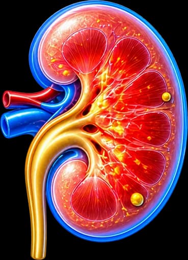
My Clinical Approach to Renal Abscess
Renal abscess is a urological emergency — the patient typically presents with high fever, severe flank pain, and clinical signs of sepsis. Delayed diagnosis, or managing a patient with antibiotics alone without imaging, is one of the most common errors in handling this condition.
At my urology practice in Rhodes, management is based on immediate CT with contrast, targeted antibiotic therapy guided by cultures, and — where indicated — percutaneous drainage under CT/ultrasound guidance. The underlying cause is investigated and treated simultaneously.
- CT with IV contrast as the investigation of choice for precise abscess mapping.
- Blood and urine cultures before starting antibiotic therapy.
- Percutaneous drainage for abscesses >3–5 cm or failing to respond to antibiotics.
- Identification and treatment of the underlying cause: stone, UPJ stenosis, obstruction.
What is a Renal Abscess
A renal abscess is a localised pus collection that forms within the renal parenchyma (intrarenal abscess) or in the perinephric space between the kidney and Gerota's fascia (perinephric abscess).
It typically develops as a complication of inadequately treated pyelonephritis or from haematogenous spread of bacteria from a distant focus. Without timely treatment it leads to urosepsis, rupture, and permanent loss of renal function.
Prevalence
Relatively uncommon but associated with significant morbidity. High-risk groups: diabetics, immunocompromised patients, those with nephrolithiasis or undertreated urinary tract infection.
Causes & Pathophysiology
Two main pathogenic routes:
Ascending Route (most common)
Complication of pyelonephritis — usually Gram-negative bacteria (E. coli, Klebsiella, Proteus). Predisposed by: nephrolithiasis, ureteric obstruction, UPJ stenosis, neurogenic bladder, or anatomical anomalies.
Haematogenous Route
Bacteraemia — typically Staphylococcus aureus from a skin focus, endocarditis, or intravenous drug use. The abscess usually localises to the renal cortex without involvement of the collecting system.
Risk Factors
Diabetes mellitus (most important), immunosuppression, chronic nephrolithiasis, undertreated UTI, pregnancy, anatomical urological anomalies.
Symptoms & Clinical Presentation
The clinical picture typically includes:
Fever & Rigors
High fever (>38.5°C) with severe rigors — the cardinal symptoms. The fever is sustained and does not adequately resolve with oral antibiotics.
Flank & Loin Pain
Unilateral severe flank or loin pain with renal angle tenderness. In a perinephric abscess the pain may be dull, persistent, and radiate to the abdomen or groin.
Absence of Classic Lower UTI Symptoms
Important feature: many patients have no dysuria or urinary frequency. This frequently delays diagnosis — a high index of suspicion is required in fever + flank pain.
Septic Presentation
In advanced disease: tachycardia, hypotension, confusion, multi-organ failure. Untreated renal abscess may rupture into the peritoneal cavity or thorax.
Untreated Renal Abscess Leads to Sepsis & Loss of the Kidney
A patient with pyelonephritis not responding to antibiotics within 48–72 hours requires immediately:
- CT with IV contrast to exclude abscess or obstructive pyelonephritis.
- Blood and urine cultures to guide targeted antibiotic therapy.
- Urgent urological assessment — exclusion of obstruction from stone or UPJ stenosis.
Diagnosis & Imaging
Diagnosing a renal abscess requires both laboratory and imaging evaluation:
CT Abdomen with Contrast
Investigation of choice — demonstrates a thick-walled cavity with central fluid, location, size, and relationship to the collecting system. Detects perinephric abscess, gas, stones, and obstruction.
Renal Ultrasound
Useful as a first-line study — detects echogenic lesions or fluid collections, but less sensitive than CT for small abscesses. Particularly useful for guiding percutaneous drainage.
Laboratory Tests
White cell count ?, CRP ?, PCT ?, creatinine. Urine dipstick and microscopy & culture. Blood cultures (positive in ~30–50%). Renal function assessment.
MRI Kidneys
CT alternative in pregnant patients or those with contrast allergy. Excellent soft tissue differentiation — distinguishes abscess from necrotic neoplasm.
Size-Based Classification
Abscess size and location determine the therapeutic approach:
< 3 cm
SmallManagement with IV antibiotics alone in patients without a severe clinical picture. Close follow-up — repeat CT within 48–72 hours if no clinical improvement.
3 – 5 cm
ModerateUsually requires percutaneous drainage + IV antibiotics. Antibiotics alone may suffice in selected patients with close clinical and imaging monitoring.
> 5 cm
LargePercutaneous CT/ultrasound-guided drainage is almost always indicated + IV antibiotics. Surgical drainage if percutaneous approach fails.
Perinephric
ComplexPus collection in the perinephric space — extends beyond the kidney. Often from rupture of an intrarenal abscess. Drainage almost always required.
Treatment Options
Treatment aims at eradicating infection, draining pus, and addressing the underlying cause:
Intravenous Antibiotic Therapy
Empirical broad-spectrum antibiotics immediately after cultures — then targeted therapy per sensitivity results. Total treatment duration 4–6 weeks (IV followed by oral). Common choices: fluoroquinolones, third-generation cephalosporins, or carbapenems in severe disease.
Percutaneous Drainage
Placement of a drainage catheter under CT or ultrasound guidance. Analysis and culture of drained material. Treatment of choice for abscesses >3–5 cm or failure of antibiotic therapy.
Treatment of the Underlying Cause
If the cause is kidney stones or UPJ stenosis, urological management is required after infection control. Nephrostomy for decompression of obstructive infection.
Surgical Management
Surgical intervention is indicated when conservative therapy fails:
IV Antibiotic Therapy (First Line)
Empirical broad-spectrum antibiotics immediately after cultures. For small abscesses <3 cm — antibiotics alone may be sufficient with close monitoring.
Percutaneous Drainage (CT/Ultrasound-Guided)
For abscesses >3–5 cm or not responding to antibiotics within 48–72 hours. Pigtail catheter placement — drainage for 5–10 days until resolution.
Surgical Drainage (Laparoscopic / Open)
For failed percutaneous drainage, ruptured perinephric abscess, or multi-loculated abscess. Laparoscopic or open surgical drainage with debridement of necrotic tissue.
Nephrectomy (Last Resort)
For a destroyed kidney from chronic abscess or xanthogranulomatous pyelonephritis with irreversible damage — nephrectomy as the definitive solution.
Follow-up & Prognosis
After treatment of a renal abscess, structured follow-up is essential:
- CT kidneys at 4–6 weeks to confirm complete resolution.
- Urine dipstick and culture after completion of antibiotics.
- Renal function tests (creatinine, GFR) if severe disease occurred.
- Urological assessment to address the underlying cause (stone, obstruction).
- Blood glucose/HbA1c in diabetic patients — optimise glycaemic control.
Prognosis
With early diagnosis and treatment, prognosis is good — complete resolution in >90% of patients. Mortality remains low (<1–3%) with modern management but can rise to 30–40% in unresolved septic presentations.
Frequently Asked Questions (FAQ)
What is a renal abscess?
A renal abscess is a localised collection of pus within (intrarenal) or around (perinephric) the kidney. It is a serious complication of urinary tract infection or haematogenous spread and requires prompt diagnosis and treatment.
What causes a renal abscess?
Two main routes: (a) Ascending — complication of pyelonephritis, usually caused by Gram-negative bacteria (E. coli, Klebsiella), often in patients with nephrolithiasis or urinary obstruction. (b) Haematogenous — bacteraemia from a distant focus (typically Staphylococcus aureus) in intravenous drug users or endocarditis.
How does a renal abscess differ from pyelonephritis?
Pyelonephritis is diffuse inflammation of the renal parenchyma. A renal abscess is a focal, walled-off pus collection that does not respond to antibiotics alone and usually requires drainage. It typically evolves from inadequately treated pyelonephritis.
What symptoms does a renal abscess cause?
High fever (>38.5°C), rigors, severe flank or loin pain, nausea, and general malaise. Many patients lack typical lower UTI symptoms (dysuria, frequency) — this frequently delays diagnosis. A perinephric abscess may present with dull, persistent loin pain radiating to the groin.
How is a renal abscess diagnosed?
CT with intravenous contrast is the investigation of choice — it clearly delineates the cavity, size, location (intrarenal or perinephric) and any underlying cause (stone, obstruction). Ultrasound is useful as an initial study but is less sensitive for small abscesses.
Does a renal abscess always require drainage?
No. Small abscesses <3 cm without a severe clinical picture may be managed with targeted intravenous antibiotics alone. Abscesses 3–5 cm or >5 cm usually require percutaneous drainage under CT/ultrasound guidance. Surgical drainage is needed if percutaneous drainage fails.
How long is the hospital stay and treatment?
Intravenous antibiotics for 10–14 days, followed by oral antibiotics for a total of 4–6 weeks. Hospital stay typically 5–10 days. Follow-up CT at 4–6 weeks to confirm resolution. If an underlying cause (stone, UPJ stenosis) exists, urological management is required after infection control.
Can a renal abscess recur?
Yes, especially if the underlying cause is not addressed — nephrolithiasis, ureteric obstruction, UPJ stenosis, uncontrolled diabetes. Treatment must include the root cause. Regular urological follow-up after resolution is essential.
Related Topics
Book Your Appointment in Rhodes
Suspected renal abscess or recurrent urinary tract infections? Individualised urological assessment at the urology practice in Rhodes.
References – Sources
- EAU Guidelines on Urological Infections 2024 — uroweb.org
- Coelho RF, et al. Renal and perinephric abscesses. BJU Int 2007;100:1282–6.
- Siegel JF, et al. Minimally invasive treatment of renal abscess. J Urol 1996;155:52–5.
- Gardiner RA, et al. Renal and perirenal abscess. BJUI 2011;108(5):760–8.
- Rubilotta E, et al. Management of renal abscess. Urol Int 2012;89:243–7.
Medical Review

Dr. Marinos Vasilas, Urologist – Andrologist
Dr. Marinos Vasilas runs a private urology practice in Rhodes with expertise in managing renal and perinephric abscesses, complex urinary tract infections, and obstructive uropathy. He follows the EAU Guidelines 2024.
View full profile


