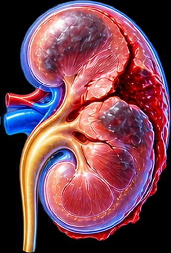
My Clinical Approach to Kidney Injury
Kidney injury is frequently managed in emergency settings — after a road traffic accident, fall from height, or sport-related trauma. A key principle: haematuria does not always reflect injury severity — serious vascular injuries may present with minimal haematuria.
At my urology practice in Rhodes, management is guided by immediate triphasic CT assessment, AAST grading, and a decision on conservative, interventional (embolisation) or surgical management based on haemodynamic status and CT findings.
- Triphasic CT as the investigation of choice for injury grading.
- Non-operative management for the vast majority of Grade I–III injuries.
- Angioembolisation for haemorrhage in Grade IV–V in haemodynamically stable patients.
- Surgery or nephrectomy only for refractory haemodynamic instability or complex injuries.
What is a Kidney Injury
A kidney (renal) injury refers to any traumatic damage affecting the renal parenchyma, renal pelvis, ureters or renal vasculature (artery/vein), as a result of blunt or penetrating trauma.
It is the most common urological injury — accounting for ~1–5% of all traumas. Blunt trauma (road traffic, falls, sport) accounts for 90–95% of renal injuries. The left kidney is more frequently injured than the right due to its anatomical position.
Epidemiology
The kidney is the most commonly injured organ of the urogenital system. High-risk patients: those with pre-existing renal pathology (horseshoe kidney, hydronephrosis, renal tumour) — who may sustain serious injury even from relatively minor force.
Mechanism & Causes
Main mechanisms of injury:
Blunt Trauma (90–95%)
This is most often caused by a road traffic accident, a fall from height, contact sport (football, boxing), or physical assault, since the kidney is compressed between the lower ribs and the spine.
Penetrating Trauma (5–10%)
This results from stab wounds or gunshot injuries. It is generally more severe, often involves multi-organ damage, and requires immediate surgical assessment.
Iatrogenic Injury
This is uncommon but important, following ureteroscopy, ESWL, percutaneous nephrolithotomy (PCNL), or renal biopsy. It is usually recognised on post-procedure imaging or at follow-up.
Pre-existing Renal Pathology
Patients with hydronephrosis, cysts, a tumour, or a horseshoe kidney are at increased risk of significant injury, even from minor trauma.
Symptoms & Clinical Presentation
Clinical findings in kidney injury include:
Haematuria
Macroscopic haematuria (visible blood in urine) is the classic symptom. However it is absent in ~25% of severe injuries — particularly in renal artery injury or pedicle avulsion.
Flank Pain & Bruising
Flank, loin, or upper abdominal pain. Bruising or tenderness at the costal/renal angle. Presence of lower rib fractures significantly raises suspicion for renal injury.
Haemodynamic Instability
In severe injury (Grade IV–V): tachycardia, hypotension, signs of shock. Haemodynamic instability requires immediate urological/surgical assessment and urgent intervention.
Urinary Extravasation
Leakage of urine into the perinephric or intraperitoneal space — a finding of renal pelvis or ureteric injury. May become apparent days after the initial trauma.
Haematuria after Trauma Always Requires Imaging
Any patient with abdominal/flank trauma and haematuria (even microscopic) or known nephrolithiasis requires immediate evaluation:
- CT abdomen/pelvis with contrast for AAST grading.
- Assessment of haemodynamic stability — in instability, immediate urological consultation.
- Particular attention in children and patients with known renal anatomical anomalies.
Diagnosis & Imaging
Diagnostic evaluation of kidney injury includes:
Triphasic CT Abdomen/Pelvis with Contrast
This is the investigation of choice. The triphasic protocol (arterial, venous, and urographic phases) assesses vascular injury, laceration extent, haematoma, and urinary extravasation, and it determines the AAST grade that directs management.
Urinalysis & Blood Tests
These include renal biochemistry (creatinine, GFR), a full haemodynamic assessment, and urine testing for haematuria. Microscopic haematuria with haemodynamic instability requires a CT regardless of the haematuria grade.
Renal Ultrasound
This is useful for initial emergency assessment and can detect perinephric haematoma, but it is insufficient to grade the injury and does not replace CT in serious trauma.
Angiography (CT-Angiography / DSA)
This is indicated when a vascular injury is suspected or to guide embolisation. CT-angiography is incorporated into the triphasic CT, while DSA is now used exclusively for therapeutic embolisation.
AAST Classification (Grade I–V)
The AAST grading scale determines therapeutic approach:
Grade I
MinorThis is a renal contusion or subcapsular haematoma without parenchymal laceration, with a non-expanding perinephric haematoma. It is managed conservatively with bed rest.
Grade II
MildThis is a renal parenchymal laceration less than 1 cm deep without urinary extravasation. It is managed conservatively, with CT follow-up.
Grade III
ModerateThis is a renal parenchymal laceration of 1 cm or more in depth without urinary extravasation or vascular injury. It is managed mainly conservatively, with embolisation if bleeding occurs.
Grade IV
SevereThis is a laceration extending to the renal pelvis with urinary extravasation, or a segmental vascular injury with contained haematoma. It is managed with angioembolisation or surgery, based on haemodynamic status.
Grade V
CriticalThis is a shattered kidney or avulsion/transection of the main renal artery or vein. It requires immediate surgical intervention, angioembolisation, or nephrectomy.
Treatment Options
Management is determined by AAST grade and haemodynamic status:
Non-operative Management (Grade I–III)
Bed rest 3–5 days, hydration, analgesia, regular haematocrit and haemodynamic monitoring. Avoid anticoagulants and NSAIDs. Repeat CT at 48–72 hours if clinical deterioration.
Angioembolisation (Grade IV–V, haemodynamically stable)
Minimally invasive technique — selective embolisation of the bleeding vessel. Allows kidney preservation and avoidance of surgery. If embolisation fails or bleeding recurs, surgery is required.
Surgical Management (Grade IV–V, haemodynamic instability)
Required for haemodynamic instability unresponsive to conservative management, vascular injury, urinary extravasation not controlled by embolisation, or shattered kidney. A nephron-sparing approach (renorrhaphy) is always preferred over nephrectomy.
Surgical & Interventional Management
Therapeutic escalation steps:
Non-operative Management (Grade I–III)
This involves bed rest, hydration, analgesia, haematocrit monitoring, and repeat CT, and it is applied to the large majority of kidney injuries.
Angioembolisation (Endovascular Intervention)
This involves selective embolisation of the bleeding branch under angiographic guidance, and it is the method of choice for haemorrhage in Grade IV–V injuries in haemodynamically stable patients.
Surgical Renorrhaphy / Partial Nephrectomy
This is an open or laparoscopic reconstruction of the kidney, used for laceration without complete shattering, and it manages urinary extravasation or vascular injury while preserving the kidney.
Total Nephrectomy (Last Resort)
Kidney removal is reserved for Grade V shattering with haemodynamic instability that cannot otherwise be controlled, or for a chronic complication with complete destruction of renal function.
Follow-up & Prognosis
After kidney injury, structured follow-up is essential:
- CT kidneys at 4–6 weeks to confirm healing.
- Urinalysis and blood tests — renal biochemistry (creatinine, GFR) at 1 and 3 months.
- Blood pressure monitoring — risk of renovascular hypertension (Page kidney from capsular compression).
- DMSA renal scintigraphy if permanent renal function loss is suspected.
- Avoid contact sports and heavy physical activity for 4–6 weeks.
Prognosis
Prognosis for Grade I–III injuries is excellent — complete recovery in >95% of patients. Grade IV–V carries a risk of permanent renal damage (~10–30%) and renovascular hypertension (~5%). Long-term renal function monitoring is required.
Frequently Asked Questions (FAQ)
What is a kidney injury?
A kidney (renal) injury is traumatic damage to the renal parenchyma or the blood vessels of the kidney, caused by blunt or penetrating trauma. It is the most common urological injury and most frequently results from road traffic accidents, falls, or physical assault.
How is a kidney injury classified?
The AAST (American Association for the Surgery of Trauma) grading scale defines 5 grades: Grade I (contusion/haematoma without laceration), Grade II (laceration <1 cm), Grade III (laceration =1 cm), Grade IV (laceration extending to the collecting system or segmental vascular injury), Grade V (shattered kidney or avulsion of the renal pedicle).
What are the symptoms of a kidney injury?
Haematuria (macroscopic or microscopic) is the classic symptom — however it is absent in ~25% of serious injuries. Additionally: flank or loin pain, bruising or tenderness in the upper quadrant, and haemodynamic instability in severe injury.
Does a kidney injury always require surgery?
No. The vast majority (>95%) of Grade I–III blunt renal injuries are managed non-operatively (bed rest, hydration, monitoring). Grade IV–V injuries and haemodynamic instability may require angioembolisation or surgical intervention.
What imaging is used for diagnosis?
Triphasic CT abdomen/pelvis with intravenous contrast is the investigation of choice — it determines the AAST grade, shows haematoma, urinary extravasation, vascular injury, and any associated abdominal injuries.
What is angioembolisation in kidney injury?
A minimally invasive procedure in which an interventional radiologist enters the vessel endovascularly and occludes the bleeding artery. It is the preferred technique for vascular haemorrhage in Grade IV–V injuries in haemodynamically stable patients.
Can a kidney injury lead to permanent consequences?
Yes. Severe injuries (Grade IV–V) may cause: chronic renal failure, renovascular hypertension (Page kidney from capsular compression), urinary extravasation, perinephric abscess, or urinoma. Long-term renal function monitoring is required.
When is nephrectomy needed for a kidney injury?
Nephrectomy (kidney removal) is reserved for extreme cases: haemodynamic instability with Grade V shattered kidney not controllable by other means, or chronic complication with complete destruction of renal function. A nephron-sparing approach is always preferred.
Related Topics
Book Your Appointment in Rhodes
Suspected kidney injury, abdominal trauma with haematuria, or urology follow-up after trauma? Specialised urological assessment at our practice in Rhodes.
References – Sources
- EAU Guidelines on Urological Trauma 2024 — uroweb.org
- Moore EE, et al. Organ injury scaling: spleen and liver. J Trauma 1995;38(3):323–4.
- Santucci RA, et al. Validation of the AAST renal injury scale. J Trauma 2001;50(2):195–200.
- Buckley JC, McAninch JW. Selective management of isolated and nonisolated grade IV renal injuries. J Urol 2006;176(6):2498–502.
- Bjurlin MA, et al. Angioembolization for renal trauma. J Trauma Acute Care Surg 2014;76(2):492–7.
Medical Review

Dr. Marinos Vasilas, Urologist – Andrologist
Dr. Marinos Vasilas runs a private urology practice in Rhodes with expertise in managing renal trauma, nephrolithiasis, and complex urological conditions. He follows the EAU Guidelines 2024.
View full profile


