My Clinical Approach
Ureteropelvic junction obstruction is one of the most interesting conditions in endourology, because its treatment is not one-dimensional — it requires careful, individualised assessment to select the appropriate method.
The core message I always give: endopyelotomy is not a smaller version of pyeloplasty — it is a different procedure, with different indications and a different long-term durability profile. Correct patient selection matters more than whether the incision is made with laser or another technology.
- I do not recommend endopyelotomy only because it is less invasive.
- I always assess stricture length, crossing vessel, renal function, and history of prior surgery.
- I explain clearly that pyeloplasty remains the primary reconstructive option for many patients.
- I do not use a single MAG3 percentage as an automatic decision criterion.
- I do not present residual hydronephrosis after treatment as automatic failure.
What Is Endopyelotomy
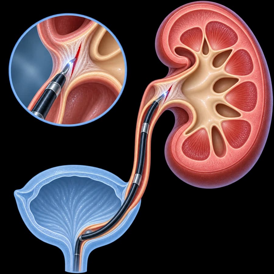
Endopyelotomy is an endoscopic procedure in which the narrowed segment at the ureteropelvic junction is controlled-incised, aiming to widen the lumen and improve drainage of urine from the renal pelvis into the ureter.
It can be performed retrograde or antegrade, depending on anatomy, any prior procedures, and the clinical context.
Endopyelotomy ≠ Pyeloplasty
Endopyelotomy is not the same as pyeloplasty.
Pyeloplasty
- • Removes or reconstructs the narrowed segment
- • Reconstructive procedure
- • Different durability profile
Endopyelotomy
- • Endoscopic incision of the narrowed segment
- • Does not perform full reconstructive re-anastomosis
- • More limited indications
What Is the Ureteropelvic Junction (UPJ)
The ureteropelvic junction (UPJ / PUJ) is the point where the renal pelvis joins the ureter.
For full information on causes, symptoms, diagnosis, and differential diagnosis of UPJ obstruction, see the page UPJ obstruction. This page focuses specifically on the diagnostic/therapeutic procedure of endopyelotomy.
Hydronephrosis ≠ Always Obstruction
Hydronephrosis is an imaging finding and does not automatically mean functionally significant obstruction.
Before treatment is decided, the following should be assessed:
- Symptoms.
- Anatomy.
- Drainage.
- Renal function.
- Progression over time.
- Recurrent infection or stone disease.
See also the page hydronephrosis. The presence of hydronephrosis alone is not an indication for endopyelotomy.
Who May Be Suitable
Endopyelotomy may be considered in selected patients with:
- A short stricture segment.
- Favourable anatomy.
- Absence of significant anatomical adverse factors (e.g. a relevant crossing vessel).
- Reasonable renal function.
- Selected primary cases.
- Selected recurrent obstruction after a prior pyeloplasty.
- Comorbidity or patient preference.
According to recent literature, the ideal candidate for endopyelotomy has a short stricture segment, absence of a crossing vessel, good ipsilateral renal function, and mild hydronephrosis — this describes the most favourable scenario, not an absolute eligibility rule.
Primary & Secondary/Recurrent Obstruction
Primary UPJ obstruction
No prior reconstructive procedure has taken place.
Secondary / recurrent
Occurs after a prior pyeloplasty, reconstructive surgery, or endoscopic treatment.
Endopyelotomy may have a different role in the two categories — outcomes should not be presented as identical.
Endopyelotomy After Failed Pyeloplasty
Recurrent UPJ obstruction after pyeloplasty can be managed on a case-by-case basis with repeat (redo) pyeloplasty, endopyelotomy, or other reconstructive strategies.
The choice depends on:
- Anatomy.
- Scar length.
- Crossing vessel.
- Renal function.
- Prior surgical approach.
- Patient factors.
Endopyelotomy is not a default salvage procedure after a failed pyeloplasty — it is assessed individually.
What If There Is a Crossing Vessel?
Vessels that cross the UPJ (lower-pole crossing vessels) may coexist with UPJ obstruction and contribute to extrinsic compression.
Endopyelotomy does not allow the UPJ to be transposed in front of or behind a crossing vessel, as reconstructive pyeloplasty can.
So the presence of a relevant crossing vessel can meaningfully affect patient selection and outcomes. According to the relevant literature, endopyelotomy outcomes tend to be worse when UPJ obstruction is associated with a crossing vessel — without this meaning every crossing vessel absolutely excludes endopyelotomy in every case.
Does Stricture Length Matter?
Yes.
Longer stricture segments generally carry a different prognosis than a short focal stricture.
There is no single universal length cutoff (e.g. 1 cm or 2 cm) that applies absolutely to every case. Some current literature describes the favourable scenario as a stricture segment shorter than approximately 2 cm, always in the context of the overall clinical picture — not as an absolute rule.
Renal Function
Treatment choice also depends on:
- Split renal function.
- Drainage.
- Preservation of cortical/parenchymal tissue.
- Trend over time.
No single split renal function percentage is used as an automatic indication or contraindication.
The decision takes into account symptoms, anatomy, function, reversibility, infection, age, the contralateral kidney, and patient preference.
What Role Does MAG3 Play
MAG3 diuretic renography can assess:
- Renal perfusion.
- Tracer uptake.
- Drainage.
- Split renal function.
T½ alone should not be used as an absolute criterion for obstruction or surgery.
Interpretation depends on hydration, bladder emptying, renal function, the drainage curve, protocol, and delayed/gravity-assisted drainage, according to current nuclear medicine standards.
CT / MR Urography
Imaging can help assess anatomy, crossing vessel, level of obstruction, stones, and alternative causes.
CTU or MRU are used selectively, according to clinical indication. Contrast CT is not mandatory for every patient.
How Endopyelotomy Is Performed
This description is deliberately kept at a general level — it is not a surgical manual with incision orientation, depth, laser settings, or balloon sizes.
Retrograde Endopyelotomy
The retrograde approach is performed through the urethra, bladder, ureter, and UPJ — without a percutaneous renal tract.
A ureteroscope and an appropriate incision technology, such as laser, may be used, depending on the clinical context.
Antegrade Endopyelotomy
The antegrade approach is performed through percutaneous renal access and may be used in selected patients.
It should not be confused with PCNL, even though it may use similar percutaneous access. If there is also a stone burden, the approach may be adapted accordingly.
Laser Endopyelotomy
Laser (e.g. Holmium:YAG, depending on availability) can be used for controlled incision of the narrowed tissue.
Success depends mainly on patient selection, anatomy, stricture length, fibrosis, renal function, presence of a crossing vessel, and any prior procedure — not on the type of incision technology.
Historically, balloon-based cutting systems (e.g. Acucise) have also been used — their current availability and use varies today, as they have largely been replaced by laser techniques.
Endopyelotomy vs Balloon Dilation
Balloon dilation
Mechanically stretches the narrowed segment.
Endopyelotomy
Makes a controlled incision through the narrowed tissue.
These are not identical procedures.
Endopyelotomy vs Pyeloplasty
| Endopyelotomy | Pyeloplasty | |
|---|---|---|
| Approach | Endoscopic | Reconstructive surgery |
| Removal/reconstruction of narrowed segment | No full reconstruction | Yes, typically |
| Skin incisions | Usually none in the retrograde approach | Yes, laparoscopic/robotic/open access |
| Managing a crossing vessel | Limited | Can be addressed anatomically |
| Recovery | Usually shorter | Longer |
| Long-term durability | Lower/selective | Generally higher in suitable patients |
| Ideal patient | Very selected | Broader reconstructive role |
We do not quote unsourced success percentages here — the comparison is described qualitatively, since real-world outcomes depend heavily on patient selection in each study.
Pyeloplasty as the Reconstructive Standard
Pyeloplasty remains the primary reconstructive treatment for many symptomatic/ functionally significant UPJ obstructions, owing to its generally higher and more stable long-term effectiveness.
Dismembered pyeloplasty according to Anderson-Hynes removes/ reconstructs the narrowed segment and can allow the UPJ to be transposed relative to a crossing vessel — this page does not analyse the technique of pyeloplasty in depth.
Endopyelotomy can be appealing because it is less invasive, has a shorter recovery, and may be suitable in selected anatomy — but it is not universally equivalent.
See the pages robotic pyeloplasty and 3D laparoscopic pyeloplasty for full information on the reconstructive procedure.
Endopyelotomy vs Robotic/Laparoscopic Pyeloplasty
Endopyelotomy
- • Less invasive
- • No full reconstruction
- • Narrower patient eligibility
Laparoscopic/Robotic Pyeloplasty
- • Reconstructive
- • Broader role
- • Generally greater durability
The robotic approach is not automatically better than an experienced laparoscopic technique — team experience matters.
Why a JJ Stent Is Placed After Endopyelotomy
The ureteral stent can:
- Maintain drainage.
- Support the area during healing.
- Limit temporary obstruction from swelling.
See the page ureteral stent (JJ) for full information.
How Long the JJ Stays In
There is no single universal duration.
It depends on the procedure, the stricture, the surgeon’s protocol, and the healing course.
JJ Stent Symptoms
There may be urgency, frequency, haematuria, bladder discomfort, or flank pain during voiding.
Fever or rigors are not normal stent symptoms.
Before Endopyelotomy
- History.
- Symptoms.
- Prior UPJ surgery.
- Renal function.
- Urinalysis/urine culture.
- Ultrasound.
- CT/MRI.
- MAG3.
- Medication review.
- Anticoagulants.
- Assessment for crossing vessels.
- Stone disease.
- History of infections.
The exact workup is individualised on a case-by-case basis.
Urinary Infection Before the Procedure
An active infection must be treated before elective upper-tract instrumentation.
We do not give a universal antibiotic regimen here. In an infected obstructed system, urgent drainage may take priority, and definitive endopyelotomy generally is not the first goal during uncontrolled urosepsis.
Fever & Hydronephrosis
Fever/rigors together with obstruction or hydronephrosis may indicate an infected obstructed system and require immediate assessment.
Possible drainage includes a JJ stent or nephrostomy, depending on the clinical situation. See the page pyelonephritis for full information.
Stones and UPJ Obstruction
UPJ obstruction may coexist with kidney stones. In selected cases, a combined strategy may be needed.
Endopyelotomy alone is not a stone treatment.
Anaesthesia
The procedure is performed in an operating theatre/endourology setting with appropriate anaesthesia — general or a selected regional approach, depending on the patient and the procedure. This is not an office-based examination.
Hospital Stay
Length of stay depends on the retrograde or antegrade approach, any concurrent procedures, the patient’s condition, infection, and complications. We do not give a universal length of stay here.
After the Procedure
Expected transient symptoms may include dysuria, haematuria, flank discomfort, and stent-related symptoms. Severe or worsening symptoms should not be considered normal.
Blood in Urine
Mild transient haematuria may occur after endoscopic instrumentation. Heavy/persistent bleeding, clots, or inability to void need assessment.
Pain
It may come from the procedure itself, swelling, the JJ stent, or ureterovesical reflux. We do not give universal analgesic doses here.
Fever After the Procedure
Fever or rigors after endopyelotomy require immediate medical assessment.
Possible causes: urinary infection, pyelonephritis, urosepsis, or a drainage problem.
Possible Complications
Infection
Requires assessment and targeted treatment.
Haematuria
Usually transient.
Pain
Mild to moderate, usually transient.
Urinary extravasation
Rare, depending on drainage management.
Ureteral/UPJ injury
Direct instrumentation can rarely cause injury.
Bleeding
Particularly if a vessel is involved near the stricture.
Stent-related symptoms
Often transient, see the relevant section.
Restenosis / recurrence
See the "Can It Narrow Again" section below.
Need for further treatment
Depends on the course and follow-up findings.
Anaesthesia-related complications
Rare, depending on the patient.
We do not quote unsourced complication rates here, including rare adjacent vascular injury — frequency depends on the anatomy and clinical context of each case.
Can It Narrow Again?
Yes.
Endopyelotomy carries a risk of recurrence, and its long-term durability may be lower than reconstructive pyeloplasty.
The risk may be influenced by:
- Stricture length.
- Significant fibrosis.
- Crossing vessel.
- Reduced renal function.
- Prior procedures.
- Anatomy.
It does not promise a permanent cure.
What Happens If It Fails
Re-evaluation
Review of anatomy, function, and cause of failure.
Repeat endoscopic treatment
In very selected cases.
Laparoscopic/robotic/open pyeloplasty
Reconstructive solution, see the relevant pages.
Repeat (redo) pyeloplasty
In selected recurrent obstruction.
Another reconstructive procedure
Depending on the case.
Nephrectomy
Only in very selected, non-salvageable cases — not a routine next step.
How Success Is Checked
Follow-up may combine symptoms, ultrasound, renal function, diuretic renography, and selected imaging, on a case-by-case basis.
Persistence of some hydronephrosis after successful treatment does not automatically mean failure.
Success should be assessed by combining symptom improvement, drainage, renal function, and the imaging trend over time.
MAG3 may be used after treatment to assess drainage and differential function, without this meaning an identical timing schedule for every patient.
“Hydronephrosis Didn’t Go Away — Did It Fail?”
Not necessarily.
Dilation can persist for some time, or even long-term, despite improved drainage. Assessment should be based on symptoms, function, drainage, and serial imaging.
Is One MAG3 Percentage Enough for a Decision?
A single split renal function percentage is not enough for a decision.
An overall interpretation is needed, combining symptoms, drainage, anatomy, trend over time, infections/stones, and patient factors.
The Patient Journey
Frequently Asked Questions (FAQ)
What is endopyelotomy?
It is an endoscopic procedure in which the narrowed segment at the ureteropelvic junction (UPJ) is controlled-incised, aiming to widen the lumen and improve drainage of urine from the renal pelvis into the ureter.
Who might be a candidate?
It may be considered in selected patients with a short stricture, favourable anatomy, absence of significant anatomical adverse factors, adequate renal function, selected primary cases, or recurrent obstruction after a prior pyeloplasty.
What is the difference from pyeloplasty?
Endopyelotomy endoscopically opens the narrowed segment without full reconstruction. Pyeloplasty removes or reconstructs the narrowed segment as a reconstructive procedure, with a different long-term durability profile.
Is endopyelotomy better because it is less invasive?
Not necessarily. In favourably selected cases it may be considered, but it should not be chosen only because it is a smaller procedure when the anatomy predicts a lower likelihood of long-term success.
Does the length of the stricture matter?
Yes. Longer strictures generally carry a different prognosis than a short focal stricture. There is no single universal length cutoff that applies absolutely to every case.
What if there is a crossing vessel?
A vessel crossing the UPJ may coexist with UPJ obstruction and contribute to extrinsic compression. It can meaningfully affect treatment choice, since endopyelotomy does not allow the UPJ to be transposed relative to the vessel the way pyeloplasty can.
What role does MAG3 play?
MAG3 diuretic renography can assess renal perfusion, tracer uptake, drainage, and split renal function. T½ alone should not be used as an absolute criterion.
Is surgery needed if there is only hydronephrosis?
Not necessarily. Hydronephrosis is an imaging finding and does not automatically mean functionally significant obstruction. It requires overall assessment of symptoms, anatomy, drainage, and renal function.
How is retrograde endopyelotomy performed?
It is performed through the urethra, bladder, and ureter up to the UPJ, without percutaneous renal access, using a ureteroscope and an appropriate incision technology such as laser.
What is antegrade endopyelotomy?
It is performed through percutaneous renal access and may be used in selected patients, particularly when anatomy or a prior procedure makes the retrograde approach more difficult.
Is laser used?
Yes, laser can be used for controlled incision of the narrowed tissue. Success depends mainly on correct patient selection, not on the type of incision technology.
Is a JJ stent needed afterwards?
A temporary ureteral stent (JJ) is often placed to support drainage and healing, but the duration is determined by the specific case and the surgeon’s protocol.
How long does the JJ stay in?
There is no single universal duration. It depends on the procedure, the stricture, the surgeon’s protocol, and the healing course.
Does it hurt afterwards?
Anaesthesia is used during the procedure. Postoperatively there may be discomfort, and stent-related symptoms can contribute to this.
What are the possible complications?
These include infection, haematuria, pain, urinary leakage, ureteral/UPJ injury, bleeding, stent-related symptoms, restenosis/recurrence, need for further treatment, and anaesthesia-related complications.
Can it narrow again?
Yes. Endopyelotomy carries a risk of recurrence, and its long-term durability may be lower than reconstructive pyeloplasty.
What happens if it fails?
Depending on the case, options include re-evaluation, repeat endoscopic treatment in very selected cases, laparoscopic/robotic/open pyeloplasty, or another reconstructive procedure.
Can it be done after a prior pyeloplasty?
Yes, in selected cases of recurrent obstruction. The choice depends on anatomy, scar length, crossing vessel, renal function, prior surgical approach, and patient factors.
How is success checked?
Follow-up may combine symptoms, ultrasound, renal function, diuretic renography, and selected imaging, on a case-by-case basis.
If hydronephrosis remains, does that mean it failed?
Not necessarily. Dilation can persist for some time, or even long-term, despite improved drainage. Assessment should be based on symptoms, function, drainage, and serial imaging.
Related Topics
Endopyelotomy or Pyeloplasty in Rhodes
The choice between endopyelotomy and pyeloplasty does not depend only on how “small” the procedure is. The location and length of the stricture, the presence of a crossing vessel, kidney function, and the history of prior treatment determine which method offers the best chance of restoring long-term drainage.
Scientific References
- Kominsky HD, Johnson BA. Percutaneous management of ureteropelvic junction obstruction. Curr Opin Urol. 2023;33(4):345–350.
- Janetschek G, Peschel R, Frauscher F. Laparoscopic pyeloplasty. Urol Clin North Am. 2000;27(4):695–704.
- Dimarco DS, et al. Long-term success of antegrade endopyelotomy compared with pyeloplasty at a single institution. J Endourol. 2006;20(10):707–712.
- Parkin J, et al. Endoluminal ultrasonography before retrograde endopyelotomy: can the results match laparoscopic pyeloplasty? BJU Int. 2003;91(4):389–391.
Meet the Doctor

Dr. Marinos Vasilas, Urologist – Andrologist
Dr. Marinos Vasilas specialises in endoscopic and laparoscopic/robotic management of upper urinary tract strictures, with emphasis on clearly informing each patient about the advantages and limitations of each approach.
Full Profile

