My Clinical Approach
Ureteroscopy with laser lithotripsy is one of the most common endourological procedures I perform — precisely because it is simple, quick, and minimally invasive. Yet the right treatment is not always the same for every patient: it depends on the location, size, and composition of the stone, the patient's anatomy, and whether there is active infection.
The first question I always ask is not “ESWL or URS?” but “is there an emergency here?”. Fever in a patient with an obstructed ureter completely changes the order of priorities — safe drainage comes first, not definitive lithotripsy.
- Assessment for signs of infected obstruction before any decision on definitive treatment.
- Stone mapping with non-contrast-enhanced CT where indicated.
- I do not use a single size cutoff as a rule — the decision is combinatorial.
- I do not place a JJ stent "out of habit" after every uncomplicated ureteroscopy.
- Counselling on URS, ESWL, RIRS and PCNL as different but complementary techniques.
What Is Endoscopic Lithotripsy (URS)
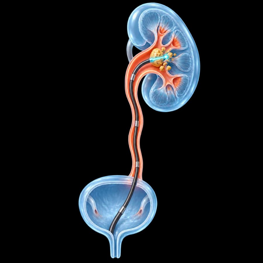
Endoscopic ureteral lithotripsy is performed via ureteroscopy (URS), in which a thin endoscope is passed through the urethra and bladder into the ureter, without any external incision. The stone is directly visualised and can be fragmented with a laser and, when needed, the fragments removed.
URS ≠ ESWL
ESWL is extracorporeal (no endoscopy) — it breaks up the stone with shock waves from outside the body.
URS ≠ PCNL
PCNL requires direct percutaneous puncture of the kidney — it is not a retrograde technique.
URS relates to RIRS
RIRS is mainly flexible retrograde intrarenal treatment, with overlaps depending on anatomy.
There are naturally overlaps depending on stone location: a stone in the upper ureter may migrate towards the kidney, so the same flexible endoscope used for URS may need to advance to intrarenal treatment (RIRS) in the same session.
How Ureteroscopy Works
No skin incision or direct kidney puncture is required, as it is in percutaneous nephrolithotomy (PCNL). Access is exclusively through the natural pathway of the urinary tract.
Semirigid vs Flexible Ureteroscopy
Semirigid Ureteroscopy
The semirigid ureteroscope is often used for stones in the distal and mid ureter, and in selected cases of the proximal ureter, depending on anatomy — without a rigid rule by location.
Flexible Ureteroscopy
The flexible ureteroscope allows access to the proximal ureter, renal pelvis, and calyces — used when access towards the kidney is required. It should not be confused with simple flexible cystoscopy, which is a different diagnostic examination.
URS vs RIRS
| URS (ureter) | RIRS | |
|---|---|---|
| Main target | Ureter | Kidney |
| Instrument | Semirigid/flexible | Mainly flexible |
| Laser | Yes | Yes |
| Basket | Selected | Selected |
| Access sheath (UAS) | Selected | More often relevant |
| JJ stent | Selected | Selected |
The main intent of URS is ureteral stone treatment, while the main intent of RIRS is retrograde intrarenal treatment of renal stones. Both use retrograde endoscopic access, and the same flexible instrument can be used in both contexts — they are not entirely unrelated techniques, but they have different primary targets.
When Endoscopic Lithotripsy Is Needed
Many ureteral stones pass spontaneously — the likelihood depends mainly on size, location, and anatomy, not on an oversimplified rule. Active treatment may be considered when:
- Spontaneous passage is unlikely based on size/location/anatomy.
- Pain persists despite appropriate analgesia.
- Obstruction is persistent.
- There is concern for renal function.
- The stone grows or persists despite conservative management.
- Conservative management has failed.
- ESWL is unsuitable or has failed.
- The patient prefers endoscopic treatment.
- It is a selected larger or impacted stone.
There is no single stone size that automatically dictates the need for surgery — the decision is made combining all of the above.
Stone + Fever = Possible Urological Emergency
Fever or rigors in a patient with an obstructed ureter may indicate an infected, obstructed kidney and a risk of urosepsis.
In this situation, priority is often urgent drainage with a JJ stent or percutaneous nephrostomy, together with antimicrobial treatment — and NOT necessarily immediate definitive laser lithotripsy. According to EAU guidelines, definitive stone treatment is usually delayed until the infection has been controlled.
For a full picture of pyelonephritis as a urological emergency, see the pyelonephritis page.
Hydronephrosis
A ureteral stone can cause obstruction and hydronephrosis, but the degree of hydronephrosis alone does not determine treatment — it is assessed together with symptoms, renal function, and the presence of infection.
Diagnosis Before the Procedure
History & symptoms
Colic, nausea, possible fever. Some patients may be asymptomatic.
Urinalysis & culture
Assessment for infection before any invasive treatment, where indicated.
Creatinine / renal function
Particularly important in a solitary kidney, bilateral obstruction, or suspected renal impairment.
Ultrasound
A first-line imaging test in many scenarios — safe, with no radiation.
Non-contrast-enhanced CT (NCCT)
A major imaging modality in many acute stone scenarios, depending on clinical context — not necessarily mandatory for every patient regardless of circumstances.
CT and Stone Characteristics
CT can provide information on size, location, number, density (Hounsfield units), anatomy, hydronephrosis, and, where relevant to ESWL, skin-to-stone distance — information that helps with treatment planning.
Urine Culture & Antibiotics
Before any endourological stone procedure, urinary infection is assessed according to current EAU guidance. Active, untreated infection can increase the risk of postoperative infection and sepsis.
A universal, fixed antibiotic regimen is not given to every patient. Prophylaxis or treatment depends on the urine culture, any resistance, the local protocol, allergies, and renal function.
Anticoagulants
Do not stop anticoagulants or antiplatelet medication on your own.
Perioperative management depends on bleeding risk, thrombotic risk, the specific medication, its indication, and the type of anaesthesia, and must be individualised in consultation with the prescribing physician.
How the Procedure Is Performed
This description is deliberately high-level — it is not a surgical technique manual.
Laser Lithotripsy
Laser energy is used for fragmentation or “dusting” of the stone. The main technologies are Holmium:YAG (Ho:YAG), the historic “gold standard”, and the newer Thulium Fiber Laser (TFL).
According to the available literature, TFL has not been proven universally superior to Ho:YAG — some studies show a shorter operative time with TFL, while others report a higher infection rate in the same group. The choice of technology depends on equipment availability and surgeon experience, not on proven universal superiority.
Fragmentation vs Dusting
Fragmentation
The stone is divided into fragments that can then be removed with a basket or forceps.
Dusting
The stone is converted into very fine particles (“dust”) that pass naturally without active extraction.
The choice depends on the stone, its location, the available laser type, and the surgeon's strategy — neither approach is universally better.
Fragment Extraction (Basket)
Selected fragments can be removed with a special stone basket through the working channel of the endoscope, under direct endoscopic visualisation.
Impacted Ureteral Stone
An impacted stone may be firmly embedded in the ureteral wall, often with local inflammation around it — which can increase the technical difficulty of the procedure.
Potential risks include ureteral injury and, later, stricture — without this meaning that every impacted stone leads to a complication.
Large/Difficult Stone & Staged Treatment
In selected cases of a large or difficult stone burden, a staged ureteroscopy, a stent followed by a second procedure, or a different treatment strategy may be required — not every procedure promises complete stone clearance in a single session.
Sometimes the safest treatment is in two stages.
Possible reasons: difficult ureteral access, significant oedema, large stone burden, infection concerns, or a long operative time. It does not mean the first procedure failed.
Pre-Stenting
In selected cases, prior placement of a JJ stent (pre-stenting) can facilitate later ureteroscopic access, passively dilate the ureter, or become necessary after a failed initial access attempt.
Routine pre-stenting is not required before every URS. According to EAU guidelines, pre-stenting appears to improve outcomes mainly for renal stones, not necessarily for ureteral stones.
Ureteral Access Sheath
The ureteral access sheath is used mainly in selected flexible ureteroscopy/RIRS, for repeated access and management of intrarenal pressure. It is not a mandatory tool in every semirigid URS or in every case — its use is individualised.
Is a JJ Stent Always Needed After Lithotripsy?
Not in every uncomplicated ureteroscopy.
A stent may be needed when there is:
- Ureteral trauma or oedema.
- Residual obstruction.
- Difficult access.
- Concern for infection.
- Residual stone fragments.
- A solitary kidney or other special clinical context.
- Other individualised indications at the surgeon’s discretion.
What Is a JJ Stent & Symptoms
A JJ stent is a thin internal tube that extends from the kidney to the bladder, allows urine drainage, and stays in place temporarily — it is usually not visible externally.
Possible symptoms include frequency, urgency, flank discomfort during urination, and mild haematuria — symptoms vary considerably between patients.
See also the ureteral stent placement page for more detail.
When Is the JJ Stent Removed
There is no single universal removal date. Duration depends on the indication for placement, the condition of the ureter, the complexity of the procedure, any infection, residual fragments, and the surgeon's protocol.
Anaesthesia, Incision, Duration, Hospital Stay
Anaesthesia
Usually performed in an operating-room setting with appropriate anaesthesia, often general, though the approach can differ by setting and patient.
Does it require an incision?
Usually not. Access is retrograde through the urethra, bladder, and ureter, without a skin incision — unlike PCNL.
Duration
There is no universal time. It depends on location, stone size and number, impaction, anatomy, laser strategy, need for flexible URS, stenting, and staged treatment.
Hospital stay
Many uncomplicated URS procedures can be performed as day-case or short-stay, depending on anaesthesia, infection, pain, and patient factors — without a guarantee of same-day discharge for everyone.
After the Procedure
Expected transient symptoms may include dysuria, urgency, frequency, mild haematuria, flank discomfort, and stent-related discomfort where a stent was placed.
Blood in the urine
Mild haematuria can appear transiently, especially with a stent. Heavy persistent bleeding, clots, inability to urinate, or systemic symptoms need assessment.
Pain
Can arise from instrumentation itself, ureteral oedema, passage of fragments, or the stent. We do not give a universal analgesic prescription here.
Fever & Sepsis After URS
Fever or rigors after ureteroscopy need immediate medical assessment because of possible urinary tract infection/sepsis — not home antibiotics.
Postoperative systemic infection is uncommon but potentially serious. Risk reduction includes appropriate infection screening before the procedure, antibiotic prophylaxis according to protocol, control of intrarenal pressure and operative factors, and avoidance of definitive treatment during uncontrolled infected obstruction.
Ureteral Injury & Stricture
Potential, uncommon injuries include mucosal injury, perforation, false passage, and, very rarely, avulsion of the ureter. Severe injuries are not common.
Ureteral stricture is a possible, rare, late complication, particularly in complex or impacted stones or significant ureteral injury during the procedure.
Stone-Free Rates: What You Should Know
There is no single universal success rate for all cases. The outcome depends on location, size, number of stones, anatomy, impaction, the imaging definition used, and follow-up duration. According to the available literature, URS tends to have a higher chance of single-session stone clearance compared with ESWL, but also a higher complication rate.
URS vs Extracorporeal Shock Wave Lithotripsy (ESWL)
| URS / Laser | ESWL | |
|---|---|---|
| Access | Endoscopic | Extracorporeal |
| Anaesthesia | Usually required | Usually less needed |
| Direct contact with stone | Yes | No |
| Single-session clearance | Often higher likelihood | May need repeat sessions |
| Invasiveness | Greater | Lower |
| JJ stent | May be needed | Not usually routine |
| Selection | Stone/anatomy dependent | Stone/anatomy dependent |
According to EAU guidelines, URS is associated with a greater chance of stone clearance without retreatment compared with ESWL, but with more adjunctive procedures, a higher complication rate, and a longer hospital stay. ESWL generally has lower morbidity.
ESWL can be an attractive option for selected ureteral stones depending on location, size, density, and patient factors. URS may be preferred when a higher chance of single-procedure clearance is desired, ESWL is unsuitable, or there is persistent obstruction/symptoms — in morbid obesity, URS is recommended as first-line therapy. No method is always superior.
URS vs RIRS vs PCNL
URS is retrograde access through the natural urinary tract. PCNL is percutaneous access directly into the kidney, and concerns mainly a large or complex renal stone burden — not a typical ureteral stone.
See also the pages on laser kidney stone lithotripsy (RIRS) and percutaneous nephrolithotomy (PCNL) for more on renal stones.
Stone Location: Upper & Lower Ureter
Stones in the upper ureter can be treated with URS or ESWL, depending on stone characteristics and anatomy. Selected stones may migrate towards the kidney and require flexible intrarenal treatment.
Stones in the lower ureter are often highly accessible endoscopically, but spontaneous passage likelihood and conservative treatment should also be considered where appropriate — not every distal ureteral stone is a mandatory indication for surgery.
Pregnancy, Solitary Kidney, Renal Impairment
Pregnancy
Requires modified imaging, radiation minimisation, and multidisciplinary evaluation (urologist, obstetrician, radiologist). URS may have a role in selected cases; we do not create a detailed pregnancy protocol here.
Solitary kidney
Obstruction in a solitary functioning kidney deserves particular attention; timing and urgency may differ, without a universal treatment rule.
Renal impairment
Renal function can influence urgency, imaging, medication, and treatment planning. Elevated creatinine alone is not an absolute contraindication to URS.
Stone Analysis, Metabolic Evaluation & Prevention
When fragments are available, stone analysis can help with composition, metabolic evaluation, and recurrence prevention, particularly in selected or recurrent stone formers.
Endoscopic lithotripsy treats the existing stone, but does not cure the underlying tendency to form stones.
Selected, recurrent, or high-risk stone formers may need stone analysis, blood tests, and 24-hour urine evaluation, according to guideline risk stratification. For a full discussion of causes, prevention, and metabolic evaluation, see the kidney stones page.
Follow-up After Endoscopic Lithotripsy
May include: symptom review, JJ stent removal where placed, imaging for residual fragments or obstruction where indicated, renal function checks, stone analysis, and metabolic evaluation in selected cases. There is no single universal CT follow-up protocol for everyone.
Small residual fragments may pass spontaneously, remain stable, or, rarely, grow and become symptomatic, depending on size, location, and composition — they should not be indiscriminately labelled “clinically insignificant” without clinical context.
The Patient's Journey
Frequently Asked Questions
What is endoscopic ureteral lithotripsy?
It is a minimally invasive procedure in which a thin endoscope (ureteroscope) is passed through the urethra and bladder into the ureter, without any external incision. The stone is directly visualised and fragmented with a laser, with possible extraction of fragments.
What is ureteroscopy (URS)?
Ureteroscopy (URS) is the endoscopic technique for accessing the ureter through the natural urinary tract. It is used mainly for ureteral stones, while its flexible variant can also advance into the kidney (RIRS) when needed.
Does it require an incision?
Usually not. Access is retrograde, through the urethra, bladder and ureter, without a skin incision — unlike percutaneous nephrolithotomy (PCNL), which requires direct percutaneous puncture of the kidney.
When is URS needed?
It may be considered when spontaneous passage is unlikely, pain persists despite appropriate analgesia, obstruction is persistent, there is concern for renal function, conservative management has failed, or extracorporeal shock wave lithotripsy (ESWL) is unsuitable or has failed — always individualised, not based on a single size cutoff.
Can the stone pass on its own?
Many ureteral stones pass spontaneously — the likelihood depends mainly on size, location and anatomy, not on an oversimplified rule such as "every stone under 5mm passes".
What is the difference between URS and RIRS?
URS targets mainly ureteral stones, while RIRS (Retrograde Intrarenal Surgery) is mainly retrograde intrarenal treatment of stones inside the kidney. Both use retrograde endoscopic access, and the same flexible instrument can be used in both contexts.
Laser or extracorporeal shock wave lithotripsy (ESWL)?
URS is more invasive, with direct endoscopic access and often a higher chance of single-session stone clearance. ESWL is less invasive, without endoscopic access, but may require repeat sessions. The best option depends on stone and patient characteristics.
What laser is used?
The main laser technologies for endoscopic lithotripsy are Holmium:YAG (Ho:YAG) and the newer Thulium Fiber Laser (TFL). No single technology has been proven universally superior in every case.
How is the stone broken up?
Laser energy can be used for fragmentation into pieces that are then extracted, or for "dusting" into very fine particles that pass naturally. The choice depends on the stone, its location, and the surgeon’s strategy.
Are all the fragments removed?
The goal is complete removal, but small residual fragments may remain, pass spontaneously, or rarely grow and become symptomatic — depending on size, location and composition.
Is a JJ stent always needed?
Not in every uncomplicated ureteroscopy. A stent may be needed in case of ureteral trauma/oedema, residual obstruction, difficult access, infection risk, residual fragments, or a special clinical context (e.g. solitary kidney).
How long does the JJ stent stay in?
There is no single universal duration — it depends on the indication, the condition of the ureter, the complexity of the procedure, any infection, and the surgeon’s protocol.
Does the JJ stent hurt?
It can cause frequency, urgency, flank discomfort during urination and mild haematuria. Symptoms vary considerably between patients.
Is blood in the urine normal?
Mild, transient haematuria can occur after the procedure, especially with a stent in place. Heavy persistent bleeding, clots, or inability to urinate need prompt assessment.
When can I return to work?
There is no single universal return date — it depends on the type of occupation, the anaesthesia used, whether a JJ stent is present, and individual recovery.
Might a second procedure be needed?
Sometimes yes. For a large or difficult stone burden, difficult access, or for safety reasons during the first procedure, staged treatment may be planned — this does not necessarily mean failure.
What are the possible complications?
These include transient haematuria/dysuria (common, mild), urinary tract infection, rarer ureteral injury or perforation, and, rarely, long-term ureteral stricture. Severe complications are uncommon but must be recognised promptly.
What happens if I have fever and a stone?
In an infected, obstructed system, priority is safe drainage (JJ stent or percutaneous nephrostomy) and control of infection — not necessarily immediate definitive laser lithotripsy, according to EAU guidelines.
Can a stone form again?
Endoscopic lithotripsy treats the existing stone, not the underlying tendency to form stones. In selected/recurrent patients, stone analysis and metabolic evaluation help with recurrence prevention.
Is stone analysis needed?
It is recommended in every patient with a first stone episode whenever material can be collected, as well as in recurrence despite pharmacological prevention or after a prolonged stone-free period.
Related Topics
Ureteral Stone Assessment in Rhodes
The choice between observation, extracorporeal shock wave lithotripsy, and ureteroscopy depends on the location and size of the stone, the likelihood of spontaneous passage, the anatomy of the urinary tract, and the presence of obstruction or infection. A proper imaging and clinical assessment allows the least invasive but effective treatment to be selected for the specific patient.
Scientific References
- EAU Guidelines on Urolithiasis 2026 — uroweb.org
- EAU Guidelines on Urological Infections — uroweb.org
- Assimos D, et al. Surgical Management of Stones: AUA/Endourology Society Guideline — auanet.org
Meet the Doctor

Dr. Marinos Vasilas, Urologist – Andrologist
Dr. Marinos Vasilas applies current ureteroscopy and laser lithotripsy techniques for the treatment of ureteral stones, in line with current EAU guidelines, with emphasis on the safe management of infected obstruction and individualised treatment selection.
Full Profile

