My Clinical Approach
Diagnostic ureteroscopy is one of the most specialised examinations in endourology — and often the most misunderstood. Many patients reach this point after a suspicious CT urogram or a positive cytology, without knowing exactly what will be examined, why it is needed, or whether it means they have cancer.
The core message I always give: the examination does not replace imaging and cytology — it complements them when they are not sufficient. The need for biopsy is assessed separately, not automatically at every examination.
- I do not recommend diagnostic ureteroscopy for every patient with haematuria.
- Biopsy is performed only when the histological information will genuinely change management.
- I always explain that grade and stage are not the same thing.
- I do not present a negative biopsy as absolute reassurance when the overall picture remains suspicious.
- A JJ stent is not placed automatically after every uncomplicated examination.
What Is Diagnostic Ureteroscopy
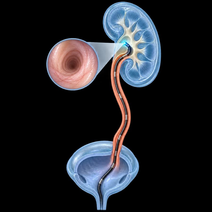
Diagnostic ureteroscopy is an endoscopic examination in which a thin ureteroscope is passed through the urethra and bladder into the ureter and, when needed, up to the renal pelvis and calyces, to directly examine the upper urinary tract.
Depending on the clinical indication, it can allow:
- Direct visual examination of the mucosa.
- Localisation of a suspicious lesion.
- Tumour mapping.
- Assessment of multifocality.
- Targeted biopsy.
- Selective upper tract cytology.
Diagnostic ureteroscopy is not a routine test — it is an invasive procedure performed when there is a specific diagnostic need.
Diagnostic ≠ Therapeutic Ureteroscopy
Diagnostic URS
Goal: diagnosis, inspection, biopsy.
Therapeutic URS
Goal: treatment, e.g. stone extraction or laser lithotripsy.
For definitive treatment of ureteral stones, see endoscopic ureteral lithotripsy (URS). This page covers exclusively the diagnostic use of the procedure.
Cystoscopy ≠ Ureteroscopy
Cystoscopy
Mainly examines the urethra and bladder.
Ureteroscopy
Advances into the ureter and, when needed, up to the renal pelvis and calyceal system.
Flexible cystoscopy is an integral part of the UTUC work-up, to rule out a concomitant bladder tumour — it is a separate examination from ureteroscopy, not an alternative to it.
Ureteroscopy ≠ RIRS
RIRS (retrograde intrarenal surgery) is essentially a flexible intrarenal procedure, mainly for therapeutic interventions inside the kidney, often for stones.
Diagnostic flexible ureteroscopy can also examine the renal pelvis and calyces when there is a diagnostic indication — without this meaning it is the same procedure as therapeutic RIRS/laser kidney stone lithotripsy.
When Diagnostic Ureteroscopy Is Needed
- Suspected upper tract urothelial carcinoma (UTUC).
- An indeterminate lesion on CT urography.
- A filling defect in the ureter or renal pelvis.
- Suspicious thickening of the ureteral/renal pelvic wall.
- Diagnosis or risk stratification not adequately resolved by imaging and cytology.
- Unexplained upper-tract obstruction in selected cases.
- Suspected ureteral stricture requiring direct assessment.
- A selected unclear upper-tract lesion.
- Targeted biopsy when histology will alter management.
Not all indications are equally common. Unexplained haematuria after a negative initial work-up and suspected UTUC are the most frequent reasons for referral.
Not Needed for Every Case of Haematuria
Diagnostic ureteroscopy is not a routine test for every patient with haematuria.
Investigation of haematuria may include, depending on risk:
- History taking.
- Urine tests.
- Upper-tract imaging.
- CT urography where appropriate.
- Cystoscopy.
- Cytology in selected cases.
Diagnostic ureteroscopy is used when there is a specific need to further investigate the upper urinary tract — not as a first step for every case of haematuria.
CT Urography & Ureteroscopy: Complementary Tests
CT Urography
- • Non-endoscopic imaging
- • Anatomical imaging
- • Assessment of kidneys/ureters
- • Masses, filling defects, wall thickening
Diagnostic Ureteroscopy
- • Direct endoscopic vision
- • Tumour mapping
- • Targeted biopsy
- • Selective cytology
The two examinations are complementary, not competing.
According to EAU UTUC guidelines, CT urography has the highest diagnostic accuracy among available imaging techniques, with a reported pooled sensitivity of around 92% and specificity of around 95% in a meta-analysis of studies. However, flat lesions without a mass effect or wall thickening are often not visible on CT — that is where ureteroscopy adds value through direct vision.
Diagnostic Ureteroscopy and UTUC
UTUC stands for Upper Tract Urothelial Carcinoma and mainly affects the renal pelvis and the ureter. See also the ureteral cancer page for symptoms, staging, risk categories, and treatment.
According to EAU guidelines, diagnostic ureteroscopy is used when imaging and voided urine cytology are not sufficient for a safe diagnosis or risk stratification of suspected UTUC.
Direct Tumour Mapping
Direct inspection can assess:
- Location of the lesion.
- Approximate size.
- Number of lesions.
- Multifocality.
- Appearance (papillary, flat, etc.).
- Involvement of the ureter / renal pelvis.
Visual appearance does not constitute a definitive diagnosis of stage or grade — histological confirmation is needed where indicated.
Biopsy During Ureteroscopy
A targeted biopsy can be taken from a suspicious lesion for histological diagnosis, grading, and risk stratification.
The small ureteroscopic biopsy has limitations.
There can be undergrading, understaging, or insufficient tissue for safe assessment. According to EAU guidelines, ureteroscopic biopsies can determine grade in over 90% of cases with a low false-negative rate regardless of sample size — however, undergrading and understaging occur compared to nephroureterectomy specimens. The small biopsy specimen does not always accurately represent the final surgical specimen.
Grade ≠ Stage
Grade
How aggressive the tumour cells appear histologically.
Stage
How deeply the tumour has invaded the tissues.
Ureteroscopic biopsy can be useful for assessing grade, but accurate pathological staging is more difficult from a small endoscopic biopsy specimen. The biopsy result should not be presented as a definitive T stage in every case.
Upper Urinary Tract Cytology
During diagnostic ureteroscopy, urine can be collected selectively from the ureter or renal pelvis (selective cytology) for cytological examination.
Selective upper tract cytology may have greater diagnostic value than voided cytology, particularly for high-grade lesions — without this meaning universal superiority in every case.
Barbotage cytology
This involves obtaining a cytology sample by washing the collecting system during the examination, usually before contrast is used for retrograde pyelography, so that specimen quality is not compromised. The exact technique is determined by the treating physician — it is described here only at a patient-friendly conceptual level.
Combining Imaging, Endoscopy, Biopsy & Cytology
The best risk assessment does not rely on a single finding.
Where appropriate, imaging, endoscopic appearance, biopsy grade, cytology, tumour size, multifocality, hydronephrosis, and the overall clinical context are combined. According to EAU guidelines, grade remains the most important single factor, while size, multifocality, and hydronephrosis have lower-quality evidence when considered individually — which is why a single variable is never used alone for the final decision.
Ureteroscopy Before Radical Nephroureterectomy
Diagnostic ureteroscopy should not be performed mechanically in every obviously high-risk tumour if the result is not going to change the diagnosis, risk stratification, or management.
The need for ureteroscopic biopsy should be assessed individually, in collaboration with the treating urologist, considering whether the information will genuinely change the treatment strategy.
Intravesical Recurrence After URS
Studies have investigated whether diagnostic ureteroscopy — and particularly ureteroscopy with biopsy before radical nephroureterectomy — may be associated with an increased risk of subsequent bladder (intravesical) recurrence.
According to systematic reviews cited in EAU guidelines, findings are not uniform: some studies associate ureteroscopy with an increased risk of intravesical recurrence, while others show that ureteroscopy alone (without biopsy) is not significantly associated with this risk — whereas biopsy performed during ureteroscopy appears to significantly increase the risk of intravesical recurrence, without a demonstrated impact on survival or recurrence outside the urinary tract.
This does not mean that ureteroscopy “spreads” cancer. It means that the decision to biopsy should be based on whether the resulting information will genuinely change management.
Why Precise Mapping Matters for Kidney-Sparing Treatment
When a kidney-sparing treatment strategy is being considered, diagnostic ureteroscopy can help assess tumour location, multifocality, appearance, and biopsy grade, allowing for better risk stratification before deciding on the treatment approach.
The final choice between kidney-sparing treatment and radical nephroureterectomy is individualised based on risk category, as described in detail on the ureteral cancer page.
Ureteral Stricture
Diagnostic ureteroscopy can, in selected cases, help establish location, appearance, lumen narrowing, or a suspicious mucosal lesion.
Anatomical assessment of ureteral stricture often also needs imaging — ureteroscopy is not a stand-alone diagnostic method.
Hydronephrosis & Obstruction
Hydronephrosis is an imaging finding, not a diagnosis by itself.
Diagnostic ureteroscopy can have a role when there is unexplained ureteral obstruction, when imaging suggests intraluminal pathology, or when there is suspicion of tumour/stricture. It is not recommended for every case of hydronephrosis.
Haematuria and Diagnostic Ureteroscopy
Visible or persistent microscopic haematuria needs structured evaluation.
Diagnostic ureteroscopy may be useful when an upper-tract lesion remains suspicious, when CT/cytology findings need clarification, or when there is a filling defect or a suspicious ureteral lesion — not as a routine test for every patient.
Ureteral Stones: An Incidental Finding
Diagnostic ureteroscopy can occasionally incidentally identify calculi, small lesions, or obstruction.
However, stones today are usually diagnosed with imaging, and when ureteroscopy is performed for stone treatment, the procedure is therapeutic, not simply diagnostic. See the endoscopic ureteral lithotripsy page for therapeutic stone management.
Retrograde Pyelography
Retrograde ureteropyelography can, in selected cases, provide an outline of the ureter, filling defects, the level of obstruction, and collecting-system anatomy, and can be combined with endoscopic evaluation.
It is not a routine requirement for every diagnostic ureteroscopy — it is used when additional anatomical information is needed.
Before Diagnostic Ureteroscopy
- History taking.
- Review of imaging.
- Urinalysis.
- Urine culture according to context.
- Renal function assessment.
- Cytology.
- Medication review.
- Anticoagulants/antiplatelets.
- Allergies.
- Anaesthetic assessment.
The exact preparation is individualised according to the indication and clinical context.
Infection & Antibiotics Before the Procedure
Instrumentation of the upper urinary tract in uncontrolled infection can increase the risk of sepsis.
Before the procedure, possible infection is evaluated, clinically significant infection is treated according to guidelines, and a perioperative antimicrobial strategy is followed based on current guidance and local protocol. We do not give a universal antibiotic regimen, brand, or duration here — these are determined individually.
Anticoagulants & Antiplatelet Medication
Patients should not stop anticoagulants or antiplatelet medication on their own.
Management depends on the specific drug, thrombotic risk, bleeding risk, whether a biopsy is planned, and the anaesthetic/surgical plan — it should always be discussed with the treating physician.
How the Examination Is Performed
Semirigid ureteroscope
Useful mainly for the distal/mid ureter, and selected proximal ureter.
Flexible ureteroscope
Allows access to the proximal ureter, renal pelvis, and calyces. Selection depends on lesion location/anatomy.
This description is deliberately high-level — it is not a surgical technique manual, a guidewire manual, or a biopsy manual.
Anaesthesia, Duration & Hospital Stay
The examination usually takes place in a surgical/endoscopic setting with appropriate anaesthesia — general, regional, or a selected alternative approach, depending on complexity, the patient, and local protocol. This is not an office-based examination.
Duration depends on unilateral or bilateral examination, lesion location, need for biopsy/cytology, anatomy, access difficulty, and the need for a stent — we do not give a universal time here.
Many uncomplicated procedures can be day-case or short-stay, depending on anaesthesia, biopsy, symptoms, infection risk, stent placement, and patient factors — we do not guarantee same-day discharge in every case.
Is a Biopsy Always Needed?
No.
Biopsy should be taken when the histological information is necessary for diagnosis, will improve risk stratification, or may change the treatment strategy, according to current UTUC guidance. It is not a mandatory step at every diagnostic ureteroscopy.
Is a JJ Stent Needed Afterwards?
Not necessarily.
A ureteral stent may be needed depending on ureteral oedema, difficult access, biopsy/trauma, obstruction, stricture, or the general clinical context. See the ureteral stent (JJ) page for complete information.
If the Ureteroscope Cannot Pass
Sometimes the ureter is too narrow for safe access. In this case it may be safer to:
- Place a temporary JJ stent.
- Allow passive dilation.
- Repeat the examination later.
Forced access is not attempted. A second procedure does not necessarily mean failure.
After the Examination
Transient symptoms may include dysuria, frequency, urgency, mild haematuria, and flank discomfort, particularly if a JJ stent was placed.
Mild, transient haematuria can occur after instrumentation or biopsy. Assessment is needed if there is heavy persistent haematuria, clots, inability to void, or worsening systemic symptoms.
Fever After the Examination
Fever or rigors after ureteroscopy need immediate medical assessment for possible infection/sepsis.
Do not start antibiotics yourself — medical assessment is needed first.
Possible Complications
Urinary tract infection
Needs assessment and targeted treatment.
Haematuria
Usually transient, especially after biopsy.
Pain
Mild to moderate, usually transient.
Ureteral mucosal injury
Direct instrumentation can rarely cause trauma.
Perforation
Rare. Managed according to severity.
Need for a JJ stent
Depending on oedema, trauma, or obstruction.
Inability to complete the examination
In a narrow ureter — see the section above.
Later stricture (rare)
Scar-related narrowing after significant trauma/instrumentation; not a common finding.
We do not quote unsourced complication percentages here — frequency depends on the complexity and clinical context of each case.
What Information the Result Can Provide
- Normal mucosa.
- Inflammatory change.
- Urothelial carcinoma.
- A papillary lesion.
- A flat suspicious lesion.
- Ureteral narrowing.
- A stone.
- A clot.
- Indirect evidence of extrinsic compression.
- Other intraluminal pathology.
Endoscopic appearance alone is not a definitive diagnosis of malignancy.
Biopsy Turnaround & a Negative Result
Turnaround time depends on the pathology laboratory, the specimen, and any additional testing (e.g. immunohistochemistry). There is no universal timeframe. The result should be integrated with imaging and cytology.
A negative or non-diagnostic small biopsy does not always exclude malignancy when imaging or endoscopic suspicion remains high.
Further management depends on specimen adequacy, imaging, cytology, clinical risk, and possible re-evaluation.
Is a Biopsy Needed If It Clearly Looks Like Cancer?
Not necessarily in every clinical scenario.
If imaging and cytology already establish a high-risk picture and histology will not change management, diagnostic ureteroscopy/biopsy may not always add enough value to justify the procedure. This is assessed individually by the treating urologist, according to current clinical guidance — it is not a universal rule.
After the Diagnosis
Benign finding
Appropriate follow-up or treatment depending on the finding.
Stricture
Determination of cause/anatomy and treatment planning.
Stone
Referral to the stone-treatment pathway.
Suspected/confirmed UTUC
Risk stratification and choice between kidney-sparing treatment and radical treatment depending on risk.
Diagnostic ureteroscopy alone is not a treatment — it informs the next treatment step.
When to Contact a Doctor Immediately
Contact a doctor immediately
- Fever or rigors.
- Severe pain that does not subside.
- Inability to urinate.
- Heavy haematuria with clots.
- Significant deterioration in general condition.
Fever after the examination should never be considered a normal symptom.
The Patient's Journey
Frequently Asked Questions
What is diagnostic ureteroscopy?
It is an endoscopic examination in which a thin ureteroscope is passed through the urethra and bladder into the ureter and, when needed, up to the renal pelvis and calyces, to directly examine the upper urinary tract.
When is it needed?
Mainly when imaging (CT urography) and urine cytology are not sufficient for a safe diagnosis or risk stratification of a suspicious upper-tract lesion. It is also used when a ureteral stricture or unexplained obstruction needs direct assessment.
Is it the same as cystoscopy?
No. Cystoscopy mainly examines the urethra and bladder. Ureteroscopy advances into the ureter and, when needed, up to the renal pelvis and calyceal system.
Is it the same as RIRS?
RIRS (retrograde intrarenal surgery) is mainly a therapeutic flexible intrarenal procedure, often for stones. Diagnostic flexible ureteroscopy can also examine the renal pelvis and calyces, but its purpose is diagnostic, not stone treatment.
Why is it needed if I have already had a CT scan?
CT urography images the upper urinary tract. Ureteroscopy, by contrast, allows direct vision and, when needed, targeted biopsy or selective cytology. Not every patient needs both tests, as they are combined only when one alone is not sufficient.
Is it needed for every case of haematuria?
No. Haematuria work-up usually includes history, urine tests, upper-tract imaging, cystoscopy, and cytology. Diagnostic ureteroscopy is used when there is a specific need to further investigate the upper urinary tract.
Can it diagnose ureteral cancer?
It can allow direct visual localisation of a suspicious lesion and targeted biopsy for histological diagnosis. The visual appearance alone is not a definitive cancer diagnosis.
Is a biopsy always taken?
No. Biopsy is taken when there is a suspicious lesion and the histological information is necessary for diagnosis, risk stratification, or treatment planning.
What does the biopsy show?
It may show normal mucosa, inflammatory change, or urothelial carcinoma with an assessment of tumour grade. It does not always provide a reliable assessment of invasion depth (stage).
Can it determine the stage?
Not reliably in every case. Small ureteroscopic biopsies can determine grade in over 90% of cases, but staging can be underestimated compared to the final surgical specimen.
What is selective cytology?
It is the collection of urine selectively from the ureter or renal pelvis during the procedure, for cytological examination. It may have greater diagnostic value than voided cytology, particularly for high-grade lesions.
What anaesthesia is needed?
Usually general or regional anaesthesia, depending on the complexity of the examination, the patient, and the local protocol. This is not an office-based examination.
Does it hurt afterwards?
There may be transient dysuria, frequency, mild haematuria, or flank discomfort, especially if a JJ stent was placed. Severe pain or fever should not be considered normal.
Is blood in the urine normal?
Mild, transient haematuria can occur after instrumentation or biopsy. Heavy persistent bleeding, clots, or inability to urinate need assessment.
Is a JJ stent always needed afterwards?
No. The need for a stent depends on ureteral oedema, difficult access, biopsy/trauma, obstruction, or stricture, and is individualised case by case.
What happens if the ureteroscope cannot pass?
In a narrow ureter it may be safer to place a temporary JJ stent for passive dilation and repeat the examination later, rather than attempt forced access. A second procedure does not necessarily mean failure.
What are the possible complications?
These include urinary tract infection, haematuria, pain, ureteral mucosal injury, rarely perforation, the need for a stent, inability to complete the examination, and rarely a later ureteral stricture.
When are the biopsy results available?
Timing depends on the pathology laboratory, the specimen, and any additional testing (e.g. immunohistochemistry). There is no single universal turnaround time.
What happens if the biopsy is negative but the CT is suspicious?
A negative or non-diagnostic small biopsy does not always exclude malignancy when imaging or endoscopic suspicion remains high. Further management depends on specimen adequacy, imaging, cytology, and clinical risk.
When should I contact a doctor immediately?
In case of fever or rigors, severe pain, inability to urinate, or heavy haematuria with clots. Fever after ureteroscopy needs immediate medical assessment for possible infection or sepsis.
Related Topics
Diagnostic Ureteroscopy in Rhodes
Diagnostic ureteroscopy is not required for every patient with haematuria or abnormal imaging. However, when CT urography and cytology are not sufficient for a safe diagnosis or risk stratification, direct endoscopic examination of the ureter and renal pelvis can provide important information and allow targeted biopsy.
Scientific References
- EAU Guidelines on Upper Urinary Tract Urothelial Carcinoma (UTUC) 2026 — uroweb.org
- EAU Guidelines on Urolithiasis 2026 — uroweb.org
- EAU Guidelines on Urological Infections — uroweb.org
Meet the Doctor

Dr. Marinos Vasilas, Urologist – Andrologist
Dr. Marinos Vasilas performs diagnostic ureteroscopy in line with current EAU guidelines on UTUC and urolithiasis, with emphasis on clearly informing each patient about the indication for the examination, the limitations of biopsy, and the next treatment step.
Full Profile

