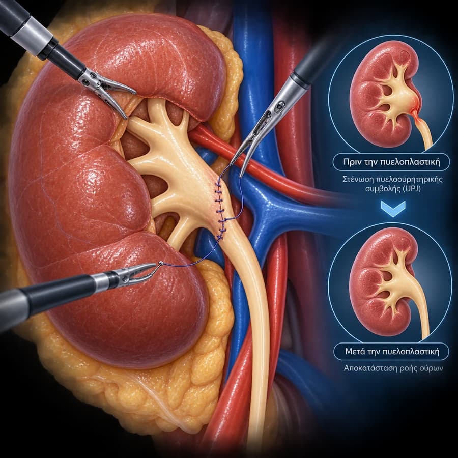
My Clinical Experience with 3D Laparoscopic Pyeloplasty
3D laparoscopic Anderson-Hynes pyeloplasty delivers outstanding results comparable to the robotic approach — the difference lies more in available technology and surgeon expertise than in the underlying technique. In specialized centers with extensive laparoscopic experience, the technique is fully reliable.
- 3D HD camera — precise anatomical identification of UPJ and crossing vessel.
- Anderson-Hynes technique: identical to robotic — excision, spatulation, anastomosis.
- DJ stent 4–6 weeks — removed cystoscopically in the office, no anesthesia.
- Hospital stay 2–3 days, early recovery.
What Is 3D Laparoscopic Pyeloplasty?
This is the surgical excision of the stenosed UPJ and anastomosis of the ureter to the renal pelvis through 3–4 incisions <12 mm — without a robotic system. The classic Anderson-Hynes technique is applied using a three-dimensional laparoscopic imaging system.
3D HD Optical System
Stereoscopic vision — precise recognition of fine structures (UPJ, ureter, crossing vessel) that demand high technical accuracy.
Anderson-Hynes Technique
Excision of the stenosed segment, spatulation of the ureter, fine anastomosis with absorbable sutures. DJ stent provides internal scaffolding.
Crossing Vessel Management
Identification of crossing vessel — anastomosis placed anterior to the vessel (transposition). A critical step that cannot be achieved with endopyelotomy.
Indications — UPJ Obstruction & Hydronephrosis
Symptomatic UPJ Obstruction
Flank pain (especially after fluid intake / Dietl’s crisis), recurrent urinary tract infections, hematuria, or hydronephrosis found on imaging.
Functional Renal Deterioration
MAG3 scintigraphy showing T½ >20 min or differential function <35% — indication for surgery even without significant symptoms.
Failed Endopyelotomy
Laparoscopic Anderson-Hynes pyeloplasty remains effective for recurrence after endopyelotomy.
For complex anatomies or patients with prior UPJ surgery, robotic pyeloplasty may offer a technical advantage due to greater instrument articulation.
Preoperative Assessment
CT Urography
Anatomical mapping of UPJ, renal pelvis, and crossing vessel. Exclusion of stones or tumor.
MAG3 or DMSA Scintigraphy
Assessment of obstructive drainage curve and differential renal function — baseline measurement for postoperative comparison.
Urine Culture
Exclusion of active infection. Treatment required before surgery if culture is positive.
How the Procedure Works
Anesthesia & Positioning
General anesthesia. Lateral decubitus. 3–4 trocars, CO₂ pneumoperitoneum. Cystoscopic DJ stent insertion before the laparoscopic phase (or intraoperatively, antegrade).
UPJ Exposure
Transperitoneal or retroperitoneal approach. Identification of renal pelvis, UPJ, and crossing vessel.
Excision & Anastomosis
Excision of the stenosed UPJ. Ureter spatulation. Fine Anderson-Hynes anastomosis with 4-0 absorbable sutures. Anterior transposition of crossing vessel if present.
Closure
Watertight anastomosis confirmed with fluid. Drain placed if needed. Wound closure.
Anesthesia, Duration & Hospital Stay
Recovery & DJ Stent
Hospital
Mobilization on the first postoperative day. Urinary catheter removal day 1–2. Oral diet within 24 hours.
DJ Stent
4–6 weeks in place. Removed cystoscopically in the office. Mild discomfort or hematuria while the stent is in — normal.
Follow-up
MAG3 scintigraphy at 3–6 months postoperatively. Annual renal ultrasound for 2 years.
Advantages & Comparison
vs. Endopyelotomy
Success rate 93–96% vs 65–75%. Ability to manage crossing vessel. Definitive repair versus frequent recurrence with endourological treatment.
vs. Open Pyeloplasty
Equivalent results with minimal invasiveness: less pain, 2–3 vs 4–6 day hospital stay, faster recovery.
Lower Cost vs. Robotic
Laparoscopic pyeloplasty has lower cost than robotic with equivalent results in the hands of an experienced laparoscopic surgeon.
Possible Complications
Urinoma / Anastomotic Leak (2–4%)
UncommonUrine leakage from the anastomosis. Managed with prolonged DJ stent or drainage.
Stenosis Recurrence (3–5%)
UncommonParticularly in cases with prior endopyelotomy or extensive scar tissue.
DJ Stent Discomfort
CommonMild pain, hematuria, urinary frequency while stent is in place — transient, resolves with removal.
Frequently Asked Questions
What is laparoscopic pyeloplasty?
3D laparoscopic pyeloplasty is the minimally invasive surgical repair of ureteropelvic junction (UPJ) obstruction without a robotic system. It uses a 3D HD camera and standard laparoscopic instruments. The Anderson-Hynes technique is applied identically to the robotic approach, with excellent results in experienced centers.
What is the success rate of laparoscopic pyeloplasty?
In specialized centers, laparoscopic Anderson-Hynes pyeloplasty achieves a success rate of 93–96% — comparable to the robotic approach. Success is defined as symptom resolution and improved drainage curve on renal scintigraphy (T½ <10 min).
How long does the DJ stent remain after laparoscopic pyeloplasty?
The stent typically remains in place for 4–6 weeks postoperatively, and is removed cystoscopically in the office without anesthesia. While the stent is in place, mild discomfort, hematuria, or urinary frequency are expected and normal.
What are the contraindications for laparoscopic pyeloplasty?
Relative contraindications include a non-functioning kidney (under 10–15% renal function), an active urinary tract infection, which should be treated first, and a large kidney stone requiring simultaneous removal, which may be combined with PCNL or RIRS. Extensive prior abdominal surgery may also affect technical feasibility.
What if there is a kidney stone alongside UPJ stenosis?
Coexisting urolithiasis and UPJ obstruction is best managed together: laser lithotripsy (RIRS) or PCNL first followed by pyeloplasty, or in selected cases simultaneous management in a single procedure.
When is renal scintigraphy performed after laparoscopic pyeloplasty?
MAG3 renal scintigraphy is performed at 3–6 months postoperatively to verify effective repair, with annual renal ultrasound for 2 years to monitor anatomical recovery.
Can laparoscopic pyeloplasty be performed after a failed endopyelotomy?
Yes. Laparoscopic (and robotic) pyeloplasty remains feasible and effective even after failed endopyelotomy or balloon dilation — with slightly increased technical difficulty due to scar tissue.
Which type of pyeloplasty is recommended for a crossing vessel?
Both robotic and laparoscopic Anderson-Hynes pyeloplasty effectively manage crossing vessels with anterior transposition. Open endopyelotomy is an inadequate treatment for crossing vessel-related UPJ obstruction.
Related Topics
Book an Appointment in Rhodes
Hydronephrosis or UPJ obstruction detected? Receive a specialist evaluation and personalized treatment plan at our urology practice in Rhodes.
Bibliography & References
- EAU Guidelines on Obstructive Uropathy 2024 — uroweb.org
- Rassweiler JJ, et al. Laparoscopic Pyeloplasty: The Experience of the German Working Group. Eur Urol 2008;53:228–236.
- Inagaki T, et al. Laparoscopic Dismembered Pyeloplasty: Transperitoneal vs Retroperitoneal Approaches. Urology 2005;66:501–504.
- Moon DA, et al. Laparoscopic Pyeloplasty: Evolution of a New Gold Standard. Urology 2006;67:932–936.
Medical Review

Dr. Marinos Vasilas, Urologist – Andrologist
Dr. Marinos Vasilas performs 3D laparoscopic and robotic pyeloplasties for the management of hydronephrosis, following current EAU 2024 protocols.
Full Profile


