My Clinical Approach
Ureterocele requires individualized management: goals are renal function preservation, addressing obstruction, and preventing UTIs with minimally invasive therapy whenever possible.
According to EAU/ESPU Pediatric Urology Guidelines 2024 and AUA Pediatric Urology:
- Complete imaging workup: U/S + VCUG + DMSA ± MR urography.
- Screen for coexisting vesicoureteral reflux (50%).
- Transurethral endoscopic puncture with holmium laser as first-line.
- Upper pole heminephrouretrectomy only if non-functional upper pole (<10% function on DMSA).
- Full reconstruction in complex cases (large ureterocele + severe VUR).
- Prophylactic antibiotic coverage until definitive treatment.
What is a ureterocele
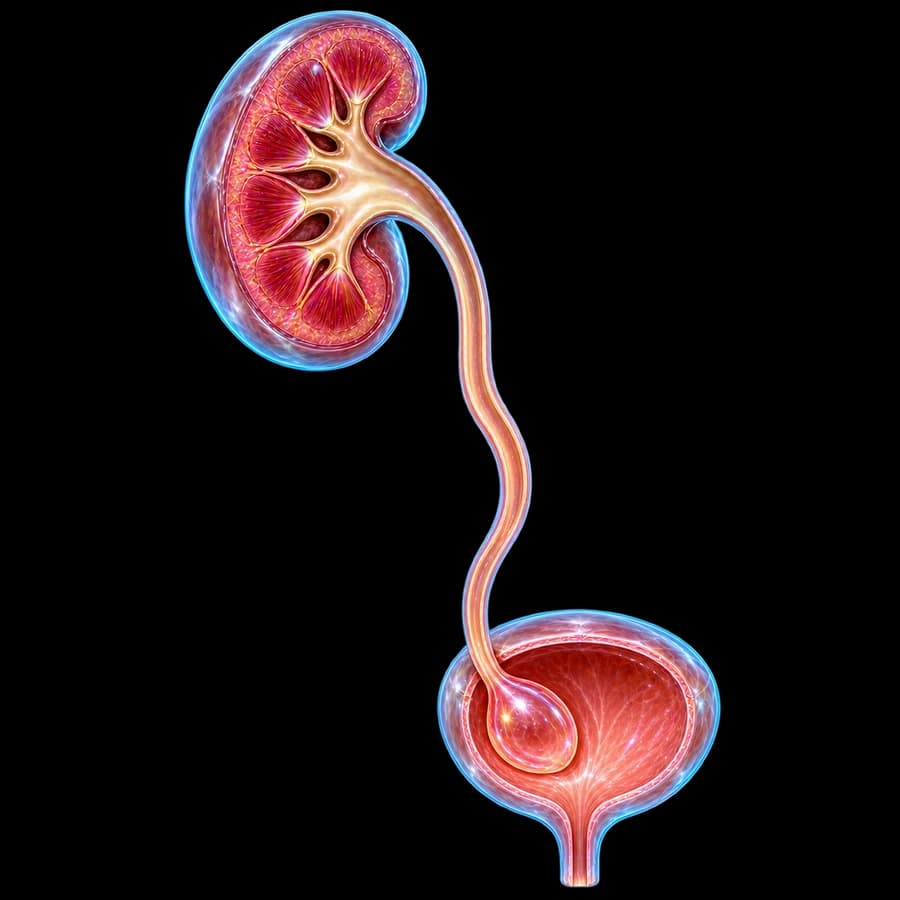
A ureterocele is a cystic dilation of the distal intravesical ureter due to congenital stenosis of its orifice. Incidence is estimated at 1:4,000 births. More common in girls (4:1) and Caucasian. Associated with complete duplex system in 80% of pediatric cases (Weigert-Meyer rule: upper pole ureter inserts more inferomedially and develops the ureterocele).
Important: In adults, most ureteroceles are orthotopic in single systems and discovered incidentally or due to lithiasis within the cystic structure.
Stephens classification (AAP 1984)
Orthotopic (Intravesical / Adult-type)
The entire ureterocele lies within the bladder. It is more common in a single system and in adults, and is usually small, asymptomatic, or stone-forming.
Ectopic
Part of the ureterocele extends below the bladder neck into the urethra. It is almost always found in the upper pole of a duplex system in children, and may prolapse externally.
Sphincteric / Sphincterostenotic
This is an ectopic ureterocele whose orifice lies within or beyond the urethral sphincter. It causes bladder neck obstruction or obstructs both ureters.
Cecoureterocele
This rare form shows extensive submucosal extension into the urethra. Distended ureteroceles of this type can cause retention or incontinence.
Symptoms & clinical presentation
Newborns / Infants
Prenatal hydronephrosis (50%+), febrile UTI, urosepsis, palpable cystic suprapubic mass, failure to thrive.
Children
Recurrent UTIs, dysuria, incontinence or dribbling, psychomotor delay from chronic infection.
Adolescent girls
Prolapsing cystic mass from vagina/urethra — urgent urologic evaluation. Acute urinary retention, rare.
Adults
Usually orthotopic: stones within ureterocele, hematuria, recurrent cystitis, or incidental finding on CT.
Diagnosis (U/S, VCUG, DMSA)
Renal-bladder ultrasound
This first-line test shows the characteristic "cobra head" cystic structure within the bladder, along with upper pole hydronephrosis in a duplex system. It can also assess parenchymal thickness.
Voiding cystourethrogram (VCUG)
This reveals a filling defect from the ureterocele, coexisting vesicoureteral reflux (in 50% of lower pole cases), any pseudo-ureterocele, and bladder neck obstruction in ectopic cases.
DMSA scintigraphy
This assesses upper pole function. Function above 10-15% favors kidney-sparing therapy, while function below 10% leads to upper pole heminephrectomy.
MR urography
This provides detailed anatomic imaging of duplication, orifice locations, ureteral dilation, and renal dysplasia. It involves no radiation, so it is preferred in children.
Cystoscopy
This confirms the diagnosis and allows simultaneous endoscopic treatment. It also rules out malignancy in adults.
Urodynamic study
This is used when bladder neck dysfunction or a coexisting neurogenic bladder is suspected, but it is not performed routinely.
Treatment options
1. Transurethral endoscopic puncture
First line for most cases. Cystoscope + holmium laser or Bugbee electrode, with low horizontal incision at the ureterocele base ("watering can" technique). Immediate decompression. Complications: de novo VUR ~30%, often managed later with Deflux.
2. Upper pole heminephrouretrectomy
When upper pole has minimal function (DMSA <10%) and lower pole is normal. Laparoscopic or robotic approach. Lower pole preservation.
3. Total bladder reconstruction
Ureterocele excision + trigone reconstruction + ureteral reimplantation by Cohen or Politano-Leadbetter. Indicated in large ureterocele with severe lower pole VUR or failed endoscopic management.
4. Ipsilateral pyelo/uretero-ureterostomy
In duplex systems with functional upper pole, anastomosis of upper pole pelvis to lower pole ureter (pyelo-pyelostomy or uretero-ureterostomy). Preserves all function.
5. Emergency decompression
In urosepsis or ureterocele prolapse from urethra: immediate endoscopic puncture and IV antibiotic coverage (cephalosporin + aminoglycoside).
Follow-up & prognosis
- Renal-bladder U/S at 4-6 weeks, 3, 6, and 12 months postoperatively.
- VCUG or RNC 3-6 months after endoscopic puncture — check for de novo VUR.
- DMSA at 12 months — assess parenchymal scars.
- Annual blood pressure + creatinine + proteinuria check.
- Continuous antibiotic prophylaxis until negative VCUG.
In summary: Prognosis is excellent in >90% with timely diagnosis and individualized treatment. Lifelong urologic follow-up ensures preservation of renal function.
Frequently Asked Questions (FAQ)
What is a ureterocele?
A ureterocele is a congenital cystic dilation of the intravesical portion of the ureter, caused by stenosis of its opening into the bladder. It is more common in girls (4:1) and is usually associated with a complete duplex collecting system in 80% of cases.
How is it classified?
The Stephens (AAP 1984) system distinguishes two main types. An orthotopic (intravesical) ureterocele lies entirely within the bladder and is usually seen in adults with a single system. An ectopic ureterocele extends below the bladder neck and is almost always associated with the upper pole of a duplex system in children.
What are the symptoms?
Many ureteroceles are asymptomatic and are diagnosed prenatally through hydronephrosis seen on fetal ultrasound. In children, they typically cause UTIs, fever, and failure to thrive. A large ectopic ureterocele can prolapse from the urethra and cause acute retention or incontinence, while in adults it can cause stone formation within the ureterocele, hematuria, and infections.
How is it diagnosed?
A renal-bladder ultrasound is the first-line test, showing the cystic "cobra head" within the bladder and upper pole hydronephrosis in a duplex system. A voiding cystourethrogram (VCUG) reveals coexisting vesicoureteral reflux (in 50% of cases) and any pseudo-ureterocele. DMSA scintigraphy assesses upper pole function, and MR urography provides the full anatomy when needed.
When is treatment needed?
Treatment is always needed when the ureterocele causes obstruction, UTIs, reflux, or stones. A small, asymptomatic orthotopic ureterocele in adults can be observed, but an ectopic ureterocele in children usually requires surgical intervention.
What are the surgical options?
Transurethral endoscopic puncture or incision is minimally invasive, using a cystoscope with a holmium laser or Bugbee electrode to decompress the ureterocele and improve drainage, though it carries a secondary VUR risk of about 30%. Upper pole heminephrouretrectomy is used if the upper pole is non-functional, while total bladder reconstruction (excision with reimplantation per Cohen or Politano-Leadbetter) is reserved for complex cases.
What is the role of prenatal diagnosis?
Over 50% of ureteroceles are diagnosed prenatally, through hydronephrosis or a cystic mass seen on second-trimester fetal ultrasound. This allows immediate postnatal ultrasound confirmation within 48 hours, prophylactic antibiotics (amoxicillin), a VCUG at 4-6 weeks, and organized multidisciplinary care.
What is the prognosis?
The prognosis is excellent with proper diagnosis and treatment, and renal function is preserved in 90% of cases with timely therapy. The most common long-term issue is persistent or de novo reflux after endoscopic puncture, which requires VCUG or RNC follow-up, along with lifelong monitoring of renal function and blood pressure.
What other urologic conditions are associated?
Ureteroceles are commonly associated with renal duplication (80%), vesicoureteral reflux of the lower pole (50%), ectopic ureter, lower pole hydronephrosis from compression, and stones within the ureterocele. They are rarely associated with VATER syndrome or renal dysplasia.
Related topics
Suspected ureterocele?
Contact us for full pediatric or adult urologic evaluation: U/S, VCUG, DMSA and personalized plan — from endoscopic laser puncture to total reconstruction.
Scientific literature
- EAU/ESPU Guidelines on Paediatric Urology — Ureterocele (2024) — uroweb.org
- Stephens FD. Caecoureterocele and concepts on the embryology and aetiology of ureteroceles. Aust N Z J Surg 1971;40(3):239-48 — pubmed.ncbi.nlm.nih.gov
- Coplen DE. Management of the neonatal ureterocele. Curr Urol Rep 2001;2(2):102-5 — pubmed.ncbi.nlm.nih.gov
- Byun E, Merguerian PA. A meta-analysis of surgical practice patterns in the endoscopic management of ureteroceles. J Urol 2006;176(4):1871-7 — pubmed.ncbi.nlm.nih.gov
- Chertin B, Mohanan N, Farkas A, Puri P. Endoscopic treatment of vesicoureteral reflux associated with ureterocele. J Urol 2007;178(4 Pt 2):1594-7 — pubmed.ncbi.nlm.nih.gov
Medical Editorship

Dr. Marinos Vasilas, Urologist – Andrologist
Dr. Marinos Vasilas treats ureteroceles in children and adults with modern approach: from endoscopic puncture with holmium laser to laparoscopic/robotic upper pole heminephrouretrectomy and total bladder reconstruction per EAU/ESPU 2024.
View full profile


