My Clinical Approach
Ureteral stricture is a progressive disease that can lead to silent renal function loss if undiagnosed. Modern urology offers a comprehensive spectrum of treatment options — from temporary JJ stent to complex robotic reconstruction using bowel segments or bladder flaps.
Per EAU 2024 and AUA-SUFU 2017 Ureteral Stricture Guideline:
- Comprehensive anatomic imaging: CT urography or MR urography + retrograde pyelography if needed.
- Functional assessment: MAG-3 renogram to differentiate obstruction from dilatation.
- Length and location characterization: short (<2cm) vs long (>3cm) strictures.
- Exclude malignancy: ureteroscopy + cytology or biopsy if UTUC suspected.
- Treatment selection based on length, location, etiology and renal function.
- Tailored robotic reconstruction for definitive cure.
What is ureteral stricture
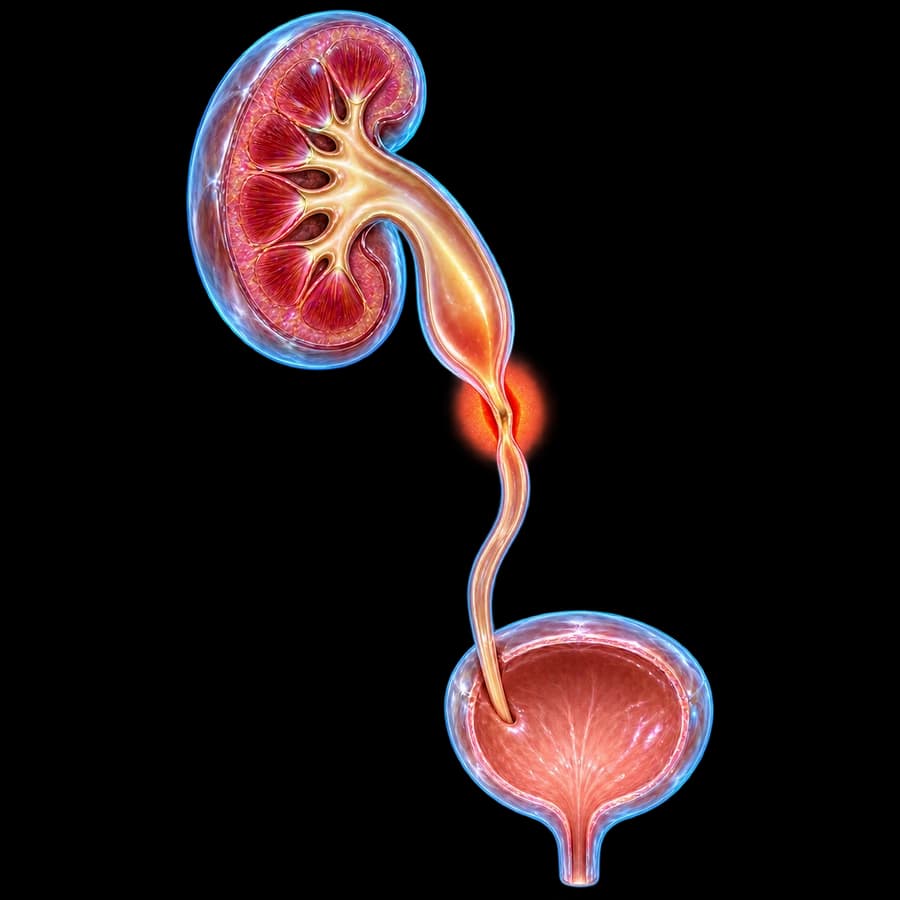
Pathologic narrowing of the ureteral lumen by fibrous scar tissue. Reduced urine passage causes progressive hydronephrosis, slower drainage, recurrent infections, and ultimately loss of renal function in the affected kidney.
Classification by location: proximal (UPJ-mid), middle, distal (mid-UVJ). By length: short (<2cm), intermediate (2-3cm), long (>3cm). By etiology: iatrogenic, ischemic, radiation, infectious, neoplastic, congenital, idiopathic.
Important: Early diagnosis is critical — untreated obstruction leads to renal cortical atrophy within 4-6 weeks.
Causes (iatrogenic, ischemic)
Iatrogenic (60-80%)
Post-URS with access sheath, impacted stone + thermal laser injury, gynecologic procedures (hysterectomy, C-section, endometriosis excision), colorectal surgery, anastomotic strictures after nephrectomy/anastomosis.
Radiation
After pelvic radiation (prostate, cervical, rectal cancer). Usually long strictures, present 6 months-10 years post-treatment. Difficult management due to fibrotic radiation injury of adjacent tissue.
Ischemic / stone-induced
Prolonged stone impaction (>1 month) with local inflammation and ischemia. Post-vascular surgery (aortic aneurysm repair).
Malignant / neoplastic
Primary UTUC (urothelial), extrinsic compression from pelvic malignancy (ovarian, cervical, rectal cancer, lymphoma). Always exclude with cytology + ureteroscopy + biopsy.
Inflammatory / infectious
Genitourinary tuberculosis, schistosomiasis, IgG4-related disease, retroperitoneal fibrosis (Ormond), sarcoidosis.
Congenital / idiopathic
Congenital UPJ or UVJ stricture (commonly pediatric), idiopathic (after exclusion of extrinsic causes).
Symptoms & clinical features
Silent
Up to 40-50% incidental diagnosis on imaging for other reasons. Gradual renal atrophy without symptoms — may lead to silent loss of renal function.
Pain / infections
Dull flank pain (after diuresis/fluid intake), recurrent pyelonephritis, hematuria, hypertension via renin-angiotensin activation.
Dietl crisis
Intermittent flank colic with nausea/vomiting and post-crisis polyuria — intermittent obstruction syndrome (more common in UPJ stricture).
Bilateral / solitary kidney
Anuria, acute kidney injury, hyperkalemia, uremia — mandates emergency decompression.
Diagnosis (CT/MR urography, MAG-3)
Renal ultrasound
This first-line test detects hydronephrosis using SFU grading from 0 to IV, at low cost and with no radiation.
CT urography
This is the gold standard for anatomic imaging. It shows the length, location, and hydronephrosis grade, excludes a stone or mass, and also stages any oncologic disease.
MR urography
This is an alternative for young patients, pregnancy, or iodinated contrast allergy, offering excellent soft tissue resolution.
MAG-3 renogram
This functional test measures split renal function, where below 40% indicates significant impairment. A T1/2 after Lasix above 20 minutes confirms obstruction, 10-20 minutes is equivocal, and below 10 minutes means no obstruction.
Retrograde pyelography (RGP)
This is used in equivocal cases or for preoperative imaging, and it is combined with ureteroscopy for cytology or biopsy if UTUC is suspected.
Whitaker test
This percutaneous pressure measurement at a fixed flow of 10 ml/min confirms obstruction above 22 cmH₂O. It is a rare test, used only when the MAG-3 result is equivocal.
Treatment options
Choice depends on length, location, etiology, renal function, and patient clinical status.
1. JJ stent (palliative)
Immediate decompression and patency maintenance. Exchange every 3-6 months. Ideal for inoperable patients, malignancy, bridge to definitive treatment. Complications: LUTS, infections, encrustation.
2. Percutaneous nephrostomy
Immediate decompression when JJ stent not feasible. Useful in sepsis/pyonephrosis. Creates antegrade access for complex endoscopic procedures.
3. Balloon dilation
Endoscopic dilation with 4-8mm balloon × 5min. Success 50-70% in short (<2cm) non-ischemic strictures. Lower yield in radiation and long strictures.
4. Endoureterotomy (laser/cold-knife/Acucise)
Incision via holmium laser or cold knife followed by JJ stent for 6-8 weeks. Success 50-70% in short strictures. More effective for distal ureter strictures.
Open/robotic reconstruction
Robotic reconstruction is the modern definitive treatment for strictures that failed endoscopic methods or are >2cm.
Ureteroureterostomy (UU)
Stricture excision + end-to-end anastomosis. Ideal for short strictures (<3cm) of mid-ureter. Success >90%.
Psoas hitch + Boari flap
For distal ureter strictures. Bladder fixation to psoas muscle + bladder flap to bridge gap. Success 85-95%.
Ileal ureteral substitution
Replacement of long ureteral segments (>10cm) with ileal segment. Success 80-90%. More morbid — requires intact renal function (eGFR >40), monitoring of metabolic disturbances.
Buccal mucosa graft
Specialized option for long upper/mid ureteral strictures. Use of buccal mucosa graft. Success 80-90%.
Renal autotransplantation
Last resort for very extensive strictures in young patients with solitary kidney. Kidney moved to pelvis with bladder anastomosis.
Follow-up & prognosis
- JJ stent removal 4-6 weeks after reconstruction.
- U/S + creatinine at 3, 6, 12 months.
- MAG-3 renogram at 3-6 months to confirm obstruction resolution.
- Annual surveillance for 5 years — recurrence may occur up to 2 years post-op.
Summary: Modern robotic ureteral reconstruction has excellent prognosis with success rates >85-95% in expert centers. Early recognition and decompression preserves renal function.
Frequently Asked Questions
What is a ureteral stricture?
A ureteral stricture is partial or complete narrowing of the ureteral lumen by fibrotic scar tissue. It causes obstruction to urine flow, hydronephrosis, and progressive loss of kidney function if untreated.
What are the most common causes?
Most cases are iatrogenic (60-80%), caused by post-ureteroscopy (URS) injury, gynecologic surgery (hysterectomy), pelvic radiation, or anastomotic strictures after nephrectomy. Other causes include impacted stones, ischemia, malignancy (UTUC), tuberculosis, retroperitoneal fibrosis, congenital factors, or an idiopathic origin.
What are the symptoms?
The condition is often silent, found incidentally in 40-50% of cases. When symptoms occur, they include dull flank pain, recurrent pyelonephritis, hematuria, and hypertension. In a bilateral or solitary kidney, it can cause decreased urine output and acute kidney injury.
How is it diagnosed?
CT urography or MR urography are the gold standard, visualizing location, length, and hydronephrosis. A MAG-3 renogram confirms obstruction when the T1/2 is above 20 minutes. In equivocal cases, a retrograde pyelography or Whitaker test (a pressure-flow study) is used.
When is treatment needed?
Treatment is needed when there is functionally significant obstruction (a MAG-3 T1/2 above 20 minutes or split renal function below 40%), symptomatic hydronephrosis, recurrent infections, or deteriorating renal function. An asymptomatic stricture without functional impact may simply be observed.
How is it treated?
The choice depends on length, location, and etiology. Options include a JJ stent for palliation, balloon dilation (50-70% success in short strictures under 2 cm), endoureterotomy with laser or Acucise, or open, laparoscopic, or robotic reconstruction (ureteroureterostomy, psoas hitch with Boari flap, or ileal ureter).
What is the success rate of reconstruction?
Robotic ureteroureterostomy achieves over 90% success in mid-ureteral strictures under 3 cm. Psoas hitch with Boari flap for distal strictures succeeds in 85-95% of cases, while ileal ureteral substitution for long strictures succeeds in 80-90% of cases, though it is more morbid.
How long does a JJ stent last?
A JJ stent usually lasts 3-6 months before it needs exchanging. It is not a definitive solution, since it can cause discomfort (LUTS, hematuria), infections, and encrustation, but it is useful as a bridge to definitive treatment or in inoperable patients.
Can stricture be prevented?
After URS, using an access sheath, avoiding mucosal injury, and placing a JJ stent afterward can help. After gynecologic surgery, identifying and protecting the ureter intraoperatively is key. With stones, prompt management of impacted stones (within 1 month) reduces ischemic risk.
Related Topics
Have ureteral stricture or hydronephrosis?
Contact us for comprehensive evaluation: CT/MR urography, MAG-3 renogram, and individualized treatment plan — from JJ stent to robotic reconstruction.
Scientific References
- EAU Guidelines on Urological Trauma (2024) — uroweb.org
- Wolf JS Jr, et al. Best practice policy statement on urologic surgery antimicrobial prophylaxis (AUA). J Urol 2008 — auanet.org
- Tracey AT, Eun DD, Stifelman MD, et al. Robotic-assisted laparoscopic repair of ureteral injury: an evidence-based review of techniques and outcomes. Minerva Urol Nefrol 2018;70(3):231-241 — pubmed.ncbi.nlm.nih.gov
- Engel O, Rink M, Fisch M. Management of iatrogenic ureteral injury and techniques for ureteral reconstruction. Curr Opin Urol 2015;25(4):331-5 — pubmed.ncbi.nlm.nih.gov
- Kozinn SI, Canes D, Sorcini A, Moinzadeh A. Robotic versus open distal ureteral reconstruction. J Endourol 2012;26(2):147-51 — pubmed.ncbi.nlm.nih.gov
Meet the Doctor

Dr. Marinos Vasilas, Urologist – Andrologist
Dr. Marinos Vasilas treats ureteral strictures with a comprehensive range: from JJ stent or nephrostomy decompression, endoscopic balloon dilation and laser endoureterotomy, to complex robotic reconstruction (ureteroureterostomy, Boari flap, ileal interposition) per EAU 2024 and AUA-SUFU 2017.
View full profile


