My Clinical Approach
Ureteral diverticulum is a rare entity that requires a high index of suspicion. My goal is precise diagnosis, exclusion of malignancy, and individualized reconstruction with preservation of renal function.
In line with EAU Guidelines 2024 and international literature (J Urol, BJU Int):
- Complete imaging workup: CT urography + retrograde pyelography.
- Ureteroscopy with biopsy to exclude urothelial carcinoma.
- Conservative observation of small asymptomatic diverticula.
- Laparoscopic/robotic diverticulectomy + anastomosis as first-line for symptomatic cases.
- Preservation of maximum ureteral length and tension-free anastomosis (Boari flap if needed).
- Evaluation for coexisting stones or stricture.
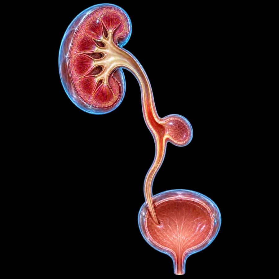
What is a ureteral diverticulum
A ureteral diverticulum is a sac-like outpouching of the ureteral wall, connected to the inner lumen by a narrow or wide neck. When it contains all layers of the wall (mucosa, muscle, and serosa), it is called a true (congenital) diverticulum. When only the mucosa herniates through a weak muscular point, it is called a pseudo-diverticulum (acquired).
Congenital forms arise from incomplete development of the ureteric bud (abortive bifid ureter). Acquired forms are secondary to ureteral stricture, chronic stones, instrumentation, or inflammation.
Important: Due to chronic urinary stasis, diverticula predispose to stone formation, recurrent infections, and — rarely — to urothelial carcinoma. Histologic confirmation is always required at the time of excision.
Culp classification & morphology
Abortive bifid ureter (blind-ending)
An incomplete bifid ureteral system, where one branch ends in a blind sac. It is usually found in the proximal or mid ureter and has a congenital cause.
Multiple small saccular diverticula
Multiple small mucosal outpouchings, typically acquired after chronic vesicoureteral reflux, inflammation, or chemo-/radiotherapy.
Solitary true diverticulum
A rare, solitary diverticulum containing all layers of the ureteral wall. It is usually found in the mid or distal third of the ureter and may reach several centimeters in size.
Pseudo-diverticulum (post-stricture)
Secondary to ureteral stricture or stone obstruction — only mucosa herniates through a weak muscular point.
Symptoms & clinical presentation
Pain
Chronic or intermittent flank pain, colicky when stones form within the diverticulum or when ureteral obstruction occurs.
Infections
Recurrent urinary tract infections from urinary stasis. May progress to pyelonephritis or perinephric abscess.
Hematuria
Gross or microscopic — from mucosal irritation, stones, or rarely urothelial carcinoma within the diverticulum.
Asymptomatic
Many are incidental findings on CT for other indications. Small diverticula without stones/obstruction are observed.
Red flags: urosepsis, hematuria with mass on CT, rapidly progressive pain, hydronephrosis — urgent urological evaluation.
Diagnosis (CTU, RPG, URS)
CT urography (CTU)
This is the gold standard test. It shows the cystic outpouching from the ureteral wall, its communication with the lumen, any stones within the diverticulum, and hydronephrosis. It also allows 3D reconstruction.
Retrograde pyelography (RPG)
This confirmatory test provides a detailed radiographic depiction of the neck, size, and communication with the ureter. It is useful for surgical planning.
Ureteroscopy (URS)
Direct visualization of the diverticular orifice. Allows biopsy to exclude urothelial neoplasia — mandatory in hematuria or suspicious imaging.
MR urography
This is used in children, during pregnancy, in renal failure, or with an iodinated contrast allergy. It provides anatomical detail without radiation.
Renal scintigraphy
This is a MAG3 or DTPA diuretic renography scan, used to assess the functional impact, such as obstruction and split kidney function.
Urine culture + cytology
This is always performed before surgery, to exclude active infection and evaluate cytology for high-grade urothelial cells.
Diagnostic pitfalls: Diverticulum may be confused with ureterocele, periureteral abscess, ovarian cyst, or ureteral cancer with central necrosis. RPG and URS with biopsy are critical for differential diagnosis.
Treatment options
1. Conservative observation
For small asymptomatic diverticula without stones, infections, or obstruction. Annual US/CT and urine culture.
2. Laparoscopic/robotic diverticulectomy + ureteroureterostomy
First-line for diverticula in the proximal and mid ureter. Complete excision + end-to-end anastomosis with JJ stent. Excellent results >95%.
3. Ureteral reimplantation
For distal third diverticula: excision + reimplantation by Cohen or Politano-Leadbetter, with antireflux mechanism. Boari flap or psoas hitch if length is needed.
4. Endoscopic management
For selected small diverticula with a wide neck: ureteroscopy + holmium laser ablation or electrofulguration of the mucosa. Less invasive — higher recurrence rate.
5. Nephroureterectomy
In confirmed urothelial carcinoma within the diverticulum or in a non-functioning kidney from chronic obstruction.
Follow-up & prognosis
- JJ stent removal at 4-6 weeks postoperatively.
- Renal US + creatinine at 3, 6, 12 months, then annually.
- CT urography at 1 year — check anastomotic patency.
- Cystoscopy + cytology annually × 5 years if confirmed or suspicious urothelial pathology.
- Immediate evaluation for new hematuria, pain, or infection.
In summary: A rare anatomical anomaly with excellent prognosis after complete excision and reconstruction. Proper preoperative workup and exclusion of malignancy are critical.
Frequently Asked Questions (FAQ)
What is a ureteral diverticulum?
A ureteral diverticulum is a rare anatomical anomaly: a sac-like outpouching of the ureteral wall. It can be congenital (true), containing all layers of the ureteral wall, or acquired (false, also called pseudo-diverticulum). The acquired form involves only mucosal herniation through a weak muscular point, usually following stricture, stones, or chronic inflammation.
How is it classified?
Ureteral diverticula are classified using the Culp classification (1947), which defines three types. Type 1 is an abortive bifid ureter, a blind-ending branch usually found in the proximal ureter. Type 2 is multiple small saccular diverticula, often acquired after inflammation. Type 3 is a solitary true diverticulum, which is rare and found in the mid or distal ureter.
How common is it?
Ureteral diverticulum is very rare, with fewer than 100 cases reported in the literature. Autopsy studies estimate an incidence of about 0.01-0.5%. It is roughly twice as common in men as in women. It can appear at any age: the congenital form is usually found in children, while the acquired form is more common in adults over 50.
What are the symptoms?
Ureteral diverticulum is often asymptomatic and found incidentally on imaging. When symptoms occur, they can include flank pain, recurrent urinary tract infections, and blood in the urine (hematuria). Urinary stasis within the diverticulum can also cause stone formation and a palpable mass on imaging. Rarely, the mass effect leads to obstruction with hydronephrosis, a swelling of the kidney.
How is it diagnosed?
CT urography (CTU) is the gold standard test. It shows the cystic outpouching and its narrow neck connecting to the ureter. Retrograde pyelography (RPG) confirms this connection and measures its dimensions. Ureteroscopy (URS) allows direct visualization and biopsy if malignancy is suspected. MR urography is used in children to avoid radiation exposure.
When is surgery needed?
Surgery is needed for symptomatic diverticula, meaning those causing recurrent infections, stones, significant pain, hematuria, obstruction, or suspected malignancy. Small, asymptomatic diverticula are instead monitored with annual ultrasound or CT scans.
What are the surgical options?
The main treatment principle is diverticulectomy with ureteral reconstruction. For the mid or proximal ureter, this means resection followed by end-to-end ureteroureterostomy. For the distal ureter, it means ureteral reimplantation (Cohen or Politano-Leadbetter technique), with a psoas hitch or Boari flap if extra length is needed. These procedures are usually performed laparoscopically or robotically. In selected small diverticula, endoscopic management with a holmium laser is also an option.
What is the prognosis?
The prognosis is excellent after complete excision and reconstruction, with a success rate above 95%. Kidney function is preserved when there was no chronic obstruction beforehand. Possible complications include anastomotic stricture (under 5% of cases), urinary tract infection, and, rarely, malignant transformation (urothelial carcinoma) in chronically irritated diverticula.
Related Topics
Suspected ureteral diverticulum?
Contact us for complete urological assessment: CT urography, retrograde pyelography, ureteroscopy and an individualized reconstructive plan.
Scientific References
- EAU Guidelines — uroweb.org
- Culp OS. Ureteral diverticulum: classification of the literature and report of an authentic case. J Urol 1947;58(5):309-21 — pubmed.ncbi.nlm.nih.gov
- Sarajlic M, Drabbe R. Ureteral diverticula: a review and contemporary management. BJU Int 2018;121(6):837-43 — pubmed.ncbi.nlm.nih.gov
- Wegner HE, Pichlmeier U, Loy V, Dieckmann KP. Ureteric diverticula. Br J Urol 1995;75(4):524-7 — pubmed.ncbi.nlm.nih.gov
- Macejko AM, Stein E, Zampini A, et al. Robotic-assisted laparoscopic ureteral diverticulectomy. J Endourol 2008;22(11):2469-72 — pubmed.ncbi.nlm.nih.gov
Meet the Doctor

Dr. Marinos Vasilas, Urologist – Andrologist
Dr. Marinos Vasilas manages ureteral diverticula with a modern laparoscopic and robotic approach: diverticulectomy, end-to-end anastomosis, and ureteral reimplantation per EAU Guidelines.
See full profile


