My Clinical Approach
Retroperitoneal fibrosis is rare but threatening to renal function. Management is multilevel: urgent kidney rescue through decompression + immunosuppressive therapy + surgical repair where needed.
According to EAU 2024 (ureteral obstruction), ACR criteria 2020 for IgG4-RD and ESH 2023:
- Early decompression with JJ stents or PCN in bilateral hydronephrosis.
- Contrast CT/MRI + serum IgG4 + ESR/CRP for diagnostic staging.
- CT-guided biopsy for histologic diagnosis and exclusion of malignancy.
- First-line corticosteroids (prednisolone 1 mg/kg taper).
- Tamoxifen / MMF / Rituximab in relapse or IgG4-RD.
- Ureterolysis with omental wrap laparoscopically/robotically in failure.
- Long-term follow-up with CT/MRI every 6-12 months + renal function.
What is retroperitoneal fibrosis
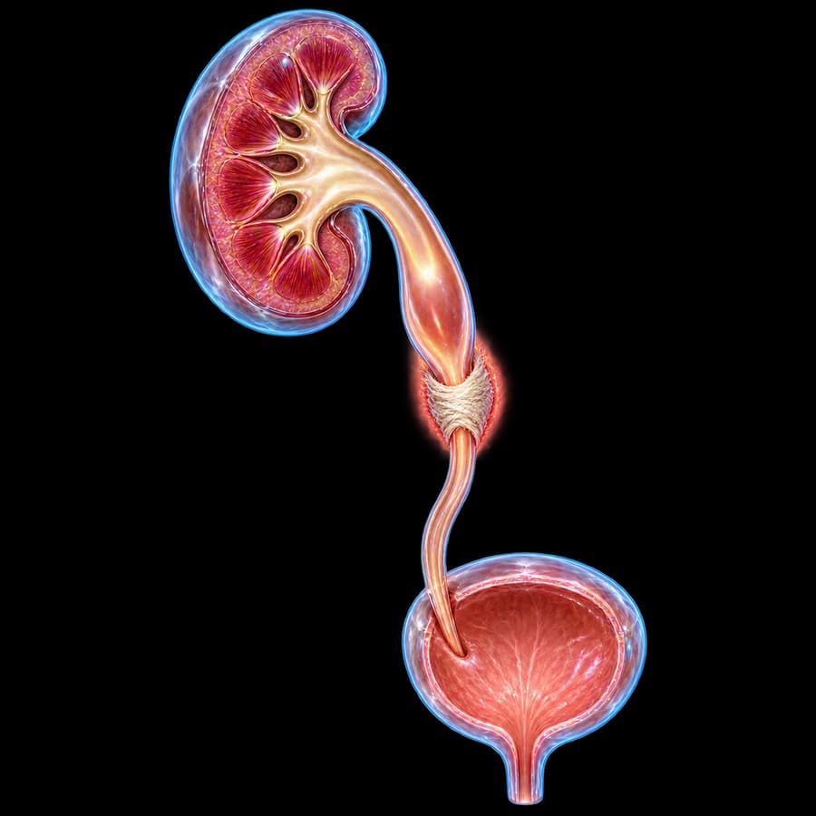
Retroperitoneal fibrosis (RPF — Ormond's disease) is characterized by accumulation of fibrous tissue that encases the descending abdominal aorta, IVC and ureters, without displacing the vessels. First described by Albarran (1905) and Ormond (1948).
Incidence: ~1:200,000-500,000 annually. Men:women = 3:1. Most common diagnostic age: 40-60 years. In 30% of cases there is systemic autoimmune disease (IgG4-RD, ANCA-associated vasculitis, rheumatoid arthritis).
Important: RPF does not displace the aorta anteriorly, a feature that distinguishes it from retroperitoneal lymphoma or sarcoma.
Causes (idiopathic vs secondary)
Idiopathic (2/3 — 70%)
- • IgG4-related disease (up to 50%)
- • Autoimmune inflammatory
- • Ormond's disease
- • Association with abdominal aortic aneurysms (inflammatory aneurysm)
Secondary (1/3 — 30%)
- • Drugs: methysergide, ergotamine, β-blockers, methyldopa, hydralazine, amiodarone
- • Malignancy: lymphoma, metastases (breast, prostate, lung), carcinoid
- • Infections: tuberculosis, actinomycosis, histoplasmosis
- • Radiotherapy of abdomen/pelvis
- • Surgery retroperitoneal / trauma
Symptoms & clinical presentation
Classic triad:
- Chronic dull flank / abdominal pain (95%) — non-specific, lasting weeks to months.
- Bilateral hydronephrosis (75%) — gradual ureteral obstruction.
- Elevated ESR/CRP/creatinine — laboratory findings.
Systemic
Fatigue, weight loss, low-grade fever, anorexia, night sweats.
From IVC compression
Lower limb edema, deep vein thrombosis (DVT), testicular weight (bilateral varicocele).
From acute kidney injury
Oliguria/anuria, hyperkalemia, nausea/vomiting, hypertension, fluid overload.
From arterial compression
Intermittent claudication, severe renovascular hypertension from renal artery stenosis (rare).
Diagnosis (CT/MRI, biopsy, IgG4)
Laboratory
ESR and CRP are elevated in 80% of cases. Creatinine, eGFR, urinalysis, and serum IgG4 (above 135 mg/dL is suspicious), along with ANA, ANCA, and rheumatoid factor, help exclude systemic autoimmune disease.
Renal ultrasound
This first-line test shows bilateral hydronephrosis and may reveal a retroperitoneal mass.
Contrast-enhanced abdominal CT
This is the main diagnostic test. It shows a characteristic retroperitoneal mass that encases the aorta, IVC, and ureters without displacing them, which distinguishes it from malignancy, since RPF does not displace the aorta anteriorly.
Abdominal MRI
This offers better soft tissue resolution. On T2 sequences, active RPF shows a high signal from edema and inflammation, while chronic, inactive disease shows a low signal from fibrosis.
PET-CT (FDG)
This assesses disease activity through increased FDG uptake in inflammatory areas, helps monitor treatment response, and helps differentiate the disease from malignancy.
CT-guided biopsy
This confirms the diagnosis by showing lymphoplasmacytic infiltrates, increased IgG4+ plasma cells (more than 10 per high-power field in IgG4-RD), and storiform fibrosis, while also excluding malignancy such as lymphoma or sarcoma.
DMSA / scintigraphy
This assesses renal function before and after decompression.
Immediate decompression (emergency)
Sepsis or AKI = emergency. Decompression within 24 hours. Undiagnosed hyperkalemia >6.5 mmol/L → immediate hemodialysis.
JJ stent (first line)
Endoscopic placement under fluoroscopy. May fail in severe ureteral encasement by fibrosis. Exchange every 3-6 months until definitive treatment.
Percutaneous nephrostomy (PCN)
When endoscopic JJ stent placement fails. Immediate kidney decompression, monitoring of diuresis. Gradual transition to JJ stent when fibrosis is achieved.
Post-decompression care
IV fluids + electrolyte monitoring (often post-decompression diuresis), antibiotics (cephalosporin + aminoglycoside in sepsis), acidosis correction.
Medical therapy
1. Corticosteroids (first line)
Prednisolone 1 mg/kg/day is given for 4 weeks, then tapered to a 5-10 mg/day maintenance dose for 6-12 months. The response rate is 75%, and monitoring includes ESR, CRP, and a CT mass check every 3 months.
2. Tamoxifen
Tamoxifen 20 mg twice daily is an alternative for patients with contraindications to steroids, or it can be combined with them. Its response is similar to low-dose corticosteroids (van Bommel 2009).
3. Mycophenolate Mofetil (MMF)
MMF 1 g twice daily is combined with low-dose prednisolone for relapse or resistance to corticosteroid monotherapy, allowing a steroid-sparing approach for chronic immunosuppression.
4. Rituximab (anti-CD20)
Rituximab, given as 1 g IV for 2 doses, 2 weeks apart, is highly effective in IgG4-related disease (Khosroshahi 2013) and is indicated after failure or relapse on other therapies.
5. Methotrexate / Cyclophosphamide
These are alternative immunosuppressives for difficult cases, though they are now often replaced by MMF or rituximab due to a better safety profile.
Surgical management
Indications: failure of conservative therapy ≥6 months, re-obstruction after stent removal, multiple relapses, intolerable side effects of immunosuppressants.
Ureterolysis with omental wrap
This is the standard technique: the fibrous tissue around the ureters is removed, the ureters are transposed intraperitoneally, and they are wrapped with omentum as an anti-fibrotic barrier. A laparoscopic or robotic approach is preferred, with a success rate of 80-90%.
Ureteral substitution
For extensive ureteral damage, options include plasty with buccal mucosa, ileal interposition, renal autotransplantation, or a Boari flap.
Permanent nephrostomy
This is used in patients unfit for surgery or after failed ureterolysis. Alternatives include Resonance metallic stents, which last about 12 months, or renal autotransplantation to preserve function.
Frequently Asked Questions (FAQ)
What is retroperitoneal fibrosis?
Retroperitoneal fibrosis (Ormond's disease) is a rare chronic inflammatory disease characterized by deposition of fibrous tissue in the retroperitoneal space. This tissue encases the aorta, inferior vena cava, and ureters, causing progressive obstruction and hydronephrosis. Incidence is about 1 in 200,000 annually, and it is more common in men (3:1), typically between ages 40 and 60.
What are the causes?
About two-thirds of cases are idiopathic, often related to autoimmune or IgG4-related disease. The remaining third are secondary, caused by drugs (methysergide, ergotamine, β-blockers, methyldopa), malignancy (lymphoma, metastasis), infection (tuberculosis, actinomycosis), radiotherapy, or abdominal aortic aneurysms.
What are the symptoms?
The classic triad is chronic flank pain, bilateral hydronephrosis, and elevated creatinine, ESR, and CRP. Systemic symptoms include fatigue, weight loss, and low-grade fever. In advanced disease, patients may develop acute kidney injury (AKI) symptoms, lower limb edema from IVC compression, hypertension, or anuria.
How is it diagnosed?
Contrast-enhanced CT or MRI of the abdomen shows a retroperitoneal mass encasing the aorta, IVC, and ureters, without displacing the vessels, which differs from malignancy. PET-CT assesses disease activity, and CT-guided biopsy confirms the diagnosis while excluding malignancy or IgG4-related disease. Serum IgG4, ESR, CRP, and renal function are also checked.
How is acute obstruction managed?
Immediate decompression is achieved with an endoscopic JJ stent, or percutaneous nephrostomy (PCN) if the endoscopic approach fails. Sepsis or AKI is treated as an emergency, initially with IV fluids, antibiotics, and electrolyte monitoring for post-decompression diuresis.
What is the medical treatment?
Corticosteroids are first-line: prednisolone 1 mg/kg/day for 4 weeks, tapered over 6-12 months. Tamoxifen 20 mg twice daily is an alternative or can be combined with steroids. Mycophenolate mofetil 1 g twice daily is used in relapse or resistance, and rituximab is used in IgG4-related disease, given as 1 g IV for 2 doses.
When is surgery indicated?
Ureterolysis with omental wrap is indicated when conservative therapy fails for 6 months or more, when re-obstruction occurs after stent removal, or in relapse. A laparoscopic or robotic approach is preferred, with a success rate of 80-90%.
What is the prognosis?
5-year survival is 90% with the idiopathic form. Renal function recovers in 70-80% of patients with timely decompression, though relapse occurs in 30%, so long-term follow-up with CT/MRI and renal function testing is required every 6 months, then annually. Secondary forms depend on the underlying cause.
What is the relationship with IgG4-related disease?
Up to 50% of idiopathic cases belong to the spectrum of IgG4-related disease (IgG4-RD), a systemic autoimmune disease marked by elevated serum IgG4 and lymphoplasmacytic infiltrates on biopsy. It often coexists with sialadenitis, type 1 pancreatitis, or cholangitis, and rituximab is highly effective for it.
Related topics
Suspected retroperitoneal fibrosis?
Contact us for full urologic and immunorheumatologic evaluation: CT/MRI, IgG4, biopsy and personalized plan — from urgent decompression to immunosuppressive therapy and ureterolysis.
Scientific literature
- EAU Guidelines on Urological Trauma & Ureteral Obstruction (2024) — uroweb.org
- Vaglio A, Salvarani C, Buzio C. Retroperitoneal fibrosis. Lancet 2006;367(9506):241-51 — pubmed.ncbi.nlm.nih.gov
- van Bommel EFH et al. Tamoxifen vs prednisone for treatment of idiopathic retroperitoneal fibrosis. Eur Urol 2009;55(6):1448-55 — pubmed.ncbi.nlm.nih.gov
- Khosroshahi A et al. Rituximab therapy leads to rapid decline of serum IgG4 levels and prompt clinical improvement in IgG4-related disease. Medicine 2013;92(2):82-91 — pubmed.ncbi.nlm.nih.gov
- Fenaroli P et al. Mycophenolate mofetil and prednisone vs prednisone monotherapy for idiopathic retroperitoneal fibrosis: an RCT. Lancet Rheumatol 2021;3(7):e483-e491 — pubmed.ncbi.nlm.nih.gov
Medical Editorship

Dr. Marinos Vasilas, Urologist – Andrologist
Dr. Marinos Vasilas treats retroperitoneal fibrosis with modern multilevel approach: urgent endoscopic decompression, pharmacologic immunoregulation (corticosteroids/tamoxifen/MMF/rituximab) and surgical ureterolysis with omental wrap laparoscopically/robotically per EAU 2024.
View full profile


