My Clinical Approach
Microlithiasis is one of the most misunderstood findings in urology. Patients often arrive at the office frightened after an ultrasound report mentioning "multiple microcalcifications". My first job is to make clear that this is not cancer.
Strategically, I follow the 2024 EAU risk-stratification framework: I assess for coexisting factors (history of cryptorchidism, atrophy <12 ml, infertility, family or personal history of germ cell tumor). In high-risk cases I schedule annual ultrasound and teach monthly self-examination.
Patient education is the most powerful "tool": early detection of any new mass by the patient himself is the essence of surveillance.
What Is Microlithiasis?
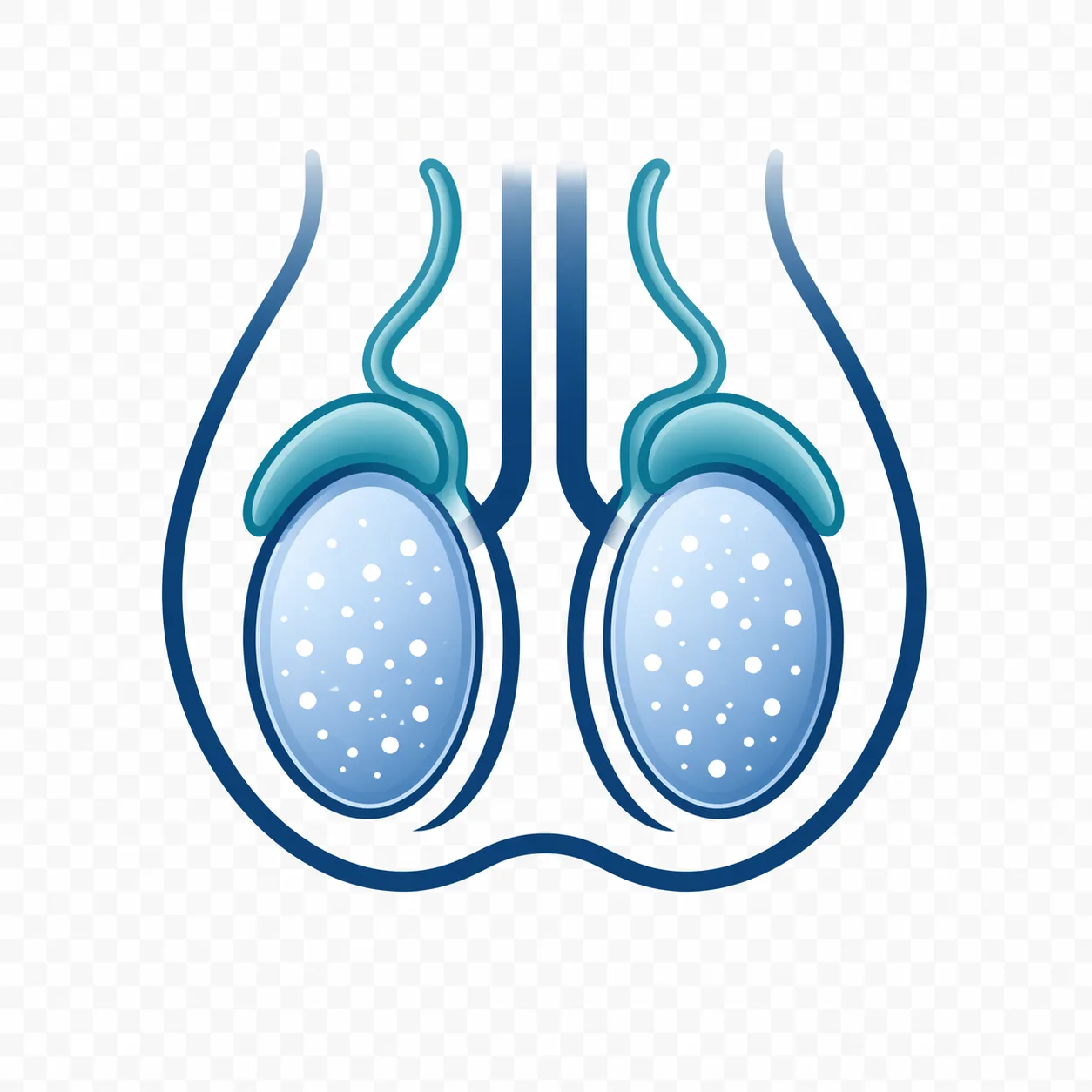
Testicular microlithiasis (TM) is the presence of multiple calcific foci <3 mm in diameter within the seminiferous tubules. On ultrasound they appear as small hyperechoic specks without acoustic shadowing.
- Classic microlithiasis (CTM): ≥5 calcifications in a single sonographic plane
- Limited microlithiasis (LTM): <5 calcifications in a single plane
- General population prevalence: 2.4–5.6%
- Prevalence in infertile men: 8–20%
Symptoms
Microlithiasis is asymptomatic. It is discovered incidentally on scrotal ultrasound performed for another reason — usually infertility workup, pain, hydrocele or varicocele evaluation.
If new symptoms appear (palpable mass, size change, heaviness, pain), the workup is no longer about microlithiasis but about possible tumor — evaluation is immediate.
Risk Factors for Tumor in Men with Microlithiasis
- Cryptorchidism (undescended testes) — strongest association
- Testicular atrophy (volume <12 ml)
- Infertility or abnormal semen analysis
- Personal history of germ cell tumor
- Family history in a first-degree relative
- GCNIS on previous biopsy
- Klinefelter syndrome
The 2024 EAU and ESUR (European Society of Urogenital Radiology) emphasize: microlithiasis without coexisting risk factors does not significantly increase germ cell tumor risk in the general population.
Diagnosis
Scrotal ultrasound
The only essential tool. Multiple small hyperechoic foci (1–3 mm) without acoustic shadowing, diffusely distributed in the parenchyma. Includes assessment of volume and parenchymal homogeneity.
Semen analysis
In men of reproductive age with microlithiasis, especially if accompanying clinical findings exist (varicocele, atrophy).
Hormonal workup
Testosterone, LH, FSH in cases of infertility or atrophy.
Management & Surveillance
Low risk (isolated microlithiasis)
Reassurance. Self-examination teaching. No need for routine ultrasound follow-up.
High risk (1+ coexisting factors)
Annual scrotal ultrasound plus monthly self-examination until age 55 (when tumor risk significantly decreases).
Very high risk
In rare cases (microlithiasis + cryptorchidism + atrophy + infertility), diagnostic biopsy for GCNIS detection is considered.
Fertility
Microlithiasis correlates with reduced sperm count in some studies, without a clear causal relationship. In men with fertility problems and concomitant microlithiasis, a complete andrological evaluation (semen analysis, hormonal workup) is the right approach.
Prognosis
In isolated microlithiasis, long-term prognosis is excellent and quality of life is unaffected. In the high-risk group, annual surveillance ensures early diagnosis of any tumor at a fully curable stage.
Frequently Asked Questions (FAQ)
Is testicular microlithiasis cancer?
No. Microlithiasis refers to small calcified deposits within the seminiferous tubules and is not malignant in itself. However, it correlates with an increased risk of germ cell tumor when combined with other risk factors.
When does it require follow-up?
When other factors coexist, such as cryptorchidism, testicular atrophy, infertility, personal or family history of germ cell tumor, or GCNIS. In isolated microlithiasis without risk factors, the 2024 EAU guidelines do not recommend intensive surveillance.
Do I need a biopsy?
Not routinely. Testicular biopsy is considered only in highly selected high-risk cases (e.g., microlithiasis with cryptorchidism, infertility and atrophy) where there is strong suspicion of GCNIS.
Does it affect fertility?
Microlithiasis itself does not cause infertility, but it correlates statistically with abnormal semen analyses. A semen analysis is recommended in men with fertility issues who also have microlithiasis.
How often should I have an ultrasound?
High-risk group: annual scrotal ultrasound plus monthly self-examination. Low-risk: self-examination without routine imaging.
Book Your Appointment in Rhodes
If your ultrasound found microlithiasis, do not panic — but do request specialist evaluation so you are correctly classified by risk and receive an individualized surveillance plan.
References
- EAU Guidelines on Testicular Cancer, 2024 update — uroweb.org
- ESUR-ESPR position statement on testicular microlithiasis. Eur Radiol 2015 — PMID: 25516431
- Pedersen MR et al. Testicular microlithiasis and testicular cancer risk. World J Urol 2018 — PMID: 29354878
Medical Review

Dr. Marinos Vasilas, Urologist – Andrologist
Dr. Marinos Vasilas runs a private urology practice in Rhodes, with particular focus on imaging-based risk stratification and andrological care of patients with testicular microlithiasis findings.
See full biography


