My Clinical Approach
For hydroceles I follow a selective approach: I do not operate on every hydrocele. Indication must be clear (symptoms, size, cosmetic concern).
I always rule out underlying pathology (tumor, infection) with ultrasound before proposing intervention. In patients >40 years with new hydrocele, I increase risk-stratification for underlying pathology.
My technique of choice is the Jaboulay technique (eversion of the tunica vaginalis) with recurrence rate <5%, consistent results and secure closure.
What is Hydrocele?
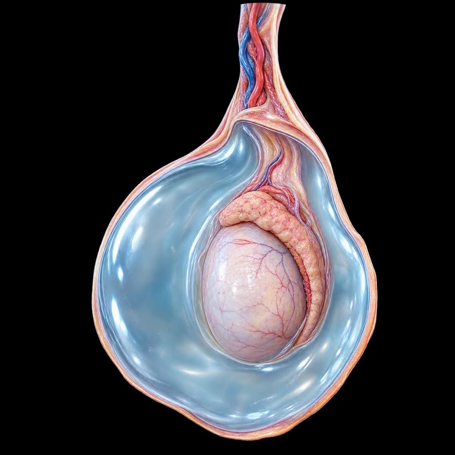
Hydrocele is the abnormal collection of serous fluid in the tunica vaginalis surrounding the testis. Normally, a minimal amount of fluid allows testicular movement.
- Frequency: 1% of adult men
- 80-90% of newborns have communicating hydrocele
- Usually unilateral
- Primary (idiopathic) in adults
- Secondary after infection, trauma, tumor
- Painless and slowly progressive
Types of Hydrocele
Communicating Hydrocele
In neonates/infants, due to patent processus vaginalis. Associated with inguinal hernia. Often spontaneously resolves by 1-2 years.
Non-communicating (Primary)
In adults, no communication with peritoneal cavity. From imbalance of fluid production/absorption.
Secondary Hydrocele
After epididymitis, orchitis, trauma, testicular cancer, surgery. Often treating cause resolves the problem.
Spermatic Cord Hydrocele
Fluid collection along the spermatic cord, separate from the testis.
Symptoms
- Painless scrotal swelling
- Sensation of heaviness or pulling
- Gradual increase in size (months/years)
- Increased size at end of day
- Difficulty palpating the testis
- Difficulty with clothing/sexual activity (large hydroceles)
- Cosmetic concern
- Rarely pain (if secondary)
Diagnosis
- Clinical examination: elastic, painless mass around testis
- Transillumination: positive (light passes through fluid)
- Scrotal ultrasound (essential)
- • Confirmation of fluid collection
- • Exclusion of tumor, infection
- • Assessment of testicular texture
- Doppler to exclude torsion
- For suspected communicating: Valsalva, standing/sitting examination
For new hydrocele in adults (especially >40 years), ultrasound is essential to rule out underlying testicular tumor.
Treatment
Observation
In neonates/infants up to 1-2 years (spontaneous resolution). In asymptomatic adults with small hydrocele.
Hydrocelectomy - Jaboulay Technique
- • Eversion of the tunica vaginalis
- • Scrotal incision 4-5 cm
- • Local or general anesthesia
- • Duration: 30-45 minutes
- • Recurrence: <5%
Lord Technique (plication)
For smaller hydroceles, with less trauma to the spermatic cord. Same recurrence rates.
Sclerotherapy (selected cases)
Aspiration + sclerosing agent injection (tetracycline, polidocanol). Applied in high surgical-risk patients. Recurrence 10-30%.
Inguinal Approach (communicating)
In children: closure of patent processus vaginalis through inguinal incision.
Recovery
- Same-day discharge
- Ice packs first 24 hours
- Scrotal support (special underwear)
- Return to light activities in 3-7 days
- Return to work in 1 week
- Sports activities in 3-4 weeks
- Sexual activity in 2-3 weeks
- Transient scrotal swelling 2-4 weeks
Complications & Recurrence
- Hematoma: 1-3%
- Wound infection: 1-2%
- Transient swelling: almost in all
- Recurrence: <5% with Jaboulay/Lord
- Testicular/epididymal injury: <1%
- Chronic pain: <2%
- Fertility impact: insignificant
Frequently Asked Questions (FAQ)
When is hydrocele surgery needed?
In adults, surgical management (hydrocelectomy) is indicated when it causes discomfort, pain, large size, cosmetic concern, or suspicion of underlying pathology. In neonates/infants, most hydroceles resolve spontaneously by 1-2 years of age.
Is hydrocele dangerous?
Hydrocele itself is not dangerous and does not affect fertility. However, it may mask underlying pathology (testicular tumor, infection, trauma). Therefore ultrasound examination is essential.
Can aspiration be done instead of surgery?
Percutaneous aspiration provides short-term relief but recurs in 100% of cases. Not recommended. Sclerotherapy is useful only in selected high surgical-risk patients.
How long is recovery?
Return to light activities in 3-7 days, work in 1 week, sports in 3-4 weeks. Scrotal swelling lasts 2-4 weeks.
What is the difference between hydrocele and varicocele?
Hydrocele is fluid collection around the testis (transilluminates). Varicocele is dilation of testicular veins ("bag of worms"). Ultrasound easily differentiates them.
Book Your Appointment in Rhodes
Hydrocele diagnosis with ultrasound and safe surgical management with Jaboulay/Lord technique and low recurrence rates.
References – Sources
- EAU Guidelines on Paediatric Urology, 2024 update — uroweb.org
- Lord PH. A bloodless operation for the radical cure of idiopathic hydrocele. Br J Surg 1964 — PMID: 14129420
- Ku JH, et al. The excisional, plication and internal drainage techniques. BJU Int 2001 — PMID: 11578439
Medical Review

Dr. Marinos Vasilas, Urologist – Andrologist
Dr. Marinos Vasilas runs a private urology practice in Rhodes specializing in surgical conditions of the scrotum and testis.
View full CV


