My Clinical Approach
In daily clinical practice, testicular cancer is a disease where time matters, but panic is rarely justified. The vast majority of young men presenting with a testicular mass will have an excellent prognosis if assessed promptly and correctly.
A small, localized tumor discovered on self-examination is very different from a delayed presentation with metastatic disease. Unfortunately, many young men delay seeking medical attention out of embarrassment, fear, or hope that the lump will resolve on its own. It will not.
My personal approach rests on three pillars:
- Rapid diagnostic workup: scrotal ultrasound and hormonal/oncological evaluation within 24 to 72 hours.
- Radical inguinal orchiectomy as the first therapeutic step — never a transscrotal biopsy.
- Multidisciplinary decision-making after pathology results, based on EAU and ESMO protocols.
Every newly diagnosed patient is offered sperm cryopreservation before any oncological treatment. This is a non-negotiable EAU recommendation.
What Is Testicular Cancer?
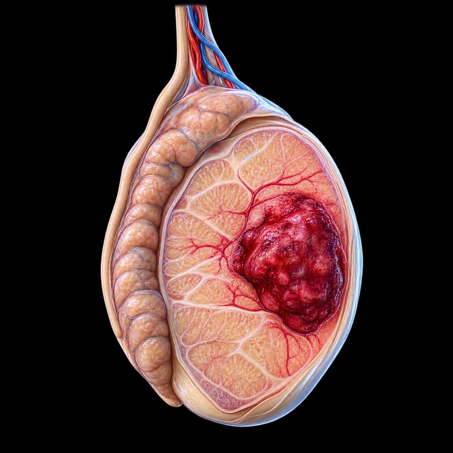
Testicular cancer is the development of a malignant tumor from cells of the testis. More than 95 percent of cases are germ cell tumors (GCTs), classified into two major histological categories with distinct treatment approaches:
Seminomas (~50%)
Slower growth, exquisitely sensitive to radiotherapy and cisplatin-based chemotherapy. More common between ages 30 and 45. Rarely elevate AFP.
Non-Seminomatous (NSGCT, ~50%)
More aggressive, faster growth, and more often metastatic at diagnosis. Includes embryonal carcinoma, choriocarcinoma, yolk sac tumors and teratoma. Often elevate AFP and/or beta-hCG.
Less common (<5 percent) are sex cord-stromal tumors (Leydig, Sertoli) and testicular lymphomas, which mainly occur in older men.
Incidence & Epidemiology
According to the EAU Guidelines on Testicular Cancer (2024), incidence in Europe is 7 to 10 cases per 100,000 men annually, with a steady rise over the past 50 years. Mortality nonetheless remains low (<0.4/100,000) thanks to modern therapies.
Symptoms & Early Detection
The hallmark sign is a painless, firm lump or asymmetric enlargement in one testicle, noticed on self-examination or incidentally (e.g., in the shower). Other possible signs include:
- Sensation of heaviness or fullness in the scrotum
- Dull ache in the lower abdomen or groin
- Sudden pain (10–20% — may mimic epididymitis)
- Breast enlargement (gynecomastia) due to beta-hCG production
- Fluid collection in the scrotum (reactive hydrocele)
- Symptoms of metastases: back pain, cough, lymphadenopathy — in advanced disease
Monthly testicular self-examination — ideally after a warm shower when the scrotum is relaxed — remains the most effective early detection tool in young men, particularly those with risk factors.
See a Urologist Without Delay
Immediate evaluation is required when you notice:
- A new firm lump or testicular swelling (even painless)
- Asymmetry or change in shape / consistency
- Persistent pain or heaviness in the scrotum
- Breast enlargement without an obvious cause
Delay is the single factor that can change the prognosis of an otherwise highly curable disease.
Causes & Risk Factors
The exact causes of testicular cancer are not fully understood. It is believed to originate from undifferentiated intratubular germ cells (GCNIS) already present in fetal life, which evolve into cancer decades later. The EAU recognizes specific risk factors:
- Cryptorchidism (undescended testes) — increases risk 4 to 8-fold, even after orchidopexy
- Family history of testicular cancer (brother, father)
- Personal history of cancer in the contralateral testicle (2–5% risk for the second)
- Klinefelter syndrome and other chromosomal abnormalities
- Testicular atrophy or hypoplasia
- Infertility and abnormal semen analysis
- Germ Cell Neoplasia In Situ (GCNIS) — a true precancerous condition
Important
Testicular microlithiasis on its own is NOT considered precancerous. The 2024 EAU guidelines clarify that close follow-up is only warranted when additional risk factors coexist (cryptorchidism, atrophy, infertility, family history).
Diagnosis & Staging
The diagnostic workflow is well-defined, rapid, and should be completed within a few days:
1. Clinical examination
Palpation of both testicles by an experienced urologist to identify the mass, assess size, consistency, and location, and palpate the epididymis and spermatic cord.
2. High-resolution scrotal ultrasound
The investigation of choice with sensitivity above 95 percent. Differentiates intra- from extra-testicular masses, solid from cystic lesions, evaluates vascularity and the contralateral testis. Per EAU, ultrasound is mandatory for any testicular mass.
3. Serum tumor markers
Before and after orchiectomy: AFP (alpha-fetoprotein), beta-hCG (chorionic gonadotropin) and LDH. Critical for diagnosis, IGCCCG staging, follow-up and early relapse detection.
4. Radical inguinal orchiectomy
Both diagnostic and therapeutic. Always performed via an inguinal approach — never transscrotal — to avoid disrupting normal lymphatic drainage. Transscrotal biopsy is contraindicated.
5. Postoperative staging
CT of chest, abdomen and pelvis to detect retroperitoneal lymph nodes and pulmonary metastases. Brain MRI or bone scan only if clinically indicated. Tumor markers are repeated according to their half-life kinetics.
TNM & IGCCCG Classification
Staging combines the anatomical TNM (8th AJCC/UICC edition) with marker levels (S0–S3). In advanced disease, the IGCCCG (Good / Intermediate / Poor prognosis) classification is applied to guide chemotherapy intensity.
Treatment
Treatment is individualized based on histologic type (seminoma vs NSGCT), stage, tumor markers and IGCCCG risk group. All decisions are taken in a multidisciplinary tumor board.
Stage I (localized disease)
After orchiectomy, three evidence-based strategies with equivalent overall survival are available:
- Active surveillance — preferred option for low-risk patients
- Adjuvant chemotherapy (1 cycle of carboplatin for seminoma, 1 cycle of BEP for high-risk NSGCT)
- Radiotherapy (seminoma only — used less often today due to long-term toxicity)
Stage II (regional lymph nodes)
For seminoma IIA/IIB: radiotherapy or chemotherapy (3 cycles BEP or 4 EP). For NSGCT: usually 3–4 cycles of BEP followed by retroperitoneal lymph node dissection (RPLND) for residual masses.
Stage III (distant metastases)
Systemic cisplatin-based chemotherapy: 3 cycles BEP (good prognosis) or 4 cycles BEP/VIP (intermediate/poor). Surgical resection of residual masses >1 cm after chemotherapy is standard in NSGCT.
Salvage Therapy
For relapsed or primary refractory disease, regimens such as TIP, VeIP or high-dose chemotherapy with autologous stem cell transplant are used. Even at this stage, treatment can be curative.
Organ-Sparing Surgery
In selected cases (small tumors <2 cm in a solitary testis, bilateral tumors, or hypogonadal patients), the EAU permits partial orchiectomy to preserve hormonal function — provided it is performed in specialized centers.
Fertility & Sperm Preservation
Many patients are diagnosed during their reproductive years. The EAU is unequivocal: every patient should be offered sperm cryopreservation BEFORE any treatment — orchiectomy, radiotherapy, chemotherapy, or RPLND.
Key facts the patient should know:
- Many patients already have abnormal semen parameters at diagnosis — the disease is associated with infertility.
- Removal of one testicle generally does not impair fertility when the other is normal.
- Cisplatin can cause transient or permanent azoospermia.
- RPLND can cause retrograde ejaculation — modern nerve-sparing techniques greatly reduce this risk.
- Recovery of spermatogenesis can take 1 to 4 years after completion of treatment.
In young patients undergoing bilateral orchiectomy, or those developing hypogonadism after treatment, testosterone replacement therapy is considered.
Follow-up & Prognosis
Post-treatment surveillance is close and long-term. The aim is early detection of relapse (which in germ cell tumors remains curable) and recognition of long-term treatment-related effects.
Years 1 to 2
Clinical exam plus tumor markers every 2 to 3 months, abdominal CT/MRI every 4 to 6 months, chest evaluation.
Years 3 to 5
Visit every 6 to 12 months with markers, scrotal ultrasound and selected imaging.
Beyond 5 years
Annual or biennial follow-up. Emphasis on long-term cardiovascular and metabolic monitoring and screening for second malignancies.
Prognosis: In stage I, disease-specific 5-year survival exceeds 99 percent. In IGCCCG good prognosis metastatic disease, 5-year survival reaches 92 percent. Even in the poor prognosis group, survival is around 67 percent — figures rarely seen in other malignancies.
Frequently Asked Questions (FAQ)
At what age does testicular cancer most commonly occur?
Testicular cancer is the most common solid malignancy in men aged 15 to 35. However, it can occur at any age, and a smaller second peak is seen after the age of 60.
How will I know if I have testicular cancer?
The most common sign is a painless, firm lump or swelling of the testicle, usually noticed on self-examination. Less commonly there may be heaviness or a dull ache in the scrotum. Any new testicular mass requires prompt urological evaluation.
Is testicular cancer curable?
Yes. Testicular cancer is one of the most curable solid tumors in men. Cure rates exceed 95 to 99 percent for localized disease, and overall survival remains very high even in advanced stages thanks to modern cisplatin-based chemotherapy regimens.
Will I lose my fertility?
Removal of one testicle (radical orchiectomy) usually does not significantly affect fertility or testosterone levels, since the remaining healthy testicle compensates. Nonetheless, the EAU explicitly recommends sperm cryopreservation before any treatment (chemotherapy, radiotherapy, or RPLND).
What are germ cell tumors?
More than 95 percent of testicular malignancies are germ cell tumors (GCTs). They are divided into two major histological categories: seminomas and non-seminomatous germ cell tumors (NSGCT). This distinction guides treatment and prognosis.
Book Your Appointment in Rhodes
If you have noticed a lump or swelling in the testicle, or if you need a second opinion on a testicular cancer diagnosis, prompt urological assessment is critical. Our practice offers rapid diagnostic workup with scrotal ultrasound, hormonal and oncological evaluation, and clear guidance on the next therapeutic step.
References
- EAU Guidelines on Testicular Cancer, 2024 update — uroweb.org
- ESMO Clinical Practice Guidelines: Testicular seminoma and non-seminoma, 2022 — esmo.org
- NCCN Guidelines: Testicular Cancer, Version 2.2024 — nccn.org
- International Germ Cell Cancer Collaborative Group (IGCCCG) Update Consortium — J Clin Oncol 2021 — pubmed.ncbi.nlm.nih.gov
Medical Review

Dr. Marinos Vasilas, Urologist – Andrologist
Dr. Marinos Vasilas runs a private urology practice in Rhodes, with particular focus on uro-oncology and andrology. He provides rapid evaluation of testicular masses, guides accurate staging, and collaborates with reference oncology centers for comprehensive patient care.
See full biography


