My Clinical Approach
For testicular cysts I follow a conservative approach. I do not propose surgery for every cyst incidentally found on ultrasound.
I meticulously distinguish simple from complicated cysts. For suspicious ultrasound findings (solid content, mixed lesion, internal vascularity) I order MRI or perform orchidectomy with intraoperative frozen section.
When surgical enucleation is needed, I use microsurgical technique to preserve as much normal testicular parenchyma as possible.
What is a Testicular Cyst?
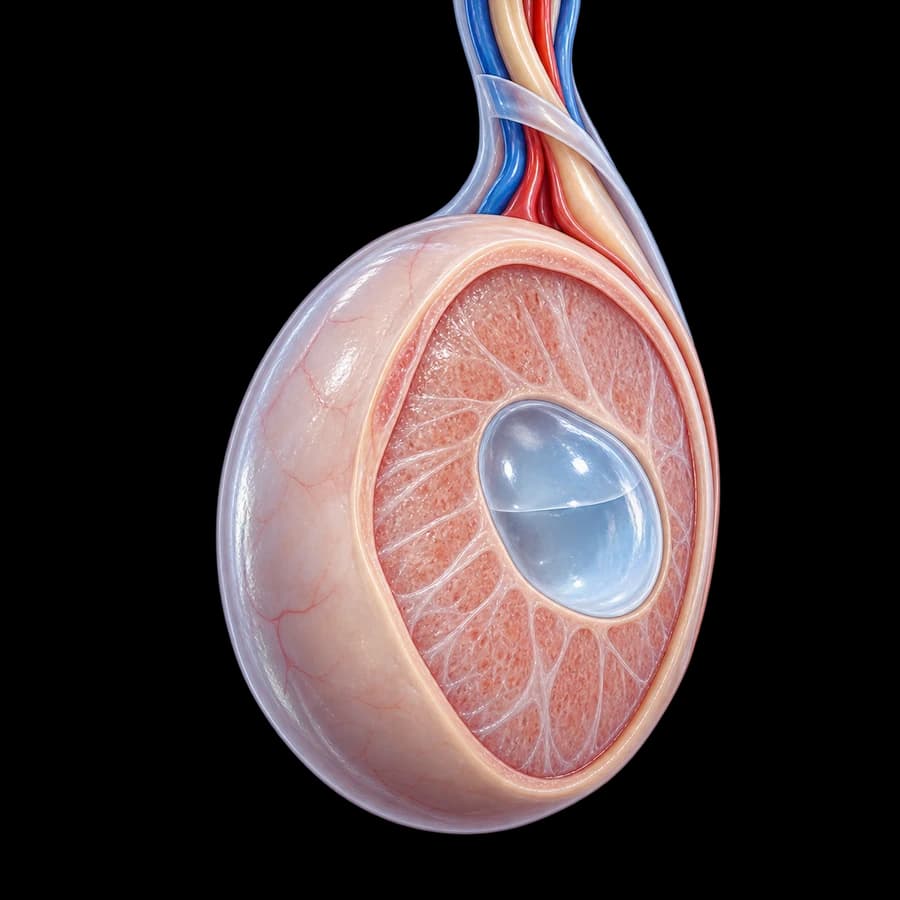
A testicular cyst is a fluid collection within a thin wall, located either in the testicular parenchyma (intratesticular) or in the tunica albuginea. Often an incidental ultrasound finding.
- Frequency: 8-10% on ultrasound in adults
- Age: 20-50 years most commonly
- Usually asymptomatic
- Often bilateral
- Benign in the majority
- Size: from millimeters to several centimeters
Types of Cysts
Tunica Albuginea Cyst
Palpable mass on the surface of the testis, 2-5 mm in size. Felt as a small hard nodule. Benign.
Simple Intratesticular Cyst
In the testicular parenchyma, anechoic, with thin wall. Usually small and asymptomatic.
Epidermoid Cyst
Benign cyst with keratin. On ultrasound it has a characteristic «onion ring» appearance. Can be treated with enucleation.
Rete Testis Cyst (Tubular Ectasia)
Multiple small cystic dilations of rete testis tubules. Always benign. Often in elderly patients.
Symptoms
- Usually asymptomatic
- Palpable nodule (tunica albuginea cyst)
- Sensation of scrotal heaviness (large cysts)
- Rarely mild pain
- Incidental ultrasound finding
- Increase in testicular size (in large intratesticular)
Diagnosis
- Clinical examination: palpation of nodule
- Scrotal ultrasound with Doppler (cornerstone)
- • Anechoic content
- • Thin smooth wall
- • Posterior acoustic enhancement
- • Absence of vascularity
- Tumor markers (if suspicion of malignancy): AFP, β-hCG, LDH
- MRI (in diagnostic dilemmas)
Caution: any solid component within a cyst or asymmetric increase in size should be investigated to rule out cystic teratoma or other tumor.
Treatment
Monitoring (first option)
For simple, asymptomatic cysts. Annual ultrasound.
Enucleation (testis-sparing surgery)
- • For symptomatic, large or complicated cysts
- • Microsurgical technique with parenchyma preservation
- • Intraoperative frozen section if suspicion of malignancy
- • Inguinal approach
Radical Orchidectomy
For suspicion of malignancy or positive frozen section. Inguinal approach (Chevassu).
Monitoring
- Simple cyst <1 cm: annual ultrasound
- Simple cyst 1-2 cm: ultrasound every 6 months initially
- Complicated cyst: every 3-6 months
- Monthly testicular self-examination
- Annual tumor markers if complicated
- Immediate reassessment for size increase
Complications
- Rarely increase in size (<5%)
- Rare rupture
- Very rare malignant transformation
- Surgical complications: hematoma, infection (<3%)
- Loss of testicular parenchyma after enucleation: minimal
- Orchidectomy: only for confirmed malignancy
Frequently Asked Questions (FAQ)
Is a testicular cyst dangerous?
Simple testicular cysts are benign and asymptomatic in the majority of cases. They do not progress to cancer. However, every intratesticular lesion should be evaluated with ultrasound and clinical follow-up.
Is a testicular cyst different from a spermatocele?
Yes. A testicular cyst is located within the parenchyma or the tunica albuginea of the testis. A spermatocele is found in the epididymis and contains sperm. Ultrasound differentiates them.
Is surgery needed?
In most cases no. Surgical removal is performed only when the cyst is large, symptomatic, or there is suspicion of malignancy on ultrasound.
How often should it be monitored?
For small simple cysts: annual ultrasound. For complicated cysts: every 6 months initially and if stable, annually.
Does it affect fertility?
Small simple cysts do not affect fertility. In rare cases of large intratesticular cysts there may be an effect on sperm production.
Book Your Appointment in Rhodes
Diagnosis and monitoring of testicular cysts with ultrasound and microsurgical management when needed.
References – Sources
- EAU Guidelines on Testicular Cancer, 2024 update — uroweb.org
- Dogra VS, et al. Sonography of the scrotum. Radiology 2003 — PMID: 12563126
- Shah A, et al. Testicular epidermoid cysts. AJR 2007 — PMID: 17242237
Medical Review

Dr. Marinos Vasilas, Urologist – Andrologist
Dr. Marinos Vasilas runs a private urology practice in Rhodes specializing in conditions of the scrotum and testis.
View full CV


