1. My Clinical Approach to Prostate Biopsy
The decision for a prostate biopsy is never based on a single PSA value alone. In my practice, I always evaluate the full clinical picture — PSA, PSA density, digital rectal examination, age, family history, and, where indicated, multiparametric prostate MRI — before recommending a transrectal biopsy.
In practice, I always:
- Explain that an elevated PSA does not equal a cancer diagnosis.
- Use MRI before the biopsy when supported by current guidelines and available.
- Clearly inform patients about the real infection risk of the transrectal approach.
- Honestly discuss the comparison with transperineal biopsy, without presenting either technique as universally better.
2. What Is Transrectal Prostate Biopsy
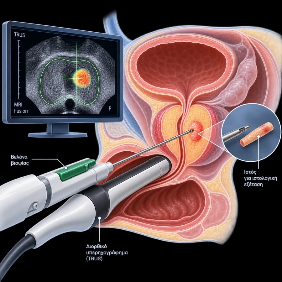
Transrectal prostate biopsy (TRUS-guided transrectal prostate biopsy) is the procedure of taking small cylindrical tissue samples (cores) from the prostate, guided by transrectal ultrasound. The ultrasound probe is inserted into the rectum, images the prostate in real time, and the biopsy needle passes through the rectal wall to reach the gland.
The samples are sent for histological examination, which determines whether cancer is present, what extent of each sample is involved, and, in case of malignancy, the grade of aggressiveness (Gleason Score / ISUP Grade Group).
Transrectal biopsy differs from transperineal biopsy, in which the needle passes through the perineal skin rather than the rectal wall. Also, MRI-targeted/fusion sampling is not a separate “procedure” — it can be performed through the same transrectal access when that is the centre’s protocol.
3. The Role of MRI & PI-RADS Before the Biopsy
Modern investigation often uses multiparametric prostate MRI (mpMRI) before a first or repeat biopsy, depending on current clinical guidelines, the clinical picture, and availability. MRI can identify suspicious lesions, provide a PI-RADS score, help plan targeted biopsy, and add information about prostate volume and anatomy.
What PI-RADS Means
PI-RADS 1–2
Lower suspicion of clinically significant cancer.
PI-RADS 3
Equivocal finding — interpretation is individualised.
PI-RADS 4–5
Increasing suspicion of clinically significant cancer.
A PI-RADS 4 or 5 is not, on its own, a histological diagnosis of cancer — it remains an imaging finding that guides the biopsy, not a substitute for histological examination.
4. How the Procedure Is Performed
Preparation
The patient is prepared according to the centre’s protocol (antimicrobial prophylaxis, rectal preparation where applicable).
Positioning
The patient is positioned appropriately for transrectal ultrasound examination.
TRUS Probe Insertion
The transrectal ultrasound probe is gently inserted and the prostate is imaged in real time.
Measurement & Anatomy
Prostate volume, margins, and anatomy are assessed.
Analgesia
An appropriate local analgesic technique is used, often a periprostatic nerve block.
Biopsies
The biopsy needle passes through the rectal wall into the prostate and cylindrical tissue cores are taken.
Targeted Sampling
If an MRI target exists, targeted sampling can be performed using fusion software or another appropriate technique.
Systematic Sampling
Where indicated, systematic biopsies are also taken from predetermined areas of the gland.
Histology
The samples are sent for histological examination.
5. Systematic & Targeted/Fusion Biopsy
Systematic Biopsy
Samples are taken from defined areas of the prostate, aiming to sample different zones of the gland. The exact number of cores is individualised depending on prostate size, clinical context, and biopsy strategy — there is no single universal core count.
Transrectal MRI Fusion Biopsy (Targeted)
The mpMRI images are imported into a fusion platform and registered with real-time TRUS, allowing targeted sampling of the identified PI-RADS lesions.
Full Guide to MRI Fusion BiopsyWhy they are often combined: MRI does not have 100% sensitivity — there may be clinically significant lesions that are not visible on MRI. This is why, in many patients, targeted biopsies are combined with systematic ones, to maximise diagnostic information.
6. Transrectal vs Transperineal Biopsy
Transrectal Approach
The biopsy needle passes through the rectal wall. It is the established, widely used approach, with extensive available experience.
Transperineal Approach
The needle passes through the perineal skin, without contact with the rectum. It requires specific equipment and training.
| Parameter | Transrectal | Transperineal |
|---|---|---|
| Access route | Through the rectal wall | Through the perineal skin |
| TRUS guidance | Yes | Yes |
| MRI Fusion compatibility | Yes | Yes |
| Infection risk | Real, due to contact with bowel flora | Often lower in comparative studies |
| Antibiotic prophylaxis | Usually required | Often less needed, depending on protocol |
| Anaesthesia | Usually local | Local or, more often, with added sedation/anaesthesia |
| Anterior zone sampling | Potentially more limited | Often easier access |
| Logistics / equipment | Widely available, established workflow | Requires specific equipment/training |
EAU 2026 guidelines give a Strong recommendation (jointly with the EAU Urological Infections Panel) for the transperineal approach as the preferred biopsy route, primarily due to its near-zero sepsis risk and better antibiotic stewardship. Data on superior cancer-detection rates are mixed — we do not present the transrectal approach as inferior or obsolete, but as a technique with a higher — though not negligible — infection risk compared with transperineal, which is increasingly adopted as first choice where available.
Why the Transrectal Approach Is Still Used
In many centres, transrectal biopsy is still used due to established workflow, wide operator experience, easy integration with TRUS, the ability to perform it as an outpatient procedure, available fusion equipment, and the centre’s familiarity with infection-prevention protocols. These reasons reflect practical organisational considerations — they are not a claim of clinical superiority over the transperineal approach.
7. When a Biopsy Is Needed
The need for a biopsy is assessed through a combination of factors — PSA alone does not equal a cancer diagnosis:
Elevated or rising PSA
Elevated PSA density
Suspicious digital rectal examination
Suspicious lesion on MRI (PI-RADS)
Age & life expectancy
Family/genetic history
Previous biopsies
Overall estimated cancer risk
8. Preparation, Antibiotics & Anticoagulants
Pre-procedure Evaluation
Depending on the protocol, this may include history, medications, allergies, PSA, MRI, urinalysis/urine culture where indicated, coagulation assessment, full blood count where relevant, antibiotic planning, and anaesthesia assessment where needed. Not every test is mandatory in every case.
Antimicrobial Prophylaxis
Because the needle passes through the rectum, there is a risk of transferring intestinal bacteria. Depending on current clinical guidelines and local protocol, antimicrobial prophylaxis may be required — increasing antimicrobial resistance affects the choice of regimen. Fluoroquinolone use for biopsy prophylaxis has been restricted under EU regulation. The exact medication and protocol are determined by the physician based on current clinical practice, not a fixed, universal regimen.
Is Bowel Preparation Needed?
Protocols differ between centres. Depending on guidelines and the protocol, rectal preparation or local antisepsis may be used. This is not a mandatory practice for every patient — follow the specific instructions given to you.
Rectal Swab & Targeted Prophylaxis
In some protocols, particularly in the context of increasing antimicrobial resistance, a rectal swab with culture may be used so that antimicrobial prophylaxis is culture-guided. This is not a universal, mandatory practice at every centre.
Anticoagulants / Antiplatelet Medication
Patients should not stop anticoagulant or antiplatelet medication on their own before the biopsy. The decision depends on the medication, its indication, thrombotic risk, and bleeding risk, and often requires coordination with the cardiologist, haematologist, or treating physician.
9. Does a Transrectal Biopsy Hurt?
The procedure may cause temporary discomfort or a sensation of pressure. An appropriate local analgesic technique is used, often a periprostatic nerve block, which significantly reduces discomfort. Experience varies from patient to patient — the procedure should not be described as “completely painless”.
10. Duration, Number of Cores & Hospital Stay
How Long Does the Procedure Take?
Duration is affected by preparation, analgesia, the strategy (systematic and/or targeted), the number of targets, fusion registration, and patient anatomy. No exact time can be given for every patient.
How Many Samples Are Taken?
The number depends on the systematic strategy, the number of MRI targets, prostate volume, previous biopsies, the fusion protocol, and clinical suspicion. Any commonly cited core count is indicative, not an absolute rule for every patient.
Is Hospitalisation Needed?
In many cases, transrectal biopsy is performed as a short outpatient (day-case) procedure, but this depends on the protocol, anaesthesia, patient risk, complications, and the centre. Same-day discharge cannot be promised for every patient.
11. Infection & Sepsis Risk
Because the needle passes through the rectum, there is a real risk of transferring intestinal bacteria. Possible infectious complications include urinary tract infection, prostatitis, fever, bacteraemia, and, more rarely, sepsis. Antimicrobial prophylaxis and appropriate preparation aim to reduce this risk, without eliminating it entirely.
Fever and chills after a transrectal biopsy require immediate medical evaluation. Increasing antimicrobial resistance is one of the reasons why several current clinical guidelines carefully examine the role of transrectal versus transperineal biopsy.
12. Red Flags After the Biopsy
Seek Immediate Medical Evaluation If You Develop:
13. Blood in Urine, Semen & Rectal Bleeding
Haematuria
Mild haematuria (red or pink urine) may appear temporarily after the biopsy and is usually considered expected in small amounts. Significant or prolonged bleeding needs medical evaluation — we do not give an absolute number of days here, as this depends on the individual case.
Haematospermia
Blood in the semen is a common finding after prostate biopsy. It may appear brown, dark, or reddish and often persists longer than haematuria. It usually resolves without treatment, though a fixed, guaranteed timeframe cannot be given.
Rectal Bleeding
Minor rectal bleeding may occur after the needle passes through the rectal wall. Significant, ongoing, or haemodynamically significant bleeding needs immediate evaluation.
14. Urinary Retention
Acute urinary retention is a possible, though less common, complication after biopsy. The risk may be affected by prostate volume, pre-existing obstruction, the number of cores taken, tissue oedema, and pre-existing lower urinary tract symptoms (LUTS).
15. After the Biopsy
Good hydration, where permitted
Avoiding strenuous physical activity for as long as the doctor recommends
Monitoring for haematuria, haematospermia, or rectal bleeding
Continuing prescribed medication as instructed
Immediate contact in case of fever or chills
Regarding sexual activity, haematospermia may persist for some time. Instructions for resuming sexual activity are individualised by the physician — there is no fixed timeframe for every patient.
16. When Results Are Available & What the Histology Shows
The timing of results depends on the pathology laboratory and any possible need for additional sections, immunohistochemistry, or a second review — a fixed date cannot be promised without knowledge of the actual laboratory workflow.
The histology report includes whether cancer is present, the Gleason Score / ISUP Grade Group, the number of positive cores, and the extent of cancer in each core, as well as possible benign or precursor findings where relevant.
Important: The biopsy provides histological grading, but does not, on its own, constitute complete staging of prostate cancer.
Gleason Score / ISUP Grade Group
The ISUP Grade Group system ranges from 1 to 5. In general, a higher Grade Group corresponds to more aggressive histological behaviour. However, treatment planning is not based on Grade Group alone — PSA, staging, cancer volume, and patient characteristics are also taken into account.
17. Negative Biopsy & Sampling Limitations
What Does a Negative Transrectal Biopsy Mean?
A negative biopsy means that no cancer was detected in the samples examined — it does not mean that the future probability of cancer is zero. The biopsy is a sampling procedure, not a complete examination of the entire gland.
A focus of cancer may be missed because it was not sampled, is small, is located in a difficult anatomical position, was not visible on MRI, or due to a sampling limitation itself. The biopsy does not have 100% accuracy.
If elevated PSA, high PSA density, PI-RADS 4–5, a suspicious digital rectal examination, or MRI progression persist, re-evaluation, a new MRI, targeted/fusion biopsy, repeat biopsy, or a different access route may be needed, depending on the case.
In patients with a previous negative systematic biopsy but persistent clinical suspicion, multiparametric MRI combined with targeted biopsy can play an important role in the next investigation.
18. Prostatitis & Fertility
Biopsy & Prostatitis
Any active infection should be assessed before the procedure. In rare cases, bacterial prostatitis can be a complication after transrectal biopsy. The biopsy should not be considered a typical cause of chronic prostatitis syndrome.
Chronic ProstatitisBiopsy & Fertility
The procedure may temporarily cause haematospermia, and should not be confused with procedures such as radical prostatectomy. Specific fertility concerns require an individualised discussion with the urologist — it cannot be claimed that there is never any effect, without data for the specific case.
19. The Patient Journey
20. The Doctor

Dr. Marinos Vasilas, Urologist – Andrologist
Dr. Marinos Vasilas specialises in the diagnosis and treatment of prostate cancer, following the clinical guidelines of the European Association of Urology (EAU).
Meet the Doctor21. Frequently Asked Questions
What is a transrectal prostate biopsy?
It is the procedure of taking small cylindrical tissue samples (cores) from the prostate, guided by transrectal ultrasound (TRUS). The needle passes through the rectal wall to reach the gland. The samples are examined histologically for possible prostate cancer.
Is a biopsy always needed after elevated PSA?
Not automatically. PSA can be elevated for many reasons other than cancer (benign prostatic hyperplasia, prostatitis, infection). The decision for biopsy is made after combined evaluation of PSA, PSA density, digital rectal examination, MRI/PI-RADS, age, and history — not from a single result.
Should an MRI be done before the biopsy?
According to modern clinical practice, multiparametric prostate MRI is often used before a first or repeat biopsy, depending on current guidelines and availability. It helps identify suspicious areas and plan targeted sampling, without always replacing the biopsy.
Does a transrectal biopsy hurt?
It may cause temporary discomfort or a sensation of pressure. Appropriate local analgesia (often a periprostatic nerve block) is used, which significantly reduces discomfort. Experience varies from patient to patient — it should not be described as "completely painless".
Is antibiotic prophylaxis needed?
Because the needle passes through the rectum, there is a risk of transferring intestinal bacteria. Depending on current guidelines and local protocol, antimicrobial prophylaxis may be used, and targeted (culture-guided) prophylaxis may be preferred. Note that fluoroquinolone use for biopsy prophylaxis has been restricted under EU regulation. For transperineal biopsy, EAU 2026 notes that prophylaxis may be safely omitted in patients without additional risk factors. The exact regimen is determined by the physician, taking antimicrobial resistance into account.
Should I stop anticoagulants before the biopsy?
You should not stop anticoagulant or antiplatelet medication on your own. The decision depends on the medication, its indication, thrombotic risk, and bleeding risk, and is made in coordination with the urologist and, where needed, the cardiologist or haematologist.
How many samples are taken?
There is no single universal number. The number of samples depends on the strategy (systematic and/or targeted), the number of suspicious findings on MRI, prostate volume, previous biopsies, and the clinical picture.
Is blood in the urine or semen normal after the biopsy?
Mild haematuria and haematospermia are common findings after transrectal biopsy and usually resolve over time without treatment — haematospermia may persist longer. Significant, prolonged, or worsening bleeding needs medical evaluation.
What is the risk of infection?
Because the access route is through the rectum, there is a real risk of urinary tract infection, prostatitis, or, more rarely, sepsis. Antimicrobial prophylaxis and appropriate preparation aim to reduce this risk, without eliminating it. Fever or chills after the biopsy need immediate evaluation.
What is the difference between transrectal and transperineal biopsy?
In transrectal biopsy, the needle passes through the rectal wall, while in transperineal biopsy it passes through the perineal skin. EAU 2026 guidelines give a Strong recommendation for the transperineal approach, mainly due to lower infection/sepsis risk and better antibiotic stewardship. Transrectal remains an acceptable choice, particularly where transperineal is not yet available — neither is universally "better" for every patient and centre.
What does Gleason Score / ISUP Grade Group mean?
It is the histological grading system for the aggressiveness of any prostate cancer found, ranging from Grade Group 1 (lower risk) to Grade Group 5 (higher risk). It is used together with other data (staging, PSA, imaging) for treatment planning — it does not determine treatment on its own.
What happens if the biopsy is negative but PSA remains elevated?
A negative biopsy means that no cancer was detected in the specific samples — it does not mean zero future probability. If PSA/PSA density remain elevated or there is a suspicious MRI, re-evaluation, a new MRI, targeted/fusion biopsy, or repeat biopsy may be needed, depending on the case.
Related Topics
Book an Appointment in Rhodes
Elevated PSA, high PSA density, a suspicious MRI, PI-RADS 3 with additional risk factors, PI-RADS 4–5, a previous negative biopsy, or the need for a second opinion? The decision for a prostate biopsy is based on a combination of PSA, PSA density, MRI/PI-RADS, clinical examination, and overall individual risk – not a single test.
Scientific References
- EAU Guidelines on Prostate Cancer. European Association of Urology — uroweb.org
- EAU Guidelines on Urological Infections. European Association of Urology — uroweb.org
- AUA/SUO Guideline: Early Detection of Prostate Cancer. American Urological Association — auanet.org
- Turkbey B, et al. Prostate Imaging Reporting and Data System Version 2.1 (PI-RADS v2.1). Eur Urol 2019;76:340–351.
- Epstein JI, et al. The 2014 International Society of Urological Pathology (ISUP) Consensus Conference on Gleason Grading of Prostatic Carcinoma. Am J Surg Pathol 2016;40:244–252.
The content of this page is for informational purposes only and does not replace an individualised urological evaluation. The decision for a prostate biopsy is individualised. The biopsy does not have 100% sensitivity. Antibiotic prophylaxis must be determined by a physician. Anticoagulants should not be stopped arbitrarily. Fever or chills after transrectal biopsy require immediate medical evaluation. Medical review by Dr. Marinos Vasilas.


