My Clinical Experience with MRI Fusion Biopsy
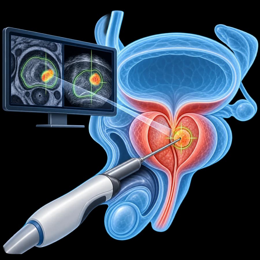
MRI-targeted fusion biopsy has transformed prostate cancer diagnosis over the last decade. At my practice in Rhodes, I perform all prostate biopsies using the fusion technique with transperineal access (TREXIT protocol), combining MRI-identified lesions with real-time ultrasound targeting.
The transperineal route eliminates rectal contamination and reduces infectious complications to near zero — a critical safety advantage that has made this the EAU-recommended standard since 2024, now a Strong recommendation under EAU 2026.
- Detects clinically significant cancer 46% more accurately than random systematic biopsy (PRECISION trial).
- Avoids unnecessary detection of low-risk, indolent tumours.
- Transperineal route: sepsis risk 0–0.1% vs 1–4% transrectal.
- Combined targeted + systematic cores: maximum sensitivity.
What Is MRI-Targeted Fusion Biopsy
MRI-targeted fusion biopsy is a two-phase diagnostic procedure for prostate cancer detection:
Multiparametric MRI (mpMRI)
A prostate mpMRI (T2, DWI, DCE sequences) is performed 3–6 weeks before biopsy. A specialist radiologist reads the images and assigns PI-RADS scores (1–5) to any suspicious lesions, defining their location, size, and probability of clinically significant cancer.
Fusion Biopsy
At the time of biopsy, MRI images are fused (overlaid) with real-time transrectal or transperineal ultrasound using dedicated software. The biopsy needle is guided electronically to the exact MRI-identified coordinates. Targeted cores are taken from each PI-RADS 3–5 lesion, plus systematic cores from the entire prostate.
PI-RADS Scoring System
PI-RADS (Prostate Imaging Reporting and Data System) is the standardized scoring framework used by radiologists to classify mpMRI lesion suspicion level and guide biopsy decisions.
PI-RADS 4–5: Prompt Biopsy Recommended
A PI-RADS 4 or 5 lesion on mpMRI carries a 50–90%+ probability of clinically significant prostate cancer. EAU 2026 guidelines recommend proceeding to biopsy without delay.
| PI-RADS Score | Suspicion Level | Probability of Significant Cancer | Recommendation |
|---|---|---|---|
| PI-RADS 1 | Very low | <5% | No biopsy; PSA surveillance |
| PI-RADS 2 | Low | <10% | No biopsy; repeat MRI in 12 months |
| PI-RADS 3 | Intermediate | 10–30% | Discuss with patient; consider biopsy based on PSA density & clinical factors |
| PI-RADS 4 | High | 50–75% | Fusion biopsy recommended |
| PI-RADS 5 | Very high | 75–90%+ | Urgent fusion biopsy recommended |
Who Needs MRI Fusion Biopsy
Elevated PSA ± Suspicious DRE (First Biopsy)
EAU 2026 recommends mpMRI before any first prostate biopsy in men with PSA >4 ng/mL or suspicious DRE. If PI-RADS ≥3, fusion biopsy follows.
Repeat Biopsy After Previous Negative Biopsy
If PSA continues to rise after a negative conventional biopsy, mpMRI + fusion biopsy increases detection of anteriorly located or small cancers missed by systematic sampling.
Active Surveillance Monitoring
Men on active surveillance for low-risk prostate cancer undergo periodic fusion biopsy to detect disease reclassification (upgrading to intermediate or high risk).
Pre-treatment Staging
MRI fusion biopsy provides precise intra-prostatic tumor localization essential for nerve-sparing decisions in radical prostatectomy and focal therapy planning.
Pre-procedure Preparation
mpMRI at Least 3–6 Weeks Before Biopsy
Recent mpMRI (ideally within 3 months) is required. If a prior biopsy was performed, wait 6–8 weeks to allow hematoma resolution before MRI.
PSA & Urine Culture
Recent PSA value (within 3 months). Urine culture to ensure no active urinary tract infection before the procedure.
Anticoagulant Management
Stop antiplatelet agents 5–7 days before. Anticoagulants: discuss bridging protocol with prescribing physician.
Antibiotic Prophylaxis
For transperineal biopsy: single-dose cephalosporin or fluoroquinolone. For transrectal: broader spectrum prophylaxis.
How MRI Fusion Biopsy Works — Step by Step
Anesthesia & Positioning
Patient positioned in lithotomy (transperineal) or lateral decubitus (transrectal). Local anesthetic injected to the periprostatic nerve plexus (prostate nerve block). Mild IV sedation offered for anxious patients.
MRI-Ultrasound Fusion & Lesion Targeting
TRUS probe positioned and ultrasound images registered with the pre-loaded mpMRI. Fusion software overlays the MRI onto live ultrasound. PI-RADS 3–5 target coordinates confirmed. 2–4 targeted cores taken per lesion.
Systematic Prostate Mapping
After targeted sampling, systematic cores taken from 10–12 pre-defined zones covering the entire prostate (both lobes, base to apex). Cores labeled and sent for histopathology.
Duration, Anesthesia & Setting
Recovery & Next Steps
Immediately After (Day 1)
Mild perineal discomfort and hematuria expected for 24–72h. Drink extra fluids. Avoid strenuous activity for 48h. Restart anticoagulants as directed.
Days 2–7
Light hematuria, hematospermia (blood in semen) and rectal spotting (transrectal) may persist for up to 2–4 weeks — this is normal. Return to normal activities and light work after 1–2 days.
Results & Follow-up (7–10 Days)
Histopathology results typically ready in 7–10 working days. Results consultation with Dr. Vasilas to review Gleason/ISUP grade, extent, and treatment options (active surveillance, robotic prostatectomy, radiation therapy).
Advantages & Clinical Evidence
PRECISION Trial (NEJM 2018)
Kasivisvanathan et al. randomized 500 men: MRI-targeted fusion biopsy detected clinically significant cancer in 38% vs 26% with systematic biopsy alone (P<0.001). Fusion biopsy also detected fewer low-risk, indolent tumours: 9% vs 22%.
PROMIS Trial (NEJM 2017)
Ahmed et al.: MRI had 93% sensitivity for clinically significant prostate cancer. MRI-based strategy would have avoided 27% of unnecessary biopsies.
Near-zero Infectious Risk (Transperineal)
Transperineal fusion biopsy (TREXIT protocol) eliminates rectal flora contamination. Sepsis rate: 0–0.1% vs 1–4% for transrectal approach. No prophylactic antibiotics needed in some protocols.
EAU 2026 Recommendation
EAU guidelines mandate mpMRI before first biopsy and give a Strong recommendation for transperineal access as the preferred biopsy route for prostate cancer diagnosis.
Frequently Asked Questions (FAQ)
What is MRI-targeted fusion biopsy and how does it differ from conventional biopsy?
MRI-targeted fusion biopsy combines a pre-biopsy multiparametric MRI (mpMRI) with real-time ultrasound to guide the biopsy needle precisely to suspicious areas identified on MRI. Conventional (systematic) biopsy samples 12 random cores without MRI guidance. The PRECISION trial (NEJM 2018) showed fusion biopsy detected clinically significant prostate cancer in 38% of patients vs 26% with systematic biopsy — a 46% improvement in detection.
Should I have an MRI before any prostate biopsy?
Current EAU guidelines (2026) recommend mpMRI before first biopsy in men with elevated PSA and/or suspicious DRE. MRI identifies suspicious areas (PI-RADS 3–5), guides targeted sampling, and reduces unnecessary biopsies (PI-RADS 1–2 may not require biopsy). The PROMIS trial (Ahmed NEJM 2017) showed MRI detected 93% of clinically significant cancers and would have avoided 27% of unnecessary biopsies.
Is fusion biopsy painful?
The transperineal approach (TREXIT protocol) used at our practice is performed under local anesthesia or mild sedation. Most patients report mild to moderate discomfort. Post-procedure perineal aching may last 1–2 days. Significant pain is uncommon.
How many biopsy cores are taken during fusion biopsy?
Typically 12–16 cores are taken: 2–4 targeted cores from each mpMRI lesion (PI-RADS 3–5) plus a systematic mapping of the entire prostate. This combined approach ensures no significant cancer is missed in non-targeted areas.
What does PI-RADS score mean?
PI-RADS (Prostate Imaging Reporting and Data System) is a standardized scoring system for mpMRI lesions: PI-RADS 1–2 (very low/low risk, <10% significant cancer), PI-RADS 3 (intermediate, 10–30%), PI-RADS 4 (high, 50–75%), PI-RADS 5 (very high, >75%). Your radiologist assigns a PI-RADS score to each suspicious area; this score guides the decision to biopsy.
What is the difference between transperineal and transrectal biopsy for MRI fusion?
Both approaches can perform MRI fusion biopsy, but transperineal has critical safety advantages: sepsis risk is 0–0.1% vs 1–4% with transrectal. Transperineal avoids rectal contamination by sampling through the perineal skin. EAU 2026 guidelines give a Strong recommendation for transperineal as the preferred route, developed jointly with the EAU Urological Infections Panel. Our practice performs all fusion biopsies via the transperineal TREXIT protocol.
When will I receive the biopsy results?
Histopathology results are typically available within 7–10 working days. Results are reviewed with you in person at a follow-up consultation where next steps (active surveillance, radical treatment, or watchful waiting) are discussed based on the Gleason/ISUP grade group.
Can fusion biopsy miss prostate cancer?
No biopsy technique is 100% sensitive. Fusion biopsy misses fewer clinically significant cancers than systematic biopsy (false-negative rate 10–15% vs 20–30% for systematic alone). Combining targeted + systematic cores minimizes this risk. If PSA continues to rise after a negative biopsy, repeat MRI and biopsy may be indicated.
Related Topics
Book an Appointment in Rhodes
Elevated PSA or suspicious MRI finding? Consultation with full evaluation and MRI-targeted fusion biopsy at our urology practice in Rhodes.
References & Sources
- EAU Guidelines on Prostate Cancer 2026 — uroweb.org
- Ahmed HU, et al. Diagnostic Accuracy of Multi-parametric MRI and TRUS Biopsy in Prostate Cancer (PROMIS): a Paired Validating Confirmatory Study. Lancet 2017;389:815–822.
- Kasivisvanathan V, et al. MRI-Targeted or Standard Biopsy for Prostate-Cancer Diagnosis (PRECISION). N Engl J Med 2018;378:1767–1777.
- Turkbey B, et al. Prostate Imaging Reporting and Data System Version 2.1: 2019 Update of PI-RADS v2. Eur Urol 2019;76:340–351.
Medical Review

Dr. Marinos Vasilas, Urologist – Andrologist
Dr. Marinos Vasilas performs MRI-targeted fusion biopsy using the transperineal TREXIT protocol according to EAU 2026 guidelines for prostate cancer diagnosis and active surveillance monitoring.
Full profile


