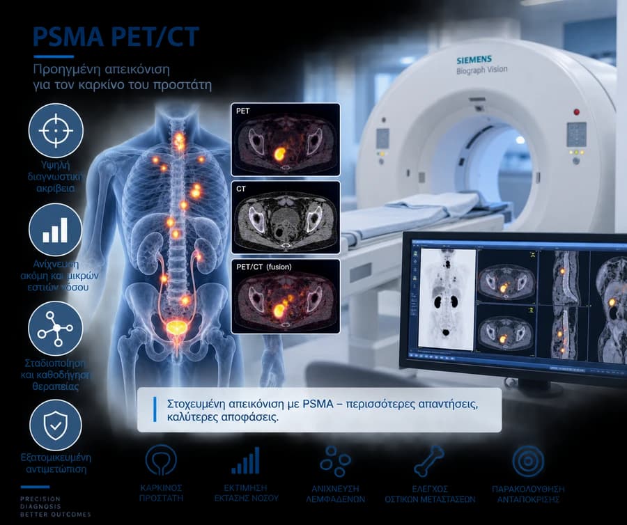
What PSMA is — and why it is not the same as PSA
PSMA stands for Prostate-Specific Membrane Antigen: a transmembrane protein expressed at high levels on many prostate cancer cells. It is easy to confuse with PSA, but these are two entirely different things:
- PSA — a blood biomarker, measured with a simple blood test
- PSMA — a molecular target on the cell surface, "seen" using a dedicated radiotracer on PET/CT
Despite its name, PSMA is not exclusively expressed in prostate cancer; physiological uptake is seen in several normal organs, and rarer uptake can occur in selected benign conditions or other malignancies. This is why PSMA uptake must always be interpreted anatomically and clinically, not as automatic "proof" of cancer.
How the scan works
The principle is straightforward:
- a radiolabeled PSMA-targeted tracer is injected intravenously
- the tracer binds to cells expressing PSMA
- PET captures the tracer distribution throughout the body
- CT provides the anatomical location of each finding
The result is a whole-body image combining molecular information (PET) with anatomical localization (CT).
PET
Shows molecular activity/uptake of the radiotracer — "where PSMA is present".
CT
Shows anatomical structures and spatially localizes the PET findings. Depending on protocol, it may be a low-dose localizing CT or a full diagnostic CT — not always with intravenous contrast.
PSMA radiotracers
Several different approved/commonly used PSMA radiotracers exist, depending on country and center:
- 68Ga-PSMA-11
- selected 18F-labeled tracers, such as 18F-DCFPyL
- 18F-PSMA-1007
- other locally approved tracers
Tracers are not identical: they differ in biodistribution (e.g., urinary excretion), which can affect interpretation in certain regions (e.g., near the bladder). There is no single "best for everyone" tracer — the choice depends on availability and the nuclear medicine center's protocol.
PSMA PET/CT vs multiparametric prostate MRI (mpMRI)
The two tests answer different clinical questions and are not interchangeable:
Prostate mpMRI
Mainly assesses prostate anatomy, morphology of intraprostatic lesions, local extent, PI-RADS before diagnosis/biopsy, and, in select cases, local recurrence.
PSMA PET/CT
Mainly assesses molecular PSMA expression, lymph nodes, distant metastases, recurrence after treatment, and whole-body disease distribution.
PSMA PET/CT vs prostate biopsy
An important distinction: PSMA PET/CT is imaging, while biopsy provides a histological diagnosis. A PSMA-positive finding does not equal a definitive histological diagnosis of cancer.
For men with suspected but undiagnosed prostate cancer, MRI plus biopsy remains the established diagnostic pathway according to current guidelines. The role of PSMA PET in primary diagnosis is evolving and being studied further, but it is not currently a routine substitute for mpMRI/biopsy.
When it is used — the 4 main clinical scenarios
PSMA PET/CT is mainly used in four scenarios:
A. Initial staging
After a prostate cancer diagnosis, in selected risk groups, to look for nodal or distant disease.
B. Recurrence after prostatectomy
Detectable/rising PSA after radical prostatectomy, when the result can change the treatment decision.
C. Recurrence after radiotherapy
Biochemical recurrence after radiotherapy, especially in candidates for salvage treatment.
D. Advanced/metastatic disease
Mapping disease extent and checking eligibility for PSMA-targeted radioligand therapy (e.g., 177Lu-PSMA).
Initial staging by risk group
The indication clearly depends on the risk group at diagnosis — it is not used the same way in everyone:
Low risk
Additional staging imaging for metastatic disease is not routinely indicated.
Favourable intermediate risk
Additional staging imaging is not routinely indicated.
Unfavourable intermediate risk
PSMA PET/CT can be performed where available, to increase staging accuracy.
High risk / locally advanced disease
PSMA PET/CT is recommended for metastatic workup, where available.
Why it isn't done for every low-risk patient
A more sensitive test does not automatically mean a better outcome, when the pre-test probability of metastatic disease is already very low. Unnecessary imaging can lead to:
- incidental findings without clear significance
- equivocal findings requiring further work-up
- patient anxiety
- radiation exposure without a clear clinical benefit
- "stage migration" without a proven treatment benefit
This is why PSMA PET/CT is not recommended routinely for every newly diagnosed low-risk patient.
Lymph node staging — what it can and cannot do
PSMA PET/CT generally has high specificity for many nodal findings, but sensitivity remains limited for very small/microscopic nodal metastases.
A negative PSMA PET/CT does not rule out microscopic nodal disease. For this reason, a negative result does not on its own override the indication for pelvic lymph node dissection (PLND) when it is indicated based on other criteria.
Bone metastases
PSMA PET/CT can generally detect skeletal metastases with greater detection ability compared with conventional bone scintigraphy, in the appropriate clinical setting.
However, not every focus of increased bone uptake is a metastasis — potential benign mimics exist (e.g., healing fractures, degenerative changes). Interpretation requires correlation of location, CT morphology, and clinical context.
PSMA PET/CT vs conventional CT and bone scan
A patient-friendly comparison:
Molecular targeting — PSMA PET/CT: yes · Conventional CT + bone scan: no
Lymph node detection — PSMA PET/CT: generally higher sensitivity · Conventional CT: size-based limitations
Bone lesions — PSMA PET/CT: high sensitivity in the right setting · Bone scan: assesses osteoblastic activity
Whole-body staging — PSMA PET/CT: yes, in one scan · Conventional: combination of several tests
Microscopic disease — PSMA PET/CT: can still be missed · Conventional: can also be missed
Basis of historical treatment trials — PSMA PET/CT: newer technology · Conventional: many historical trials were based on it
Conventional imaging is not "obsolete" in every scenario — it remains relevant where PSMA PET/CT is unavailable or not indicated.
The ProPSMA trial and "stage migration"
The prospective randomized ProPSMA trial (Hofman et al.) in high-risk men showed significantly higher diagnostic accuracy for PSMA PET/CT compared with conventional CT and bone scintigraphy in detecting metastatic disease. However, greater diagnostic accuracy does not by itself prove an improvement in overall survival.
This connects to an important concept: "stage migration". More sensitive imaging can detect disease that conventional imaging would have classified as M0. The same patient may therefore be classified differently depending on which imaging technology was used. Since many treatment trials were historically based on conventional imaging, managing low-volume disease detected only by PSMA PET often requires discussion at a multidisciplinary team (MDT) level.
Biochemical recurrence — when PSA rises after treatment
A rising PSA after treatment with curative intent may indicate biochemical recurrence. PSMA PET/CT can help locate:
- local recurrence in the prostate/prostate bed region
- pelvic lymph nodes
- distant lymph nodes
- bone foci
- distant metastatic disease
This information can guide the planning of salvage treatment.
After radical prostatectomy
According to current EAU guidance, PSMA PET/CT is indicated for PSA recurrence after radical prostatectomy when:
- PSA is > 0.2 ng/mL
AND
- the result is expected to influence the subsequent treatment decision
This does not mean the scan must always "wait" until PSA reaches 0.2 — detection probability, biochemical recurrence criteria, and timing of treatment initiation are separate concepts. The most important message: an indicated salvage radiotherapy should not be delayed simply to "wait" for a positive PSMA PET/CT.
Negative PSMA PET after prostatectomy — what it really means
A negative PSMA PET/CT does not mean the absence of recurrent prostate cancer. At low PSA levels, disease may simply be below the spatial/molecular detection threshold of the test.
A negative scan does not automatically justify:
- delaying salvage treatment
- assuming the biochemical recurrence is "false"
- stopping PSA monitoring
The treatment decision always depends on the full oncological context.
After radiotherapy
In biochemical recurrence after radiotherapy, PSMA PET/CT is particularly useful in patients who are candidates for potentially curative salvage treatment.
The complementary role of prostate MRI is also important when local salvage treatment is being considered: PSMA PET shows the distribution of disease, while MRI is valuable for local anatomical recurrence — these are not identical tests.
Can it be falsely positive?
Yes. PSMA uptake can be seen in:
- selected benign conditions
- ganglia
- healing fractures or selected bone lesions
- inflammatory findings
- selected other neoplasms
Interpretation requires correlating location, CT morphology, uptake pattern, and clinical context — not a decision based on uptake intensity alone.
Normal biodistribution — not every uptake is 'suspicious'
The tracer's normal distribution typically includes organs such as:
- salivary/lacrimal glands
- kidneys/urinary tract
- liver/spleen (depending on tracer)
- bowel
- other normal structures
This is normal biodistribution, not metastatic disease.
False negatives and heterogeneity of PSMA expression
Not every prostate cancer cell expresses PSMA to the same degree. Some tumors or metastases may show intense, heterogeneous, minimal, or essentially absent uptake.
A negative PSMA PET does not prove the absence of cancer. Possible causes of a false-negative result:
- very small lesion size
- low PSMA expression
- tumor heterogeneity
- particular tumor biology
- technical factors
No imaging method detects every microscopic focus of cancer.
In selected cases of more aggressive/dedifferentiated or neuroendocrine disease, PSMA expression may be reduced; in these cases, other imaging such as FDG PET/CT may provide complementary information in select scenarios — not as a universal rule.
What 'PSMA-avid' means and what SUV is
The term "PSMA-avid" means that a focus shows increased uptake of the PSMA-targeted tracer. It does not by itself determine: Gleason score/grade, treatment response, or absolute certainty of malignancy.
SUV (Standardized Uptake Value) quantifies tracer uptake under specific imaging conditions. There is no single, universal SUV threshold that reliably separates benign from malignant lesions — self-interpretation of SUV values by patients is not recommended.
PSMA PET and 177Lu-PSMA therapy — two different things
An important distinction:
- Diagnostic PSMA PET — an imaging test
- 177Lu-PSMA therapy (radioligand/targeted therapy) — a therapeutic intervention
The diagnostic scan can be used in selected patients with advanced prostate cancer to assess whether the disease shows sufficient PSMA expression to be a candidate for PSMA-targeted radioligand therapy. This is an example of a "theranostic" approach: the same molecular target is used first for imaging and then, in selected patients, for targeted therapy.
A positive PSMA PET does not automatically mean every patient is eligible for 177Lu-PSMA therapy — eligibility depends on disease stage, prior treatments, PSMA expression criteria, current approvals/guidelines, and the overall clinical picture.
PSMA PET/CT is not a routine surveillance test for everyone
Post-treatment imaging is generally indicated when:
- there is suspicion of recurrence/disease progression
AND/OR
- the result could change clinical management
Annual PSMA PET/CT is not recommended for every asymptomatic patient after prostatectomy with undetectable PSA.
Preparation, safety, radiation, and contrast
The exact preparation depends on the nuclear medicine center's protocol. Topics generally discussed include:
- current medications
- hydration and bladder emptying
- kidney function, if a diagnostic contrast-enhanced CT is planned
- allergy history, if iodinated contrast is planned
- previous imaging/reports and recent prostate cancer treatments
PSMA PET/CT does not automatically involve intravenous iodinated contrast — the CT component may be a low-dose localizing scan or a full diagnostic scan, depending on protocol. The test involves exposure to ionizing radiation from the tracer and the CT component — the dose varies depending on tracer, administered activity, and CT type. PSMA radiotracers are given in very small (tracer) amounts; rare injection-related reactions are a separate issue from any reaction to iodinated contrast.
Treatments such as androgen deprivation therapy may affect PSMA expression and scan appearance, depending on timing and treatment context — do not stop any prostate cancer treatment before the scan without guidance from your urology/oncology team.
What PSMA PET/CT cannot tell us
Despite its great diagnostic value, PSMA PET/CT cannot on its own reliably determine:
- exact Gleason score/ISUP grade
- histological subtype
- disease below the detection threshold
- whether every focus of uptake is definitely malignant
- which treatment is automatically "the best"
- prognosis based solely on the SUV value
An imaging result is one part of the clinical decision — not the whole decision. The correct use of PSMA PET/CT always depends on when and why it is performed, and its interpretation must be combined with PSA, histology, stage, risk group, prior treatments, and other imaging tests.


