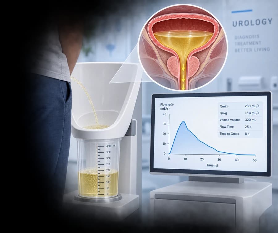
What uroflowmetry is
Uroflowmetry is a non-invasive, functional test: the patient urinates naturally into a dedicated uroflowmeter, which records the urine flow in real time.
- no catheter is required for free (spontaneous) uroflowmetry
- voiding occurs with a normal, spontaneous urge — not a forced one
- the test lasts only as long as the natural void itself
Do not confuse it with invasive pressure-flow urodynamics, which requires catheterization and additionally measures bladder pressure.
What the test measures
Uroflowmetry objectively records:
- Qmax — maximum urinary flow rate (mL/s)
- Qave — average urinary flow rate (mL/s)
- Voided volume — total volume expelled (mL)
- Flow time — the time during which measurable flow occurs (s)
- Voiding time — total duration of voiding, including any interruptions (s)
- Time to Qmax — time from flow onset to maximum flow (s)
- Flow pattern — the shape of the flow curve
If post-void residual (PVR) is measured, this is usually done through a separate measurement (ultrasound/bladder scanner) and is not produced automatically by the uroflowmeter itself.
What Qmax is
Qmax is the highest instantaneous urine flow rate recorded during voiding. It is the best-known parameter of the test, but it should never be interpreted in isolation.
Qmax is volume-dependent. A low Qmax with a very small voided volume may be unreliable and poorly representative of normal voiding.
The role of voided volume
According to current EAU guidance, uroflowmetry is best interpreted when the voided volume is sufficient for reliable assessment, with the guidance citing an approximate volume of more than 150 mL.
If the voided volume is too low:
- Qmax may appear artificially low
- the flow pattern may not represent normal voiding
- repeat testing may be useful
When repeat uroflowmetry is useful
Because Qmax shows intra-individual variation and depends on voided volume, repeating the measurement can improve reliability.
Especially consider repeat testing when:
- the voided volume is low
- Qmax is unexpectedly reduced
- the flow pattern is abnormal
- the patient reports that the test did not resemble their normal home urination
A single test is not always definitive.
Was this your normal void?
A small but high-value question that is always worth asking:
Was this urination representative of your everyday voiding?
An anxious patient, embarrassment, or an excessively forced bladder filling before the test can produce a non-representative result.
How to prepare for the test
General principles of preparation:
- arrive with a comfortably full bladder and a normal urge to void
- if instructed, avoid urinating shortly before the examination
- drink normally, rather than with excessive forced fluid loading
- tell staff if your bladder is painfully overfull
- tell the clinician if the void was not representative
There is no single universal protocol ("drink exactly 1.5 litres one hour before") unless this is the verified protocol of the specific clinic. Painful bladder overdistension should never be caused.
Why privacy matters
According to International Continence Society (ICS) principles, privacy can influence voiding.
Uroflowmetry should be performed in a setting that allows the patient to void as naturally and privately as possible. Anxiety or an inability to relax may alter the result — this should not be trivialized.
Normal, bell-shaped flow
A smooth, bell-shaped curve is commonly associated with a coordinated, unobstructed void.
However, not every non-bell-shaped curve should automatically be labeled pathological. Interpretation is always done together with sex, age, voided volume, symptoms, post-void residual, and the overall clinical context.
Plateau flow
A prolonged, relatively flat (plateau) pattern may, in the appropriate clinical context, raise suspicion of a fixed outflow resistance, such as a urethral stricture.
The curve shape alone does not establish the diagnosis. See the dedicated urethral stricture page when clinically relevant.
Intermittent flow
Interrupted flow may occur in several situations, including:
- abdominal straining
- impaired detrusor contractility
- dysfunctional voiding
- outlet obstruction
- a non-representative void
No diagnosis is made from the pattern alone.
Prolonged, low-amplitude flow
A prolonged void with low-amplitude flow may occur with:
- increased outlet resistance
- impaired detrusor contractility
- mixed dysfunction
Once again: uroflowmetry alone cannot differentiate the underlying mechanism.
Bladder outlet obstruction (BOO)
A low Qmax increases suspicion of bladder outlet obstruction in the right clinical context.
However, uroflowmetry does not directly measure bladder pressure. A low-flow void can result from:
1. increased outlet resistance
2. a weak detrusor contraction
3. a low voided volume
4. a combination of factors
Diagnostic certainty based on a single value should be avoided.
Detrusor underactivity
A weak bladder contraction may also produce low flow.
Low flow does not automatically mean an enlarged prostate.
Free uroflowmetry cannot reliably distinguish between bladder outlet obstruction (BOO) and detrusor underactivity. When understanding the mechanism matters clinically, pressure-flow urodynamics may be considered in selected patients.
BPH, BPE, and BPO — an important terminology distinction
These terms are not interchangeable — current EAU terminology is used:
BPH
Benign Prostatic Hyperplasia — a histological term.
BPE
Benign Prostatic Enlargement — describes the size of the gland.
BPO
Benign Prostatic Obstruction — describes functional outflow obstruction.
A large prostate doesn't always mean obstruction
A large prostate does not automatically prove obstruction. A smaller prostate does not exclude outlet obstruction either.
See the dedicated benign prostatic hyperplasia (BPH) page for the complete clinical picture.
Role in the work-up of BPH/LUTS before treatment
In men with:
- a weak urinary stream
- hesitancy
- intermittent flow
- abdominal straining
- a sensation of incomplete emptying
- lower urinary tract symptoms (LUTS)
uroflowmetry can be performed during the initial assessment of male LUTS and is recommended before medical or invasive treatment, according to current EAU guidance.
Before BPH treatment
Baseline uroflowmetry provides objective data before:
- medical therapy
- HoLEP
- TURiS
- Aquablation
- Rezūm
- other real BPH treatments
It does not determine the choice of treatment on its own — it is considered together with symptoms, quality of life, and the remaining clinical work-up.
After BPH treatment
Uroflowmetry can objectively document the change after treatment, together with:
- Qmax
- voided volume
- PVR
- symptoms/IPSS
A better Qmax is only one component of the treatment outcome — it does not automatically equate to complete clinical success.
Urethral stricture
Urethral stricture may cause a weak stream, prolonged flow, or a plateau-type curve.
However, uroflowmetry alone does not define the location, length, caliber, or cause of the stricture. Further investigation may include cystoscopy or urethrography, depending on the clinical context.
PVR — post-void residual
PVR (post-void residual) is the urine left in the bladder after voiding. It is usually measured separately by:
- ultrasound
- a bladder scanner
- catheterization in selected circumstances
PVR complements uroflowmetry.
PVR tells us how much urine remains — not why it remains.
High PVR
Potential causes of a high PVR include:
- bladder outlet obstruction
- detrusor underactivity
- neurological dysfunction
- mixed mechanisms
Obstruction is not diagnosed from PVR alone, and no single universal PVR threshold is used for all patients.
Qmax and PVR together
A simple interpretive framework, without automatic diagnoses:
Good flow + low PVR
Often a reassuring finding, depending on the clinical context.
Low flow + low PVR
Could reflect obstruction, a weak contraction with a small voided volume, or other factors.
Low flow + high PVR
Requires further assessment.
Good flow + high PVR
Also requires clinical interpretation — it should not be dismissed.
Uroflowmetry vs pressure-flow urodynamics
One of the most important distinctions concerning this test:
Uroflowmetry
Non-invasive, spontaneous voiding, Qmax/Qave/volume/curve — without measuring bladder pressure.
Pressure-flow urodynamic study
Invasive functional test: bladder pressure, abdominal pressure, detrusor pressure, and the pressure-flow relationship — can help distinguish BOO from detrusor underactivity.
Do I need urodynamics after uroflowmetry?
Not necessarily.
Most men with uncomplicated LUTS do not automatically need invasive urodynamics. A urodynamic study may be considered when understanding the underlying mechanism could materially change an invasive treatment decision, or in selected complex cases.
Not every abnormal flow is automatically referred for invasive urodynamics.
Neurogenic bladder
Uroflowmetry can contribute information in neurological patients who void spontaneously, but it does not reliably measure filling pressures, compliance, neurogenic detrusor overactivity, or detrusor-sphincter dyssynergia by itself.
See the dedicated neurogenic bladder page.
Normal uroflowmetry does not prove a "safe" neurogenic bladder.
Uroflowmetry in women
Men and women have different typical flow characteristics — male Qmax thresholds are not used universally for women.
In women, the test can help evaluate voiding dysfunction or incomplete emptying, but a low flow in a woman does not automatically indicate anatomical obstruction.
What can distort the result
Potential factors affecting reliability:
- a low voided volume
- an excessively overfull bladder
- anxiety
- lack of privacy
- an interrupted void
- abdominal straining
- movement during the test
- equipment artefact
- a non-representative void
Not every unusual curve should be treated as disease.
What uroflowmetry cannot tell us
Despite its great clinical usefulness, uroflowmetry cannot, on its own, tell us:
- why the flow is low (obstruction versus a weak bladder)
- the exact bladder pressure
- the location, length, or cause of a urethral stricture
- whether surgery is needed
- an "absolute" normal Qmax value for every patient
Qmax should never be interpreted in isolation — correct assessment combines the flow, the voided volume, the curve shape, the post-void residual, and the symptoms.


