What the DRE assesses clinically
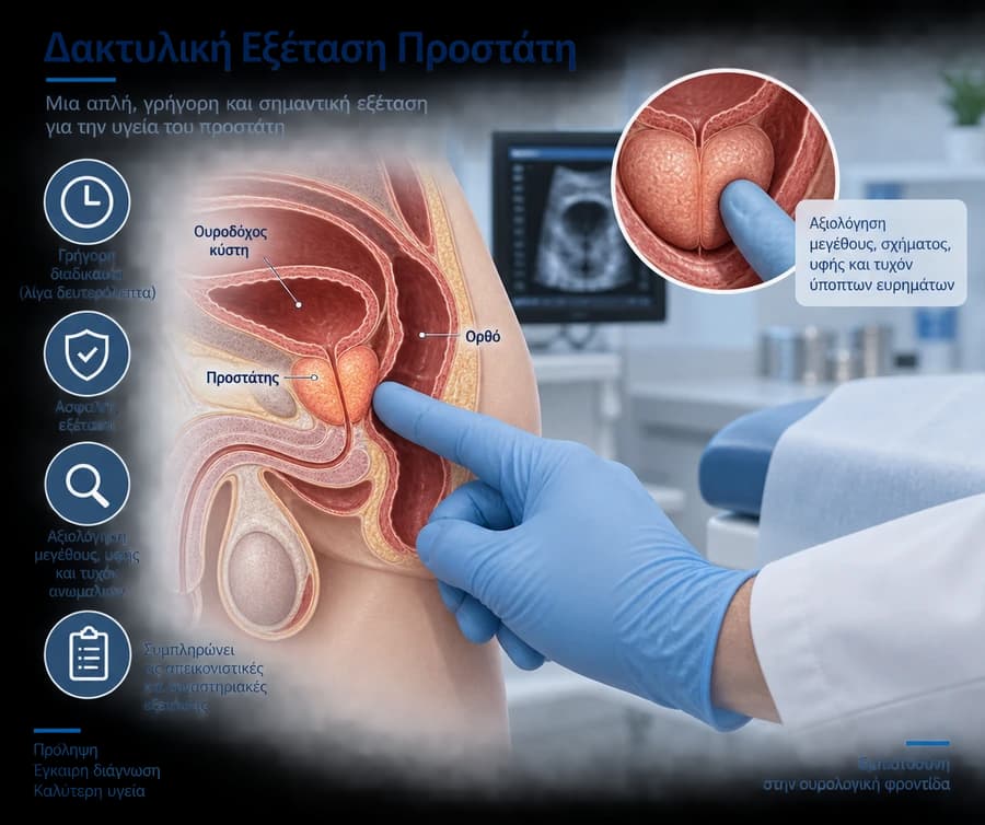
The DRE assesses:
- prostate size (approximate)
- consistency (soft/elastic versus hard)
- symmetry of the lobes
- the presence of nodules or focal hardness
- tenderness on palpation (e.g., possible inflammation)
The examination mainly assesses the posterior part of the gland, so a normal finding does not completely rule out pathology.
Low-suspicion picture
A smooth, elastic texture, with no nodules and no focal hardness.
Borderline suspicion
Mild asymmetry or an equivocal area that requires correlation with PSA and often imaging.
High-suspicion picture
A hard nodule, clear asymmetry, or a fixed/notably hard area requires immediate investigation.
When it is particularly useful
The DRE is particularly important when:
- PSA is elevated or borderline
- there are lower urinary tract symptoms in a middle-aged or older man
- an initial urological risk assessment for prostate cancer is being performed
- clinical follow-up is needed for an already known prostate condition
Using DRE together with PSA improves clinical accuracy compared with either method alone.
Technique, safety, and limitations of the method
The examination takes only a few seconds and is performed with a glove and lubricant.
- it is quick and low-cost
- it requires no special preparation
- its limitation is that it does not "see" the entire prostate
Its findings should therefore always be interpreted together with the history, PSA, and, where indicated, mpMRI or biopsy.
What it means for the next diagnostic step
An indicative algorithm:
- normal DRE + low PSA: usually monitoring
- equivocal DRE + borderline PSA: repeat PSA and risk stratification
- suspicious DRE regardless of PSA: a strong indication for advanced work-up (often mpMRI, with or without biopsy)
The key is individualization based on age, family history, and overall risk.


