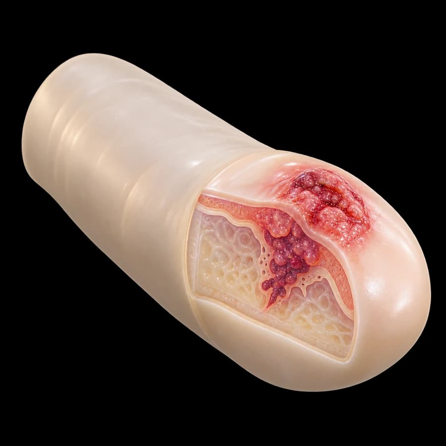
My clinical approach
In penile cancer, time to diagnosis is the most important prognostic factor. Unfortunately, many patients delay consultation due to embarrassment — resulting in advanced disease at presentation.
In my practice I follow the EAU-ASCO Penile Cancer 2024 guidelines:
- Immediate biopsy of any persistent lesion >3 weeks.
- Staging with penile MRI and inguinal imaging.
- Priority to organ-preserving therapies where technically feasible (laser, glans resurfacing, glansectomy).
- Dynamic sentinel node biopsy for cN0 in pT1b or higher stage.
- Coordinated team: urologist-oncologist-pathologist-radiation oncologist.
- Referral to specialized center for advanced/metastatic disease.
- Counseling for HPV vaccination of partners and prevention.
What is penile cancer
Penile cancer is a rare malignancy in Western countries (1/100,000 men/year), but more common in developing countries (up to 4–6/100,000). The vast majority (95%) is squamous cell carcinoma (SCC).
Located mainly on the glans (48%), foreskin (21%), coronal sulcus (6%), more rarely on the penile shaft. Median age at diagnosis 60 years, although incidence in younger men is rising due to HPV.
Precancerous lesions: erythroplasia of Queyrat (CIS of glans), Bowen's disease (CIS of penile shaft), HPV-related papillary carcinomas. Associated with 30–40% risk of progression to invasive SCC.
Risk factors
- High-risk HPV (types 16, 18, 31, 33, 45) — responsible for ~50% of cases.
- Phimosis — 10× increased risk.
- Lichen sclerosus (BXO) — associated with 2.3–10% of SCC.
- Chronic balanitis/balanoposthitis.
- Smoking — 3–4× increased risk.
- Poor hygiene and chronic irritation.
- Immunosuppression (HIV, transplant) — 8× increase.
- PUVA therapy (psoriasis) — increased SCC risk.
- Advanced age (more common >60 years).
- Multiple sexual partners, STI coinfection.
Neonatal circumcision: provides nearly complete protection from penile cancer. In population groups with near-universal circumcision (Jewish populations), incidence is <0.1/100,000.
Symptoms and clinical picture
- Persistent non-healing lesion on the glans or foreskin.
- Ulcer, plaque, wart-like growth, red or white patch.
- Hard palpable mass or swelling.
- Discharge (blood, pus), foul odor.
- Pain or itching (in advanced stages).
- Enlarged inguinal lymph nodes (inflammatory or metastatic).
- Difficulty retracting foreskin (phimotic penis hiding lesion).
- In advanced disease: bleeding, foul smell, discharge from cancerous ulcer.
Diagnosis and staging
Detailed clinical examination
Inspection and palpation of lesion (size, location, infiltration), foreskin assessment, mandatory bilateral inguinal lymph node palpation.
Lesion biopsy
MANDATORY. Incisional or excisional depending on size. Histologic type (SCC, subtypes), grade (G1-G3), lymphovascular invasion (LVI), depth of invasion.
Penile MRI with artificial erection (PGE1)
Assessment of corpus spongiosum/cavernosum and urethral invasion. Determines T stage and surgical approach (organ-preserving vs. penectomy).
Inguinal node staging
Ultrasound ± FNA in palpable nodes. In cN0 with pT1b or higher: dynamic sentinel node biopsy (DSNB) — preferred over prophylactic lymphadenectomy.
Metastatic staging
Abdominal/pelvic CT/MRI (pelvic nodes, metastases), chest CT. PET-CT for cN+ or high-risk cases.
HPV typing
HPV testing (PCR or p16 immunohistochemistry) — prognostic factor and influences follow-up.
TNM staging (AJCC 8th edition)
Tis / Ta
Carcinoma in situ (CIS) or non-invasive verrucous carcinoma.
T1a / T1b
T1a: subepithelial connective tissue invasion without LVI and low-grade. T1b: with LVI or high-grade.
T2
Corpus spongiosum invasion ± urethra.
T3
Corpus cavernosum invasion ± urethra.
T4
Adjacent organ invasion (scrotum, prostate, pelvic bones).
N0-N3
N0: no nodes. N1: 1 unilateral. N2: multiple or bilateral. N3: pelvic nodes or extranodal extension.
Treatment
Early stages (CIS, Ta, T1a)
Organ-preserving therapies: topical 5-FU cream or imiquimod (CIS), CO₂/Nd:YAG laser, glans resurfacing (epithelial removal with skin graft), circumcision (if located on foreskin). All preserve the penis.
Local disease T1b-T2
Glansectomy (glans removal with neoglans formation from skin graft) or partial penectomy with preservation of adequate length for erection and standing urination. Goal: negative margin >5mm.
Advanced disease T3-T4
Total penectomy with perineal urethrostomy. Neoadjuvant chemotherapy (TIP — paclitaxel, ifosfamide, cisplatin) in advanced/pelvic disease.
Inguinal node management
cN0 with low risk (Tis, Ta, T1a low-grade): surveillance. cN0 with high risk (pT1b or higher): DSNB. cN+: radical inguinal lymphadenectomy ± pelvic. Adjuvant chemotherapy/radiotherapy for pN2-N3 or extranodal extension.
Metastatic disease (M1)
Systemic chemotherapy (TIP), palliative radiotherapy, clinical trials (anti-PD-1 immunotherapy — pembrolizumab, EGFR inhibitors — cetuximab). Multidisciplinary approach.
Prognosis and survival
- Localized disease (T1-2, N0): 5-year survival 85–90%.
- Inguinal nodes N1: 5-year survival 70–80%.
- Inguinal N2: 5-year survival 50–60%.
- Pelvic node disease (N3): 5-year survival 20–30%.
- Metastatic disease (M1): 5-year survival <10%.
- Poor prognostic factors: high-grade, LVI, extranodal extension, pelvic nodes.
- HPV+ carcinomas: more favorable prognosis than HPV-.
- Recurrence mainly in first 2 years — intensive follow-up.
Prevention
- HPV vaccination (Gardasil-9) in boys 9–14 years — primary prevention.
- Neonatal circumcision in high-risk populations.
- Treatment of phimosis in adults.
- Surveillance and treatment of BXO with topical corticosteroids.
- Smoking cessation.
- Good hygiene, avoidance of chronic irritation.
- Condom use — partial protection from HPV.
- STI screening and treatment.
- Immediate urology referral for persistent lesions >3 weeks.
Frequently asked questions (FAQ)
What is penile cancer?
A rare malignancy (incidence 1/100,000 men/year in Western countries). 95% are squamous cell carcinoma. Strongly associated with HPV (mainly types 16/18) and lichen sclerosus (BXO). Early diagnosis allows penile preservation.
What are the early symptoms?
Persistent lesion on the glans or foreskin: ulcer, plaque, wart-like growth, red/white patch, or hard non-healing lesion lasting >3 weeks. Other: discharge, bleeding, foul odor, enlarged inguinal lymph nodes.
What are the risk factors?
High-risk HPV (types 16, 18, 31, 33, 45), phimosis, chronic inflammation/balanitis, lichen sclerosus (BXO), smoking, poor hygiene, immunosuppression, PUVA therapy, advanced age. Neonatal circumcision provides nearly complete protection.
How is it diagnosed?
Clinical examination and biopsy of the lesion (incisional/excisional) — MANDATORY. Inguinal node assessment (palpation, ultrasound ± FNA, dynamic sentinel node biopsy [DSNB]). Staging with penile MRI (cavernous invasion), abdominal/pelvic CT/MRI, chest CT for advanced stages.
How is TNM staging done?
T1a (superficial, no LVI), T1b (LVI or high-grade), T2 (corpus spongiosum invasion), T3 (corpus cavernosum or urethral invasion), T4 (adjacent organs). N1-N3 by inguinal/pelvic nodes. M1 = distant metastases.
What are the treatment options?
Early stages (CIS, T1a): CO₂/Nd:YAG laser, topical 5-FU/imiquimod, glansectomy, glans resurfacing, circumcision. Advanced: partial or total penectomy. Lymph nodes: inguinal lymphadenectomy for cN+ or pT1b. Adjuvant chemotherapy/radiotherapy for advanced disease.
What is dynamic sentinel node biopsy (DSNB)?
Modern staging technique for inguinal nodes in cN0 high-risk patients (pT1b and above). Tc-99m + patent blue dye injection, identification and removal of only the sentinel node. Significantly reduces morbidity vs. classical lymphadenectomy.
What is the prognosis?
Stage-dependent. 5-year survival: localized disease (T1-2, N0) 85–90%, inguinal nodes N1-N2 50–60%, pelvic disease (N3) 20–30%, metastatic (M1) <10%. Early referral to specialized center significantly improves outcomes.
How can it be prevented?
HPV vaccination (9-valent, Gardasil-9) in boys aged 9–14 — primary prevention. Good hygiene, smoking cessation, treatment of phimosis and BXO, condom use. Self-examination for persistent lesions — immediate urology referral.
Related topics
Suspicious lesion? Immediate evaluation
Early diagnosis allows organ-preserving therapies and high cure rates. Don't delay — contact us for an appointment.
Scientific literature
- EAU-ASCO Collaborative Guidelines on Penile Cancer (2024) — uroweb.org
- Brouwer OR, Albersen M, Parnham A, et al. EAU-ASCO Penile Cancer Guidelines 2023 Update. Eur Urol 2023;83(6):548-560 — pubmed.ncbi.nlm.nih.gov
- Hakenberg OW, Compérat EM, Minhas S, et al. EAU Guidelines on Penile Cancer. Eur Urol 2015;67(1):142-50 — pubmed.ncbi.nlm.nih.gov
- Backes DM, Kurman RJ, Pimenta JM, Smith JS. Systematic review of HPV prevalence in invasive penile cancer. Cancer Causes Control 2009;20(4):449-57 — pubmed.ncbi.nlm.nih.gov
- Pagliaro LC, Williams DL, Daliani D, et al. Neoadjuvant TIP for metastatic penile cancer. J Clin Oncol 2010;28(24):3851-7 — pubmed.ncbi.nlm.nih.gov
Meet the doctor

Dr. Marinos Vasilas, Urologist – Andrologist
Dr. Marinos Vasilas offers specialized diagnosis and treatment of penile cancer with emphasis on organ-preserving techniques (laser, glansectomy, glans resurfacing) and multidisciplinary approach per EAU-ASCO 2024 guidelines.
View full profile


