My clinical approach
Peyronie's disease is as psychologically burdensome as it is anatomical. Many patients arrive with guilt ("did I do something wrong?") or shame. Correct management combines: clear information about the natural history, individualised treatment by phase, and recognition of the psychological burden.
In my practice I follow the EAU 2024 Sexual & Reproductive Health and AUA Peyronie's Disease 2015 (reaffirmed 2023) guidelines:
- Distinguish active vs stable phase before any therapeutic decision.
- Penile Doppler with pharmaco-induction for objective curvature measurement.
- Conservative approach in the active phase — never surgery.
- Injectable CCH or verapamil in selected cases (curvature 30–90°, no hourglass).
- Surgery in stable phase: plication for milder cases, grafting for larger curvatures, prosthesis when ED coexists.
- Psychological support — we systematically discuss relationship impact.
What is Peyronie's disease?
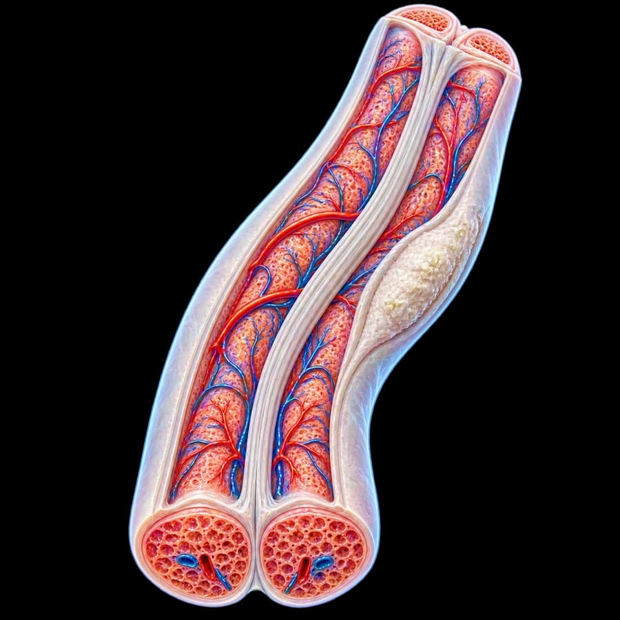
Peyronie's disease (Induratio penis plastica) is a benign fibrotic disorder of the tunica albuginea of the corpora cavernosa. It is characterised by formation of a fibrous plaque leading to:
- Penile curvature on erection (mostly dorsal).
- Pain (active phase).
- Penile shortening.
- "Hourglass" or "hinge" deformity.
- Erectile dysfunction (in 30–50% of patients).
- Difficulty or inability to have intercourse in severe cases.
Phases of the disease
Active / inflammatory phase
Duration: 6–18 months from symptom onset.
- Pain (mainly on erection)
- Progressive curvature
- Plaque formation/progression
- Increasing deformity
No surgery in this phase.
Stable phase
Usually after 12 months, no changes for 3–6 months.
- Pain resolved
- Curvature stabilised
- Mature plaque (often calcified)
- Frequent ED appearance
Surgery feasible in this phase.
Causes and risk factors
The exact aetiology remains unknown. Prevailing theory: repeated microtrauma of the tunica during intercourse in genetically predisposed individuals, with abnormal healing and fibrous plaque development.
- Diabetes mellitus (correlated with severity).
- Dupuytren's contracture — co-occurrence 15–20%.
- Plantar fibromatosis (Ledderhose disease).
- Family history.
- Autoimmune diseases.
- Radical prostatectomy — association 11–16%.
- Hypertension, hyperlipidaemia, smoking.
Symptoms
Physical
- Palpable plaque
- Erectile curvature (dorsal in 70%)
- Pain (active phase)
- Penile shortening
- "Hourglass" deformity
- Erectile dysfunction
- Inability to have intercourse
Psychosocial
- Anxiety, depression
- Reduced self-esteem
- Sexual avoidance
- Relationship strain
- Fear of irreversible damage
Diagnosis
Detailed history
Symptom onset, curvature progression, pain, erectile function (IIEF-5), psychosocial impact.
Clinical examination
Plaque palpation (location, size, number), penile length assessment.
Photographic documentation
Patient self-photographs in erection from 3 angles (top, lateral, frontal) or pharmaco-induced examination in clinic.
Penile Doppler ultrasound with pharmaco-induction
Plaque mapping, search for calcification, assessment of arterial/venous haemodynamics.
ED evaluation
IIEF-5, hormonal screen (testosterone) in selected patients.
Treatment options
Treatment is individualised based on disease phase, degree of curvature, presence of ED and impact on sexual life.
Conservative (active phase)
Observation, NSAIDs for pain. Oral vitamin E, pentoxifylline, L-carnitine — limited data, used with caution. Penile traction therapy: 30 min–8 hours/day, reduces curvature by 10–20°.
Intralesional injectable therapies
Collagenase Clostridium histolyticum (CCH, Xiapex): FDA/EMA approved, for curvature 30–90° without hourglass. Protocol: 4 cycles × 2 injections, mean curvature reduction ~17°.
Intralesional verapamil: 10mg every 2 weeks × 12 injections. Cheaper, less data, modest efficacy.
Low-intensity shockwave therapy (Li-ESWT)
For pain only in the active phase. Does not reduce curvature or plaque size. EAU: "may be offered" for pain, not for curvature.
Surgical management
Surgery is offered only in the stable phase (12+ months, no changes for 3–6 months), when curvature is >30° or impedes intercourse.
1. Tunica plication (Nesbit / Yachia / plications)
For curvature <60°, good erectile function, no hourglass. Plication of the convex side for straightening. Success rate >85%. Complication: penile shortening (1–3 cm).
2. Plaque incision/excision with grafting
For curvature >60°, hourglass or very short penis. Plaque excision or multiple incisions + graft (bovine pericardium, autologous vein or synthetic). Length preservation, but ED risk 15–20%.
3. Penile prosthesis
When severe ED coexists, refractory to medical therapy. Corrects ED + curvature simultaneously. Usually 3-piece inflatable prosthesis. Satisfaction 80–90%.
Prognosis and recovery
- Tunica plication: 1-day hospital stay, 4–6 weeks recovery, 6 weeks intercourse abstinence.
- Excision with graft: 1–2 days hospital stay, 6–8 weeks recovery, 6–8 weeks abstinence.
- Penile prosthesis: 1–2 days hospital stay, activation 4–6 weeks postoperatively.
- Natural history without treatment: ~12% spontaneous improvement, ~40% stabilisation, ~48% worsening.
- Plication success: >85% satisfaction, mild shortening 1–3 cm.
- CCH (Xiapex): mean curvature reduction ~17°.
- Penile prosthesis: 80–90% satisfaction.
Frequently asked questions (FAQ)
What is Peyronie's disease?
Peyronie's disease is a benign fibrotic disorder of the tunica albuginea of the corpora cavernosa, characterised by the formation of a hard fibrous plaque. The plaque causes penile curvature on erection, pain and — in many patients — erectile dysfunction.
How common is it?
Prevalence is estimated at 3–9% in adult men, but it is likely underdiagnosed. Peak incidence is between 50–60 years, but it can occur in younger men.
What are the main symptoms?
Palpable plaque on the penis, curvature on erection (usually dorsal), pain (mostly in the active phase), penile shortening, erectile dysfunction, difficulty or inability to have intercourse, psychological burden.
What are the causes?
The exact cause is unknown. The prevailing theory: repeated microtrauma during intercourse in genetically predisposed individuals, with abnormal healing and fibrous plaque formation. Risk factors: diabetes, Dupuytren's contracture, autoimmune disease, family history.
How is it diagnosed?
Clinical examination (plaque palpation), curvature history, patient erection self-photographs or pharmaco-induced examination. Penile Doppler ultrasound for plaque mapping, calcification and haemodynamic assessment.
What are the treatment options?
Conservative: observation, NSAIDs, vitamin E (limited evidence). Injectable: collagenase clostridium histolyticum (CCH) or intralesional verapamil. Surgical (stable phase): tunica plication (Nesbit), plaque excision/incision with grafting, or penile prosthesis when ED coexists.
When is surgery needed?
Only in the stable phase (usually 12+ months from onset), when: curvature is >30°, intercourse is impeded, or there is concomitant erectile dysfunction refractory to medical therapy. Surgery is not performed in the active phase.
Does the disease return after treatment?
Surgical correction has high success rates (>85%) in the stable phase. The fibrous plaque does not disappear, but the curvature is corrected. Possible complications: penile shortening, mild sensory loss, ED in 5–15%.
Related topics
Book an appointment in Rhodes
If you notice erectile curvature, palpable plaque or pain, timely evaluation and individualised treatment can prevent worsening and restore your sexual life.
Scientific literature
- EAU Guidelines on Sexual and Reproductive Health (2024) — Peyronie's Disease — uroweb.org
- Nehra A, Alterowitz R, Culkin DJ, et al. Peyronie's Disease: AUA Guideline. J Urol 2015;194(3):745-53 (reaffirmed 2023) — auanet.org
- Gelbard M, Goldstein I, Hellstrom WJ, et al. Clinical efficacy, safety and tolerability of collagenase clostridium histolyticum for the treatment of peyronie disease in 2 large double-blind, randomized, placebo controlled phase 3 studies. J Urol 2013;190(1):199-207 — pubmed.ncbi.nlm.nih.gov
- Mulhall JP, Schiff J, Guhring P. An analysis of the natural history of Peyronie's disease. J Urol 2006;175(6):2115-8 — pubmed.ncbi.nlm.nih.gov
- Levine LA, Burnett AL. Standard operating procedures for Peyronie's disease. J Sex Med 2013;10(1):230-44 — pubmed.ncbi.nlm.nih.gov
Meet the doctor

Dr. Marinos Vasilas, Urologist – Andrologist
Dr. Marinos Vasilas offers comprehensive management of Peyronie's disease: objective staging with Doppler ultrasound, injectable therapies, tunica plication, plaque incision with grafting, and penile prosthesis when ED coexists.
See full profile


