My clinical approach
For adrenal cysts, my goal is to distinguish the simple asymptomatic cyst that needs only confirmation from complex cases requiring surgical evaluation. Hormonal workup is always part of assessment.
What is an adrenal cyst?
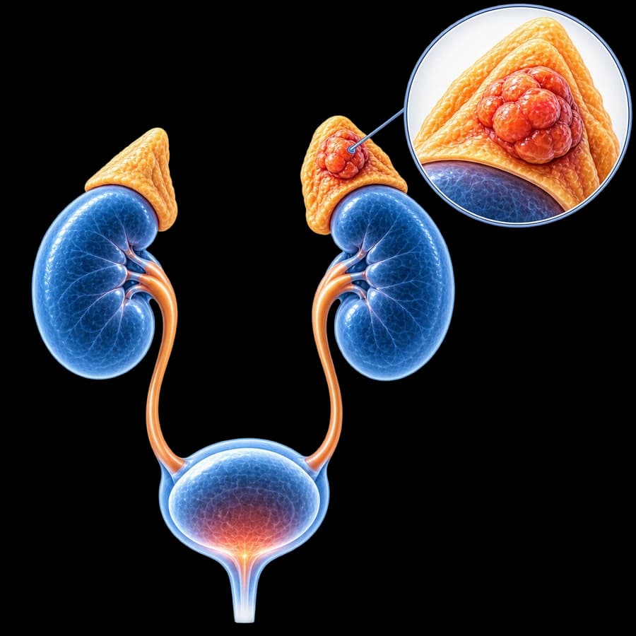
Benign fluid-filled lesion. Classification: endothelial (vascular/lymphatic origin, ~45%), pseudocysts (post-hemorrhage/trauma, ~40%), epithelial (true cysts, ~10%), parasitic (mainly echinococcal, rare). Overall autopsy incidence ~0.1%.
Symptoms
- Asymptomatic in most cases (incidental finding).
- In large cysts (> 5 cm): abdominal/flank pain, mass sensation.
- Rare: spontaneous rupture or hemorrhage.
- Parasitic complications: allergy, anaphylactic shock after rupture.
Diagnosis
- Ultrasound: anechoic lesion with posterior acoustic enhancement.
- CT: wall < 3 mm, water-density contents (0-20 HU), no enhancement after contrast.
- MRI: T1 low, T2 high signal. Differentiates hemorrhagic cysts.
- Parasitic workup (echinococcal serology) in endemic areas or characteristic imaging.
- Hormonal workup (1-mg DST, metanephrines, ARR if hypertensive) always — ESE/ENSAT 2023.
Treatment
- Observation for simple cysts < 5 cm, asymptomatic, with typical appearance.
- Laparoscopic adrenalectomy: > 5 cm, symptomatic, complex features, malignancy suspicion.
- Percutaneous drainage + sclerotherapy (ethanol): selected simple cysts.
- Parasitic: albendazole + careful surgical removal (avoiding rupture).
Follow-up
Simple small cysts follow general incidentaloma guidelines (ESE/ENSAT 2023): reassessment at 6-12 months in equivocal cases, no repeat imaging in clearly simple cysts < 4 cm.
Frequently Asked Questions (FAQ)
What is an adrenal cyst?
A rare benign fluid-filled lesion of the adrenal gland. Four types: endothelial (~45%), epithelial (~10%), pseudocysts (~40%, post-hemorrhage/trauma), parasitic (rare, echinococcal).
Is it dangerous?
Almost always no. Most small simple cysts are asymptomatic and require no treatment. Risk of hemorrhage/rupture in large (> 5 cm) or complex cysts.
How is it diagnosed?
CT/MRI: thin wall (< 3 mm), water-density contents (HU 0-20), no internal septations. Complex features (thick wall, septations, solid components, enhancement) require further evaluation.
When is surgery needed?
Symptomatic, > 5 cm, complex features, or suspicion of malignancy. Laparoscopic adrenalectomy or drainage + sclerotherapy.
How is follow-up done?
Small simple cysts (< 4-5 cm) with typical appearance follow the same incidentaloma surveillance guidelines (ESE/ENSAT 2023).
Related Topics
Book Your Appointment in Rhodes
Have an adrenal cyst finding? Let's evaluate it with proper imaging and complete hormonal workup.
References
- Fassnacht M, et al. ESE/ENSAT clinical practice guideline on the management of adrenal incidentalomas (2023) — academic.oup.com
- Erickson LA, et al. Adrenal cysts: a clinicopathologic study — Mayo Clin Proc — pubmed.ncbi.nlm.nih.gov
Medical Review

Dr. Marinos Vasilas, Urologist — Andrologist
Dr. Marinos Vasilas evaluates adrenal cysts with careful imaging and hormonal workup, avoiding unnecessary interventions.
View full profile


