My Clinical Approach
Pheochromocytoma is the textbook example of a curable but lethal tumour. The biochemical diagnosis is precise, the surgical cure rate is high — but inadequate preoperative preparation can transform a controlled operation into an intraoperative catastrophe.
In my practice three principles are non-negotiable:
- No surgery without adequate alpha-blockade — minimum 10-14 days, with target seated BP < 130/80 and orthostatic hypotension as the marker of effective vascular relaxation.
- Genetic testing for every patient — even sporadic-appearing cases. Hereditary diagnosis changes follow-up for the patient and the entire family.
- Multidisciplinary anaesthetic plan — endocrinology, anaesthesia, intensive care and the surgical team must agree on the preoperative strategy.
What Is Pheochromocytoma?
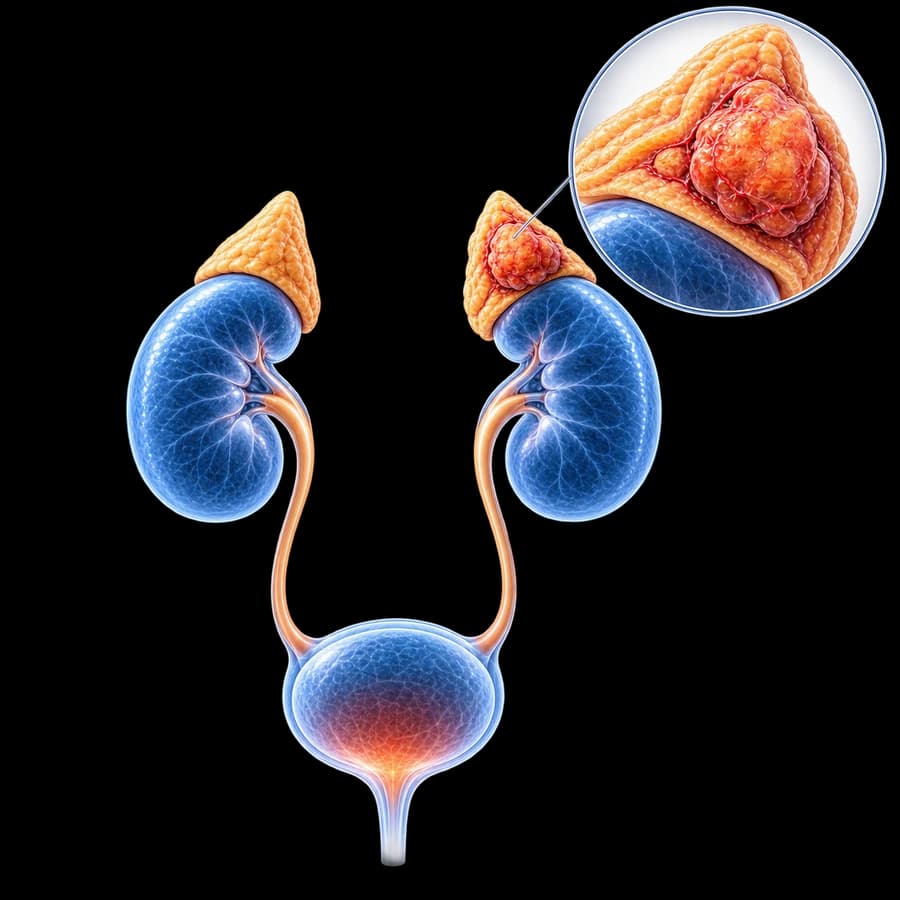
Pheochromocytoma arises from chromaffin cells of the adrenal medulla, which produce catecholamines (adrenaline, noradrenaline, dopamine). The same tumours arising in extra-adrenal sympathetic ganglia are termed paragangliomas.
Although classically considered the "10% tumour" (10% bilateral, 10% extra-adrenal, 10% malignant, 10% hereditary), modern data show much higher hereditary rates (~30-40%) and approximately 10-17% metastatic potential.
Genetics — Hereditary Forms
MEN2 (RET)
Multiple endocrine neoplasia type 2. Pheochromocytoma + medullary thyroid cancer + hyperparathyroidism (MEN2A).
VHL
Von Hippel-Lindau syndrome. Pheo + retinal/CNS haemangioblastomas + clear cell renal cancer + pancreatic NETs.
NF1
Neurofibromatosis type 1. Café-au-lait spots, neurofibromas, pheochromocytoma in 1-5%.
SDHx (B/C/D/AF2)
Mutations of the succinate dehydrogenase complex. SDHB carries the highest metastatic potential.
MAX, TMEM127, FH
Newer genes — increasingly identified through routine genetic testing.
Sporadic (~60-70%)
No identifiable mutation. Lower recurrence rate but lifelong follow-up still required.
Symptoms and Crisis
Classic triad: episodic headache, palpitations, sweating with hypertension. Other features:
- Sustained or paroxysmal hypertension.
- Pallor (rarely flushing).
- Anxiety, panic attacks, sense of impending doom.
- Tremor, nausea, abdominal pain.
- Weight loss, hyperglycaemia.
- Orthostatic hypotension between attacks.
- Cardiomyopathy, arrhythmias, myocarditis.
Pheochromocytoma Crisis
Severe hypertension (> 200/120), end-organ damage (stroke, MI, pulmonary oedema, heart failure), hyperthermia, multi-organ failure. Triggers: surgery without preparation, beta-blocker without prior alpha-blockade, contrast media, opioids, metoclopramide, anaesthetic induction. Medical emergency requiring ICU.
Diagnostic Workup
- Plasma free metanephrines (sensitivity > 96%, specificity 85-90%) — first-line test.
- 24h urinary fractionated metanephrines and catecholamines.
- Sample collection: supine, after 30 minutes rest, avoiding interfering substances (acetaminophen, tricyclics, sympathomimetics).
- Chromogranin A as additional marker.
- Plasma methoxytyramine for SDHx tumours and dopamine-secreting tumours.
Imaging — Anatomic and Functional
Adrenal CT/MRI
First-line anatomic imaging. Pheochromocytomas typically show high HU on unenhanced CT (> 10), strong enhancement, slow washout, and may show areas of necrosis or haemorrhage.
123I-MIBG Scintigraphy
Functional imaging targeting catecholamine uptake. Useful for staging, recurrence, extra-adrenal disease.
68Ga-DOTATATE PET/CT
Higher sensitivity for SDHx tumours, paragangliomas, and metastatic disease. Becoming the preferred functional modality.
FDG-PET/CT
For aggressive or metastatic tumours, particularly SDHB-related.
Preoperative Alpha-Blockade
Mandatory in every patient with biochemically positive pheochromocytoma. Standard regimen:
- Phenoxybenzamine (non-selective irreversible alpha-blocker) 10 mg twice daily, increasing every 2-3 days. Or doxazosin (selective alpha-1) 2-16 mg/day.
- Duration: minimum 10-14 days.
- Targets: seated BP < 130/80, standing BP > 90 systolic, mild orthostatic hypotension as a marker of effective vascular relaxation.
- High sodium and fluid intake from day 3-4 of alpha-blockade to expand circulating volume.
- Beta-blocker (e.g. propranolol) only AFTER complete alpha-blockade if tachycardia (> 100 bpm) or arrhythmia present. Never beta-blocker first — risk of paradoxical hypertensive crisis.
- Nicardipine, magnesium and labetalol available as adjuncts perioperatively.
Surgical Treatment
- Laparoscopic adrenalectomy is gold standard for tumours < 6-8 cm without local invasion.
- Transabdominal or retroperitoneal approach by surgeon expertise.
- Open surgery: very large tumours, local invasion, suspected malignancy.
- Cortical-sparing partial adrenalectomy in selected hereditary bilateral cases (MEN2, VHL).
- Early adrenal vein control to limit catecholamine surges, minimal tumour manipulation, close anaesthetic communication.
- ICU monitoring for the first 24 hours due to risk of post-resection hypotension and hypoglycaemia.
Long-term Follow-up
Per the ESE 2016 guideline (Plouin et al.), all patients require lifelong follow-up:
- Plasma or urinary metanephrines at 6 weeks postoperatively, then annually.
- Imaging based on biochemistry and clinical risk profile.
- Genetic testing for every patient — directs personal and family surveillance.
- Cascade testing of family members in hereditary cases.
- For SDHB mutation or known metastatic disease — more intensive imaging schedule.
Bottom line: A timely diagnosis and proper preoperative preparation transform pheochromocytoma from a deadly into a curable disease. Lifelong follow-up and genetic screening are non-negotiable.
Frequently Asked Questions (FAQ)
What is pheochromocytoma?
Pheochromocytoma is a rare neuroendocrine tumour arising from chromaffin cells of the adrenal medulla, which produces excess catecholamines (adrenaline, noradrenaline, dopamine). When the tumour arises outside the adrenal it is called paraganglioma.
How common is it?
Annual incidence ~2-8 cases per million. Up to 40% have a hereditary basis (MEN2, von Hippel-Lindau, NF1, SDHx mutations). Prevalence in adrenal incidentalomas: 1.5-7%; in resistant hypertension: 0.1-0.6%.
What are the symptoms?
The classic triad is episodic headache, palpitations, and sweating with hypertension. Other features: pallor, anxiety, tremor, weight loss, hyperglycaemia, orthostatic hypotension. Up to 10% are asymptomatic and discovered incidentally.
How is it diagnosed?
Per the Endocrine Society 2014 guideline: plasma free metanephrines or 24h urinary fractionated metanephrines as initial test (highest sensitivity). Imaging with adrenal CT/MRI; functional imaging (123I-MIBG, 68Ga-DOTATATE PET/CT) for staging or extra-adrenal disease.
Why is preoperative preparation critical?
Without adequate alpha-blockade, surgical manipulation can trigger life-threatening hypertensive crisis, arrhythmia or pulmonary oedema. Pre-op preparation with alpha-blockade for 10-14 days is mandatory to reduce perioperative cardiovascular complications.
How is it managed surgically?
Laparoscopic adrenalectomy is standard for tumours < 6-8 cm without local invasion. Transabdominal or retroperitoneal approach. Open surgery for very large or invasive tumours. Cortical-sparing partial adrenalectomy is considered in hereditary bilateral disease.
Is genetic testing required?
Yes. Per current guidelines all patients should be offered genetic counselling and testing, given the 30-40% hereditary rate. Identifying syndromes (MEN2, VHL, NF1, SDHx, MAX, TMEM127, FH) directs personal and family surveillance.
What is the long-term follow-up?
Lifelong biochemical follow-up with annual metanephrines. Risk of recurrence/metastasis ~5-15% even after complete resection. Higher risk in: extra-adrenal location, large tumours, SDHB mutation, young age. Imaging based on biochemistry and clinical risk.
Related Topics
Book Your Appointment in Rhodes
A pheochromocytoma demands precise preoperative preparation and meticulous anaesthetic coordination. We deliver the full pathway — from biochemical workup and genetic testing through laparoscopic adrenalectomy and lifelong follow-up.
References
- Lenders JW, et al. Pheochromocytoma and Paraganglioma: An Endocrine Society Clinical Practice Guideline (2014) — academic.oup.com
- Plouin PF, et al. ESE clinical practice guideline for long-term follow-up of patients operated on for a phaeochromocytoma or a paraganglioma (2016) — academic.oup.com
- Fassnacht M, et al. ESE/ENSAT Adrenal Incidentaloma Guidelines (2023) — academic.oup.com
Medical Review

Dr. Marinos Vasilas, Urologist — Andrologist
Dr. Marinos Vasilas leads the surgical management of pheochromocytoma in Rhodes, working closely with endocrinology, anaesthesia and intensive care to ensure safe perioperative management of catecholamine-secreting tumours.
View full bio


