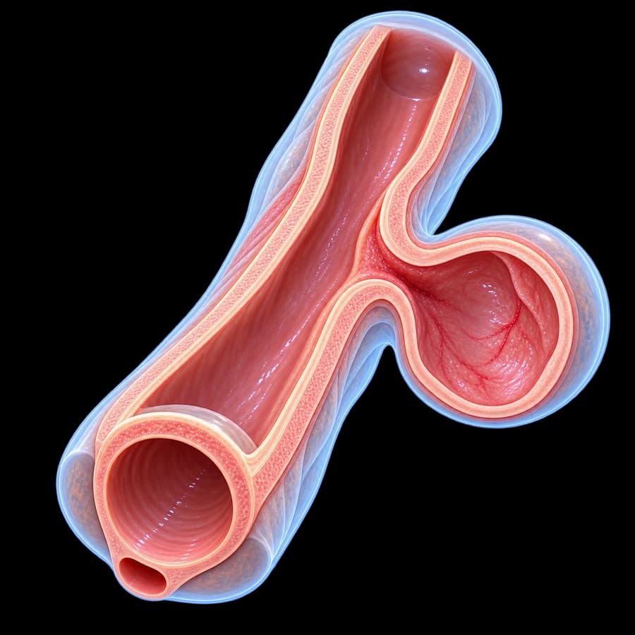
What Is a Urethral Diverticulum
A urethral diverticulum is an outpouching of the urethral mucosa through a defect in the muscular wall, forming a sac that communicates with the urethral lumen. Prevalence is 1–6% in women, peaking between ages 30–60.
Leach L/N/S/C3 classification: Location relative to the urethral neck, Number of diverticula, Size, Configuration (single/multiloculated/circumferential).
Causes & Pathogenesis
Acquired (most common)
- Recurrent infection and obstruction of Skene glands (paraurethral glands) — abscess formation that ruptures into the urethral lumen.
- Traumatic childbirth: stretching and tearing of the urethral wall.
- Iatrogenic: previous urethral surgery (sling).
- Chronic obstruction: urethral stenosis.
Congenital (rare)
Paraurethral cysts with secondary communication.
Symptoms — The 3 Ds
- Dysuria: burning and pain on urination.
- Dyspareunia: pain during intercourse — localized to the anterior vaginal wall.
- Dribbling: post-void urinary dribbling — caused by emptying of the diverticulum.
Additional features:
- Recurrent UTIs (50–80%).
- Palpable anterior vaginal wall mass.
- Pus or blood from the urethra on compression.
- Chronic pelvic pain.
- Hematuria.
- Urinary incontinence (irritative).
Diagnosis
1. Clinical examination
Vaginal examination with palpation of the anterior wall — mass, tenderness, expression of pus from the urethra on compression.
2. Pelvic MRI with endovaginal coil
Gold standard. Demonstrates the diverticulum, its neck, extent (single/multiloculated/circumferential), relationship to the sphincter, and any internal stones or malignancy.
3. Cystourethroscopy
Identifies the ostium of the diverticulum — assists in preoperative planning.
4. Double-balloon urethrography
Older method — largely replaced by MRI.
5. Urodynamics
If concomitant stress urinary incontinence is suspected.
Treatment
Transvaginal diverticulectomy
Treatment of choice. 3-layer technique:
- Layer 1: reconstruction of urethral mucosa with absorbable 4-0 vicryl sutures.
- Layer 2: periurethral fascia.
- Layer 3: vaginal wall — with offset incisions to avoid overlying suture lines (reduces fistula risk).
For large or recurrent diverticula: interposition of a Martius flap (labial fat pad) or vaginal wall flap.
Perioperative: indwelling urethral catheter for 14–21 days, suprapubic cystostomy in selected cases.
Conservative approach
Only for asymptomatic small diverticula (<1 cm) in elderly high-risk patients: observation + antibiotics for UTI flare-ups.
Complications & Prognosis
- Diverticulum recurrence (5–15%).
- Urethrovaginal fistula (5–10%) — prevented by Martius flap in complex cases.
- De novo urinary incontinence (5–10%) — from sphincter trauma.
- Urethral stricture (1–3%).
- Wound infection (1–5%).
- Postoperative chronic pain.
Prognosis: symptom resolution >90% in expert centers.
Malignancy risk: ~6% adenocarcinoma (clear cell, intestinal-type, urothelial). Always send for histopathology.
FAQ
What is a urethral diverticulum?
A saccular outpouching of the urethral mucosa communicating with the urethral lumen. Found mostly in women (1–6%), rare in men. Usually arises from obstructed and infected Skene glands.
What are the symptoms (the 3 Ds)?
Classic triad: dysuria (burning), dyspareunia (pain during intercourse), post-void dribbling (urinary leakage after voiding). Also: recurrent UTIs, palpable anterior vaginal mass.
How is it diagnosed?
Gold standard: pelvic MRI with endovaginal coil — accurately demonstrates the diverticulum, its neck and relationship to the sphincter. Adjuncts: cystoscopy, urethrography.
What is the treatment?
Transvaginal diverticulectomy with 3-layer closure (Martius flap in selected complex cases). Conservative observation only for asymptomatic small diverticula.
Is there a cancer risk?
Yes — about 6% risk of malignant transformation, primarily clear-cell adenocarcinoma. Histopathology is mandatory for every excised diverticulum.
Conclusion
Urethral diverticulum is a commonly missed cause of chronic symptoms in women. Pelvic MRI with endovaginal coil and transvaginal diverticulectomy provide definitive treatment with excellent outcomes.
Scientific References
- EAU Guidelines on Female Urology 2024 — uroweb.org
- Leach GE et al. L/N/S/C3 Classification of female urethral diverticula. Neurourol Urodyn 1993.
- Rovner ES. Urethral diverticula: a diagnostic dilemma. Curr Urol Rep 2003.
- Thomas AA et al. Urethral diverticula in 90 female patients: a study with emphasis on neoplastic alterations. J Urol 2008.
Meet the Doctor

Dr. Marinos Vasilas, Urologic Surgeon – Andrologist
Specialist in female urological surgery — transvaginal diverticulectomy with 3-layer closure and Martius flap.
View full profile


