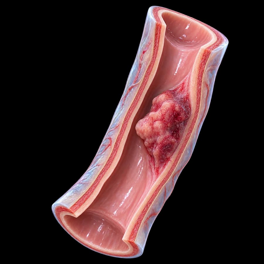
What is Urethral Cancer
Primary urethral cancer is rare (incidence ~4 per million). Represents <1% of all genitourinary malignancies. It is the only urologic cancer 2–3 times more common in women than men.
Histologic types
- Squamous cell carcinoma (~75%): most common, especially anterior urethra. HPV and chronic irritation associations.
- Urothelial carcinoma (~15%): mainly posterior urethra. Often coexists with bladder cancer.
- Adenocarcinoma (~5%): often associated with urethral diverticula in women or Cowper glands in men.
- Melanoma, sarcomas (<1%).
Risk Factors
- Chronic urethral strictures (especially post-gonococcal).
- Recurrent UTIs and chronic urethritis.
- HPV infection — especially high-risk types 16/18.
- Urethral diverticula (women) — ~6% undergo malignant transformation.
- Chronic indwelling urethral catheter.
- Lichen sclerosus (Balanitis Xerotica Obliterans).
- Prior bladder or ureteral cancer.
- Age >50 years.
- Smoking.
Symptoms
Symptoms are non-specific and often absent in early stages — leading to diagnostic delay:
- Gross hematuria or urethral bleeding (most common).
- Palpable mass in perineum, penis, or meatus.
- Obstructive symptoms — weak stream, hesitancy.
- Storage symptoms — dysuria, frequency.
- Urethral discharge.
- Pain or discomfort in perineum/penis.
- Priapism (in men, rare).
- Inguinal lymph node enlargement (metastasis).
- Painful or bloody ejaculation.
Diagnosis & Staging
1. Clinical examination
Inspection of meatus, palpation of penis/perineum, digital rectal exam (men), pelvic exam (women), inguinal lymph node palpation.
2. Urethroscopy + biopsy
Diagnostic gold standard. Direct visualization and histologic confirmation.
3. Pelvic MRI
Best imaging for local extension (T-stage), invasion of adjacent organs, modality of choice for surgical planning.
4. Chest/abdominal/pelvic CT
Detect metastases (lung, liver) and assess retroperitoneal lymph nodes.
5. TNM Staging
T1 (subepithelial connective tissue) → T2 (corpus spongiosum/prostate) → T3 (corpora cavernosa/cervix) → T4 (adjacent organs).
Treatment
Individualized by location, stage, histology, and patient expectations:
Anterior urethra — localized disease
- Endoscopic ablation/local excision: for superficial lesions (Ta, T1) with laser or TUR.
- Partial urethrectomy / penectomy: for invasive lesions with functional preservation.
- Total penectomy + perineal urethrostomy: for extensive lesions.
Posterior urethra / advanced disease
- Radical cystoprostatourethrectomy with urinary diversion (men) or anterior pelvic exenteration (women — cystourethrectomy ± hysterectomy).
- Inguinal / pelvic lymphadenectomy: for palpable or positive nodes.
Multimodal approach
- Neoadjuvant chemotherapy (cisplatin-based) for locally advanced.
- Radiotherapy: for squamous cell carcinoma — may replace surgery in selected cases (organ preservation).
- Immunotherapy (checkpoint inhibitors): for metastatic disease.
Prognosis & Follow-up
5-year overall survival depends strongly on stage:
- Stage I (T1N0M0): 70–85%.
- Stage II: 50–60%.
- Stage III: 30–40%.
- Stage IV: <15%.
Follow-up: clinical exam + urethroscopy every 3 months for 2 years, every 6 months for 5 years. Annual CT imaging.
FAQ
What is urethral cancer?
A rare malignant neoplasm arising from the urethral epithelium. Represents <1% of all urologic cancers and is the only urologic cancer more common in women than in men.
What are the risk factors?
Chronic irritation: recurrent infections, urethral strictures (especially post-gonococcal), urethral diverticulum (women), HPV-16/18, chronic indwelling catheter, lichen sclerosus.
What are the symptoms?
Gross hematuria, urethral bleeding, palpable mass in perineum/penis/meatus, progressive obstruction, inguinal lymphadenopathy. Symptoms are often delayed — leading to diagnostic delay.
How is it diagnosed?
Urethroscopy + biopsy. Pelvic MRI for staging, chest/abdominal CT for metastases. Histology identifies subtype: urothelial, squamous (most common), adenocarcinoma, melanoma.
What is the treatment?
Individualized by type and stage: local excision for limited lesions, partial or total urethrectomy, urethrectomy with hysterectomy/prostatectomy in advanced stages. Adjuvant cisplatin-based chemotherapy and radiation depending on histology.
Conclusion
Urethral cancer is rare but demands prompt action. Early diagnosis through urethroscopy in any persistent hematuria and individualized multimodal management at a specialized center are critical for outcome.
Scientific References
- EAU Guidelines on Primary Urethral Carcinoma 2024 — uroweb.org
- Gakis G et al. EAU Guidelines on Primary Urethral Carcinoma. Eur Urol 2013;64:823-830.
- NCCN Guidelines Version 2.2024 — Bladder Cancer (includes Urethral Cancer section).
- Smith Y et al. Treatment outcomes of primary urethral cancer. J Urol 2020.
Meet the Doctor

Dr. Marinos Vasilas, Urologic Surgeon – Andrologist
Specialized in the diagnosis and oncologic surgery of rare urinary tract neoplasms — urethral, ureteral and renal pelvic cancer.
View full profile


