My clinical approach
In hypospadias, early and accurate assessment determines the plan. Parents need to be informed in detail from the first days — immediate surgical scheduling is not necessary, but circumcision should NEVER be done without prior pediatric urology evaluation.
In my clinical practice I follow the EAU Pediatric Urology 2024 guidelines:
- Complete assessment at birth: meatus location, chordee, glans configuration, foreskin, testes.
- Avoid circumcision — foreskin needed as graft material.
- Hormonal workup and karyotype in proximal hypospadias with bilateral cryptorchidism (DSD).
- Preoperative testosterone treatment for hypoplastic glans.
- Surgery at 6-18 months (optimal 6-12 months).
- Technique selection based on severity: TIP for distal, Bracka for proximal.
- Long-term follow-up to puberty for functional and psychosexual outcomes.
What is hypospadias
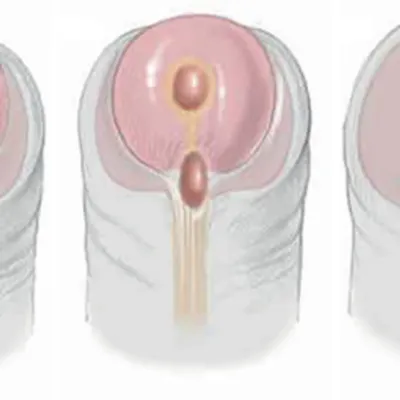
Hypospadias is a congenital penile anomaly in which the urethral meatus does not open at the tip of the glans, but ventrally (on the underside) at various positions along the penis, scrotum or even the perineum.
It is one of the most common congenital male genital anomalies with incidence 1 in 200-300 male births. The incidence shows an upward trend in recent decades, possibly due to increased exposure to endocrine disruptors.
Characteristics: abnormal meatus location, penile curvature (chordee) ventrally (in more severe forms), abnormal foreskin (hooded foreskin) — foreskin only on dorsal surface, absent ventrally.
Classification
Classification is based on the urethral meatus location after correction of any chordee:
Distal (50%)
Glanular (meatus at base of glans), coronal (in coronal sulcus). The most common and mildest form.
Mid-penile (30%)
Meatus at middle ventral surface of penis. Often accompanied by mild chordee.
Proximal penile
Meatus near base of penis. Often significant chordee, more complex repair required.
Penoscrotal / scrotal (20%)
Meatus on scrotum. Severe form with clear chordee, bifid scrotum, need for DSD evaluation.
Perineal
Meatus on perineum. The most severe form, often with DSD, requires thorough workup.
Megameatus intact prepuce (MIP)
Rare variant — wide meatus at base of glans with normal foreskin. Often diagnosed after circumcision.
Etiology
Multifactorial. In most patients no single cause is identified:
- Genetic factors — familial predisposition (8% of cases), mutations in androgen signaling genes (SRD5A2, AR).
- Hormonal disorders — defects in testosterone or DHT production/action in the 1st trimester.
- Environmental endocrine disruptors — pesticides, vinyl monomers, plasticizers, phytoestrogens.
- Prematurity — increased frequency in newborns <37 weeks.
- Low birth weight (<2500 g).
- Maternal age >35 years.
- In vitro fertilization (IVF/ICSI) — small increase in risk.
- Maternal diabetes mellitus, obesity, progesterone use.
Clinical picture
The classic triad of hypospadias includes:
- Abnormal urethral meatus location — ventral to the glans at various positions.
- Penile curvature (chordee) — ventral in more severe types, evident on erection.
- Abnormal foreskin (hooded foreskin) — present only dorsally, absent ventrally.
- Wide or narrow meatus, abnormal urinary stream (broad or downward-directed flow).
- In severe forms: bifid scrotum, microphallus, association with cryptorchidism.
- In older age (if undiagnosed): difficulty urinating standing, sexual dysfunction.
Diagnosis
Clinical examination at birth
By pediatrician/obstetrician. Inspection: meatus location, degree of chordee, foreskin shape, testicular palpation, scrotal examination.
Pediatric urology referral
Immediate referral for categorization and repair planning. Detailed parental counseling.
Workup for associated conditions
Testicular palpation (cryptorchidism in 10%), inguinal hernia screening (10%). Renal ultrasound if other anomalies coexist.
DSD evaluation (only in severe forms)
In proximal/penoscrotal/perineal hypospadias with bilateral cryptorchidism: karyotype, hormonal workup (LH, FSH, testosterone, DHT), 17-OH-progesterone.
Preoperative preparation
In hypoplastic glans or microphallus: topical (testosterone cream) or injectable testosterone therapy 1-3 months before surgery to enlarge glans.
Surgical repair
Timing of surgery
Ideally 6-18 months (optimal 6-12 months). Advantages: less psychological impact, better healing, fewer complications. Goal: completion before development of psychosexual awareness.
TIP / Snodgrass (distal hypospadias)
Tubularized Incised Plate. Midline incision of urethral plate and tubularization around catheter. Success >90% for glanular/coronal. Complications: fistula 5-10%, meatal stenosis.
Onlay flap (mid-proximal hypospadias)
Island flap from inner foreskin surface placed onto the urethral plate. Preserves urethral plate. Good outcomes in mid/proximal forms without severe chordee.
Two-stage Bracka (severe hypospadias)
Stage 1: chordee correction, buccal mucosa graft placement on ventral surface. Stage 2 (6 months later): tubularization. Preferred technique for proximal, penoscrotal, perineal hypospadias.
Postoperative care
Urethral catheterization 7-14 days. Antibiotics, analgesia. Dressing changes. Follow-up first weeks, then at 3, 6, 12 months and yearly to puberty.
Complications & prognosis
- Urethrocutaneous fistula: 5-15% — most common complication. Often requires second surgery.
- Meatal stenosis: 5-10%. Dilations or surgical correction.
- Neourethral stricture: 5-15%, more common in proximal forms.
- Wound dehiscence: 1-5%.
- Recurrent chordee: 5-10%.
- Urethral diverticulum (neourethral dilation): rare.
- Cosmetic results: satisfactory in most patients.
- Sexual function: normal in majority after successful repair.
- Fertility: usually normal (except severe DSD).
- Psychosexual: significantly better outcomes when repair done at young age.
Frequently asked questions (FAQ)
What is hypospadias?
A congenital anomaly where the urethral meatus is not located at the tip of the glans but ventrally (on the underside) at various positions. Incidence 1 in 200-300 male births. Often accompanied by penile curvature (chordee) and abnormal "hooded foreskin".
What are the forms of hypospadias?
Classified by meatus location: glanular (50%), coronal, distal penile, mid-penile, proximal penile, penoscrotal, scrotal, perineal. The more proximal the meatus, the more severe the form.
What are the causes?
Multifactorial: genetic factors (mutations in hormonal signaling genes), hormonal disorders (androgen receptor defects), environmental exposures (endocrine disruptors — pesticides, plasticizers), prematurity, low birth weight, IVF.
Is it associated with other anomalies?
Yes. 10% have coexisting cryptorchidism, 10% inguinal hernia. Severe forms (proximal-scrotal) with bilateral cryptorchidism require evaluation for disorders of sex development (DSD): karyotype, hormonal workup.
How is it diagnosed?
Clinical examination at birth by pediatrician/obstetrician. Inspection: meatus location, presence of chordee, foreskin shape. In severe forms: pediatric urology referral, hormonal workup and renal ultrasound.
When should surgical repair be done?
Ideal age is 6-18 months (optimal 6-12 months) — before development of psychosexual awareness and favorable for anatomical results. Circumcision should NEVER be done before repair (foreskin needed as graft material).
What techniques are used?
Distal hypospadias: TIP (Snodgrass) — incision of urethral plate and tubularization. Proximal/severe: two-stage (Bracka — buccal mucosa graft placement and tubularization 6 months later), island flap (Onlay flap), tubularized preputial flap.
What are the complications?
Urethrocutaneous fistula (5-15%) — most common. Meatal stenosis, dehiscence, recurrent chordee, urethral diverticulum. Psychosexual: satisfactory sexual and urinary function in most patients after successful repair.
Related topics
Suspected hypospadias? Early pediatric urology evaluation
Contact us for personalized repair planning from birth. Do not proceed with circumcision before pediatric urology evaluation.
Scientific literature
- EAU Guidelines on Paediatric Urology (2024) — Hypospadias chapter — uroweb.org
- Snodgrass W. Tubularized incised plate urethroplasty for distal hypospadias. J Urol 1994;151(2):464-5 — pubmed.ncbi.nlm.nih.gov
- Bracka A. Hypospadias repair: the two-stage alternative. Br J Urol 1995;76 Suppl 3:31-41 — pubmed.ncbi.nlm.nih.gov
- Baskin LS, Ebbers MB. Hypospadias: anatomy, etiology, and technique. J Pediatr Surg 2006;41(3):463-72 — pubmed.ncbi.nlm.nih.gov
- Springer A, van den Heijkant M, Baumann S. Worldwide prevalence of hypospadias. J Pediatr Urol 2016;12(3):152.e1-7 — pubmed.ncbi.nlm.nih.gov
Meet the doctor

Dr. Marinos Vasilas, Urologist – Andrologist
Dr. Marinos Vasilas offers personalized assessment and surgical repair of hypospadias in all forms, with TIP, Onlay and two-stage Bracka techniques per EAU Pediatric Urology 2024 guidelines.
View full profile


