My Clinical Approach to Prostate Stones
Prostatic calculi are one of the most common incidental findings I encounter in my urological practice in Rhodes. Most patients are understandably concerned when told they have "stones in the prostate". In the vast majority of cases, however, this finding requires reassurance, not intervention.
Every patient with new findings of prostatic calculi receives a PSA blood test, digital rectal examination, and — where indicated — multiparametric MRI to exclude significant pathology. The calculi themselves are then managed expectantly.
- PSA + DRE for every patient with newly identified prostatic calculi.
- Exclude concurrent pathology: BPH, chronic prostatitis, prostate cancer.
- Treatment only when calculi contribute to recurrent infection or refractory pelvic pain.
- Expectant surveillance for asymptomatic findings — without unnecessary alarm.
What Are Prostate Stones
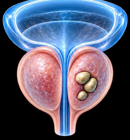
Prostatic calculi are calcified deposits that form within the ducts and glandular acini of the prostate. On transrectal ultrasound (TRUS), they appear as hyperechoic foci with posterior acoustic shadowing. They can be microscopic or form clusters several millimetres in diameter.
Their prevalence increases with age. They are found in an estimated >75% of men over 50 undergoing TRUS, with or without BPH or chronic prostatitis.
Clinical Significance
In the vast majority of cases, prostatic calculi are benign, incidental findings. Clinical significance arises only in specific contexts — primarily when associated with chronic prostatitis or recurrent urinary tract infections.
Causes & Pathophysiology
Two main mechanisms lead to prostatic calculus formation:
Primary (Endogenous) Calculi
Form through calcification of normal prostatic secretions (corpora amylacea). Typically asymptomatic and represent a natural consequence of ageing. Most prevalent in the transition zone.
Secondary (Exogenous) Calculi
Form through retrograde urine flow into prostatic ducts, or through calcification of inflammatory exudate in areas of chronic prostatitis. More likely to be associated with symptoms.
Risk Factors
Benign prostatic hyperplasia (BPH), chronic prostatitis, history of urinary tract infection, advanced age.
Symptoms & Clinical Presentation
The majority of patients with prostatic calculi are entirely asymptomatic. The condition is typically discovered incidentally on ultrasound performed for another reason.
Asymptomatic (Majority)
Incidental finding on ultrasound. No intervention required — only assessment of concurrent pathology.
Perineal / Pelvic Discomfort
Vague sensation of perineal heaviness or lower pelvic pressure, which may worsen with prolonged sitting. Often associated with concurrent chronic prostatitis rather than the calculi themselves.
Lower Urinary Tract Symptoms (LUTS)
In patients with BPH, the presence of calculi may compound LUTS. Recurrent urinary tract infections or secretory stasis may also occur.
Prostate Stones Are Not a Substitute for Cancer Screening
Finding prostatic calculi is an opportunity — not a reason for alarm, but a trigger for essential assessments:
- PSA blood test to exclude prostate cancer — regardless of age or symptoms.
- Digital rectal examination (DRE) to assess prostate texture and consistency.
- If PSA elevated or DRE suspicious: evaluation for prostate cancer with mpMRI and possible biopsy.
Classification — Types of Prostatic Calculi
Prostatic calculi are classified by anatomical location and origin:
Central Calculi
Transition ZoneOften with BPHLocated in the central/transition zone. Typically multiple, small. Usually asymptomatic. Most common type.
Peripheral Calculi
Peripheral ZoneOften with ProstatitisLocated in the peripheral zone. More likely associated with chronic prostatitis or prior infection. May be associated with perineal pain symptoms.
Ejaculatory Duct Calculi
Central / PericentralRareUncommon location — may be associated with obstructive oligospermia or ejaculatory symptoms. Requires specialist andrological assessment.
Diagnostic Evaluation
Standard assessment for a patient with newly identified prostatic calculi:
History & Clinical Examination
Assessment of urinary symptoms, history of prostatitis, UTI, or BPH. Digital rectal examination (DRE) to evaluate prostate consistency and tenderness.
PSA Blood Test
Mandatory for every patient with prostatic calculi — regardless of age or symptoms. Essential for prostate cancer exclusion.
Transrectal Ultrasound (TRUS)
Maps calculi distribution (number, size, location). Evaluates prostate volume and tissue characteristics. Measures post-void residual urine.
mpMRI (Selected Cases)
Indicated in patients with elevated PSA or suspicious DRE — to exclude clinically significant malignancy. Also considered when dense calcifications may affect ultrasound interpretation.
Treatment Approach
Treatment depends on symptoms and co-existing pathology:
Expectant Surveillance (Asymptomatic Calculi)
The vast majority of patients require no intervention. Annual urological review with PSA + DRE + ultrasound. Reassurance and patient education.
Treatment of Co-existing Pathology
If chronic prostatitis is present: prolonged antibiotic therapy (fluoroquinolones), alpha-blockers for pain relief, anti-inflammatory agents. If BPH: treatment according to LUTS severity and IPSS score.
Interventional Management (Rare)
In exceptionally rare cases where calculi are associated with refractory recurrent infections unresponsive to medical management, TURP or laser ablation may be considered to remove stone-harbouring prostatic tissue.
Ejaculatory Duct Obstruction (Selected Cases)
In young men with infertility and confirmed ejaculatory duct obstruction due to calculi: specialist andrological evaluation and potentially transurethral resection of the ejaculatory ducts (TURED).
Complications & Associated Conditions
Prostatic calculi may coexist with or contribute to:
- Chronic Prostatitis — the closest association. Calculi may act as a "bacterial reservoir" harbouring organisms resistant to antibiotic penetration, perpetuating refractory infection.
- Benign Prostatic Hyperplasia (BPH) — frequent co-existence, particularly in the transition zone.
- Male Infertility — rare, when calculi involve the ejaculatory ducts.
- In suspicious clinical contexts: mandatory exclusion of prostate cancer with PSA + mpMRI.
Follow-up & Prognosis
Follow-up for asymptomatic prostatic calculi:
- Annual urological review: PSA + DRE + ultrasound.
- If findings remain stable and PSA/DRE are normal — no further intervention.
- If new symptoms develop or PSA changes — reassessment with imaging.
Prognosis
Prostatic calculi in isolation carry an excellent prognosis and do not adversely affect lifespan or long-term health. Clinical outcome is primarily determined by any co-existing conditions (BPH, chronic prostatitis) and the effectiveness of their management.
Frequently Asked Questions (FAQ)
Are prostatic calcifications the same as prostate stones?
Yes. In clinical practice, the terms "prostatic calcifications", "prostatic calculi", and "prostate stones" are used interchangeably to describe calcified deposits within the prostate gland.
Can prostate stones cause cancer?
No direct causal link has been established. The presence of prostatic calculi does not indicate or lead to prostate cancer. However, dense or unusual calcifications can sometimes affect interpretation of multiparametric MRI. Regular PSA testing remains independently necessary.
Do prostate stones pass on their own?
No, not like kidney or ureteral stones. Prostatic calculi are fixed within the glandular tissue and do not pass through the urinary tract. They persist as stable imaging findings and are typically monitored rather than removed.
Do prostate stones always need treatment?
No. The vast majority of men with prostatic calculi require no specific treatment. When the stones are asymptomatic and no other pathology co-exists, regular surveillance is sufficient. Treatment is only considered when calculi are linked to recurrent infections or refractory chronic prostatitis.
Can prostate stones affect fertility?
In rare cases, large calculi near the ejaculatory ducts may contribute to obstructive azoospermia or oligospermia. Evaluation of male infertility includes scrotal and transrectal ultrasound (TRUS) to assess ductal anatomy.
Are prostate stones common in young men?
They are significantly more common in men over 50, especially in those with BPH or a history of chronic prostatitis. In younger men, finding prostate stones is usually an incidental finding during ultrasound performed for another reason.
Do prostate stones cause pain?
Most patients are completely asymptomatic. When symptoms occur, they typically include a vague sensation of perineal heaviness, mild pelvic discomfort, or intensification of existing chronic prostatitis symptoms. Severe pain is uncommon and should prompt investigation for other pathology.
Should I be worried if I was told I have prostate stones?
Generally no. Prostatic calculi are extremely common and usually benign findings. Concern is warranted only if they are accompanied by elevated PSA, suspicious digital rectal exam findings, recurrent infections, or unusual symptoms — all of which require further urological evaluation.
Related Topics
Book an Appointment in Rhodes
Told you have prostate stones on ultrasound? Specialist urological assessment with PSA, DRE, and TRUS at our practice in Rhodes, Greece.
References
- EAU Guidelines on Urological Infections 2024 — uroweb.org
- Sfanos KS, et al. Prostatic Calculi — Where Did They Come From? Urology 2009;73:787–791.
- de la Rosette JJ, et al. Transrectal ultrasonography in the diagnosis of prostatic disease. Br J Urol 1991;67:629–635.
- Shoskes DA, et al. Cytokine polymorphisms in men with chronic prostatitis/chronic pelvic pain syndrome. Urology 2002;59:797–801.
- NIDDK: Prostatitis — Inflammation of the Prostate — niddk.nih.gov
Medical Author

Dr. Marinos Vasilas, Urologist – Andrologist
Dr. Marinos Vasilas runs a private urology practice in Rhodes specialising in the diagnosis and management of prostate conditions, including prostatic calculi and chronic prostatitis.
Full profile


