My Clinical Approach
In a kidney tumour, the most important question before surgery is not only whether the tumour can be removed, but whether it can be removed safely while preserving the kidney.
Suitability for partial nephrectomy is not determined by size alone, but by tumour anatomy, its relationship to vessels and the collecting system, the function of both kidneys, and surgical complexity.
- I always consider first whether partial nephrectomy is technically and oncologically feasible.
- I do not use a magic cut-off in centimetres or minutes of ischaemia.
- My goal is complete tumour removal with maximal preservation of functioning kidney tissue.
- I explain clearly that the laparoscopic technique does not guarantee an outcome and does not eliminate the risk of complications.
- I do not promise unchanged kidney function or zero risk of recurrence.
What Is Partial Nephrectomy
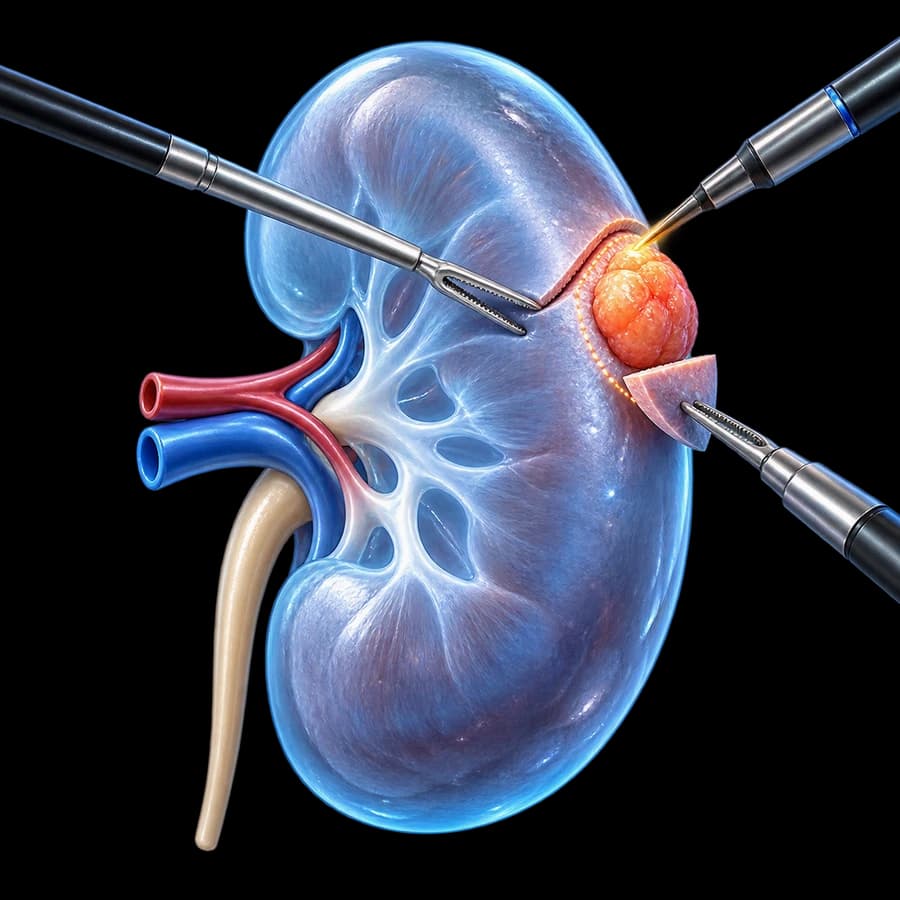
In partial nephrectomy, the kidney tumour is removed together with the necessary margin of healthy tissue, while the rest of the kidney remains in place.
The goals are:
- Complete tumour removal.
- Negative surgical margin where achievable.
- Maximum preservation of functioning renal parenchyma.
- Safe reconstruction of the kidney.
Partial Nephrectomy ≠ Biopsy
Partial nephrectomy is definitive surgical tumour excision — it is not a renal biopsy, diagnostic sampling, or ablation.
Why Preserve the Kidney
Preservation of renal parenchyma may reduce the loss of kidney function compared with radical nephrectomy.
This has particular importance in:
- Chronic kidney disease.
- Hypertension.
- Diabetes.
- Solitary kidney.
- Bilateral tumours.
- Younger patients with greater lifetime renal risk.
We do not promise that kidney function will remain exactly the same.
T1 Kidney Tumours
Partial nephrectomy is a preferred nephron-sparing surgical strategy for many T1 tumours, when technically feasible.
We do not create a rule that every T1a must always be treated with partial nephrectomy, or that every T1b always needs radical nephrectomy.
T2 Kidney Tumours
Selected larger tumours may be treated with partial nephrectomy in:
- Expert centres.
- Favourable anatomy.
- Imperative indications (solitary kidney, bilateral disease).
- Chronic kidney disease.
when oncologically/technically appropriate. We do not present partial nephrectomy for T2 tumours as a routine solution for everyone.
Size Is Not the Only Criterion
Tumour diameter alone does not determine whether partial nephrectomy can be performed.
Also relevant:
- Location.
- Exophytic/endophytic character.
- Depth.
- Proximity to the renal hilum.
- Relationship to the collecting system.
- Relationship to the vessels.
- Multifocality.
- Kidney function.
- Surgeon experience.
How Complex Is a Kidney Tumour?
Tools such as the RENAL nephrometry score and the PADUA classification can be used to describe the anatomical complexity of a kidney tumour, taking into account features such as:
- Size.
- Depth.
- Proximity to the collecting system.
- Polar location.
- Relationship to the renal hilum.
No score by itself decides whether partial or radical nephrectomy should be performed.
Exophytic vs Endophytic Tumour
Exophytic tumour
A large part of it projects outside the renal contour.
Endophytic tumour
A large part of it lies within the renal parenchyma.
Endophytic lesions may be technically more complex, but this does not automatically contraindicate partial nephrectomy.
Can Partial Nephrectomy Be Done for a Tumour Near the Vessels?
Yes, in selected patients.
Hilar tumours may be more demanding due to proximity to:
- Branches of the renal artery.
- The renal vein.
- The collecting system.
But hilar location does not automatically mean radical nephrectomy. Expertise matters.
Central/Complex Tumours
Complex anatomy may increase:
- Operative complexity.
- Bleeding risk.
- The likelihood of entering the collecting system.
- Reconstruction requirements.
- Ischaemic burden.
but does not necessarily preclude partial surgery.
Solitary Kidney
In a solitary kidney, nephron preservation has particularly great importance.
Treatment strategy must take into account:
- Baseline eGFR.
- Tumour complexity.
- Malignancy risk.
- Dialysis risk.
- Alternatives.
- Technical feasibility.
We do not write that partial nephrectomy is always possible.
Bilateral/Multifocal Tumours
Partial nephrectomy has an important role in selected bilateral or multifocal tumours, as well as in hereditary kidney cancer syndromes, with the aim of preserving kidney tissue.
Hereditary Kidney Cancer
Selected hereditary syndromes may be associated with bilateral, recurrent, or multifocal tumours and may require a long-term nephron-sparing strategy. This page is not a genetics guide — genetic assessment is individualised.
Partial vs Radical Nephrectomy
| Partial Nephrectomy | Radical Nephrectomy | |
|---|---|---|
| Tumour | Removed with a margin of healthy tissue | Removed together with the whole kidney |
| Renal parenchyma | Largely preserved | The entire kidney is lost |
| Kidney function | Greater preservation | Greater nephron loss |
| Technical complexity | Often greater | Different procedure |
| Main use | Suitable localised tumours | When partial is not appropriate |
| Oncologic goal | Complete resection | Complete resection |
Partial nephrectomy is not a “smaller oncological operation” when used correctly — it is nephron-sparing oncologic surgery.
See also the “When Is Radical Nephrectomy Needed” section on the 3D laparoscopic radical nephrectomy page.
Oncological Effectiveness
In appropriately selected localised tumours, partial nephrectomy aims for oncological control while preserving kidney tissue.
We do not make blanket claims that partial is always oncologically identical to radical, always superior, or that radical nephrectomy causes worse cancer outcomes.
Laparoscopic vs Robotic Partial Nephrectomy
| Laparoscopic Partial | Robotic Partial | Open Partial | |
|---|---|---|---|
| Minimally invasive | Yes | Yes | No |
| 3D vision | System-dependent | Standard | Direct |
| Instrument control | Direct manual | Via console | Direct manual |
| Articulated instruments | Limited | Yes | Direct manual |
| Intracorporeal reconstruction | Possible/experience-dependent | Technically facilitated | Direct |
| Complex tumours | Selected | Selected | Selected |
| Oncologic objective | Same | Same | Same |
| Best choice | Individualised | Individualised | Individualised |
The robotic platform can facilitate some technically demanding steps of intracorporeal reconstruction, but this does not mean that a properly performed laparoscopic partial nephrectomy is oncologically inferior. No approach is presented as the “winner”. See also robotic partial nephrectomy.
Laparoscopic vs Open Partial Nephrectomy
Open partial nephrectomy remains important in selected very complex tumours, extensive multifocal disease, challenging anatomy, or situations where direct exposure is advantageous. We do not present open surgery as outdated.
What Laparoscopic Surgery Means
The surgeon directly manipulates the laparoscopic instruments through small incisions, viewing the procedure on a monitor via a camera that may offer 2D or, where available, stereoscopic 3D vision.
There is no console or robotic system — every instrument movement is controlled directly by the surgeon’s hand.
3D Laparoscopy vs Robotic Surgery
3D Laparoscopy
Stereoscopic camera, conventional laparoscopic instruments, the surgeon handles the instruments directly by hand.
Robotic
Stereoscopic robotic camera, articulated robotic instruments, the surgeon controls the system via a console.
Both approaches are minimally invasive, fully controlled by the surgeon, and depend heavily on team expertise. 3D laparoscopy is not robotic surgery, and stereoscopic vision alone does not guarantee a superior oncological outcome.
Why 3D Imaging Matters in Partial Nephrectomy
Partial nephrectomy requires a combination of tumour excision, vascular management, collecting-system repair when needed, and renal reconstruction/suturing.
Stereoscopic three-dimensional vision, where available, can facilitate depth perception and identification of anatomical structures during minimally invasive reconstruction. The technology does not guarantee the outcome.
Preoperative Imaging
Contrast-enhanced multiphasic CT or MRI can assess:
- Tumour size.
- Location.
- Depth.
- Arterial anatomy.
- Venous anatomy.
- Relationship to the collecting system.
- The contralateral kidney.
- Metastatic disease where relevant.
We do not claim that every case requires identical imaging.
Is a Biopsy Needed Before Partial Nephrectomy?
Not in every patient.
Biopsy can be particularly useful when:
- Pathology may change management.
- Active surveillance is being considered.
- Ablation is planned.
- Diagnosis is uncertain.
- Systemic treatment is contemplated.
If surgery is clearly indicated for a suspicious, resectable renal mass, biopsy is not always required.
Small Tumour ≠ Automatic Surgery
Selected small renal masses may be managed with partial nephrectomy, active surveillance, or ablation, depending on the tumour, age, comorbidity, kidney function, and patient preference.
We do not give a universal growth-rate threshold or imaging interval without current verification.
How the Procedure Is Performed
This description is deliberately kept at a general level — it is not a surgical manual with clamping technique, trocar placement sequence, suturing sequence, or device details.
Why Blood Flow to the Kidney Is Temporarily Interrupted
In many partial nephrectomies, temporary clamping of the renal arterial inflow may be needed to:
- Control bleeding.
- Correctly identify the tumour.
- Allow safe reconstruction.
This creates temporary warm ischaemia.
Warm Ischaemia Time
There is no single magic “safe” number of minutes of warm ischaemia that applies to everyone. We do not use simplistic rules such as “under 20 minutes is safe” or “30 minutes is the limit”.
The modern interpretation emphasises that the kidney-function outcome depends on:
- Baseline kidney function.
- The amount/quality of preserved parenchyma.
- Tumour complexity.
- The duration of ischaemia.
- The type/extent of vascular clamping.
- Patient factors.
Ischaemia should be limited as much as reasonably feasible without sacrificing surgical safety or the quality of the excision/reconstruction.
“Every minute counts” should not be used as a marketing slogan without nuance — a shorter ischaemia time can be advantageous, but safe tumour removal, haemostasis, or reconstruction should not be sacrificed to achieve an arbitrary number of minutes.
Off-Clamp & Selective Clamping
Off-clamp partial nephrectomy
Partial nephrectomy without complete clamping of the renal artery. May have a role in selected tumours/patients, but can increase bleeding and is not suitable for every tumour.
Selective/superselective clamping
May be mentioned as a selected technique in certain cases — it is not a mandatory or standard step for every tumour.
Off-clamp is not automatically better. The goal is safe oncological surgery with maximal kidney preservation — not simply “zero minutes” of ischaemia at any cost. Evidence does not support a universal superiority of off-clamp surgery.
Resection or Tumour Enucleation
Different surgical strategies include:
- Standard resection with a narrow margin of healthy parenchyma.
- Enucleoresection.
- Tumour enucleation in selected cases.
depending on the tumour’s pseudocapsule, anatomy, pathology suspicion, and surgical strategy. We do not present a “wide centimetre margin” as a requirement.
Surgical Margins
The goal is complete tumour excision with negative surgical margins, while preserving as much healthy kidney tissue as possible.
Modern partial nephrectomy does not require an arbitrarily wide healthy-tissue margin. We do not state that a 1 cm margin, or any other specific measurement, is mandatory without supporting evidence.
What Happens If the Histology Shows a Positive Surgical Margin?
A positive surgical margin does not automatically mean completion radical nephrectomy is required.
Management may include:
- Risk assessment.
- Closer surveillance.
- Consideration of additional treatment only in selected contexts.
based on pathology, tumour biology, margin context, imaging, and recurrence risk. Frozen section is not used routinely in every partial nephrectomy — its use is selective. We do not cause unnecessary fear.
Kidney Reconstruction (Renorrhaphy)
After excision, the kidney is reconstructed — this includes control of bleeding, repair of the collecting system where required, and reconstruction of the renal tissue. We do not give suturing technique.
Urine Leak
Deep/central tumours may communicate with the kidney’s collecting system, which may require repair during surgery. Urine leak can occur postoperatively, particularly if:
- The collecting system was opened during surgery.
- The tumour was deep/central.
- Reconstruction was complex.
Management varies according to severity and may include observation, drainage, a ureteral stent in selected cases, or rarely further intervention. It is not automatically a serious complication in every case.
Delayed Bleeding & Pseudoaneurysm
Delayed haemorrhage can present with:
- Haematuria.
- Flank pain.
- Haemoglobin drop.
- Haemodynamic symptoms.
A rare cause of delayed bleeding is a renal artery pseudoaneurysm or an arteriovenous fistula, which can sometimes be treated with selective angiographic embolisation. This needs prompt medical assessment.
Could the Whole Kidney Need to Be Removed After All?
Yes, rarely/in selected circumstances.
Intraoperative findings may require conversion to radical nephrectomy for:
- Oncological safety.
- Uncontrollable bleeding.
- Unexpected anatomy.
- Inability to preserve a functioning kidney safely.
This is a safety/oncological decision, not automatically a surgical failure.
Conversion to Open Surgery
Likewise, conversion to open surgery may be required for bleeding, difficult anatomy, oncological concerns, or safety. Not a failure.
Kidney Function After the Procedure
Postoperative kidney function depends on:
- Baseline eGFR.
- The amount of preserved functioning parenchyma.
- Tumour complexity.
- Ischaemic injury.
- Age.
- Diabetes.
- Hypertension.
- Pre-existing CKD.
We do not promise unchanged creatinine/eGFR.
Parenchymal Preservation
The amount and quality of preserved kidney parenchyma is one of the most important factors for long-term kidney function.
We do not present ischaemia time as the sole determinant of kidney function.
Chronic Kidney Disease
In patients with pre-existing CKD, nephron preservation is particularly valuable, perioperative renal assessment is essential, and nephrology involvement may be appropriate. We do not make blanket statements.
Might I Need Dialysis?
After unilateral partial nephrectomy, dialysis is uncommon in patients with adequate baseline renal function.
The risk can be relevant in a solitary kidney, advanced CKD, severe bilateral disease, or perioperative renal injury. We do not give a zero-risk guarantee.
Preoperative Assessment
- Multiphasic CT/MRI.
- Tumour complexity.
- Kidney function.
- Contralateral kidney.
- Full blood count.
- Biochemical/metabolic profile.
- Urinalysis when appropriate.
- Medications.
- Anticoagulants.
- Anaesthetic review.
- Cardiovascular risk.
- Diabetes/hypertension.
- Previous surgery.
- Biopsy where clinically relevant.
Anticoagulants & Anaesthesia
Do not stop anticoagulants or antiplatelets on your own.
The perioperative plan must be individualised according to the specific drug, indication, and bleeding/thrombotic risk.
Laparoscopic partial nephrectomy is performed under general anaesthesia, in an operating-room setting.
Duration & Hospital Stay
There is no universal operating time. It depends on tumour complexity, hilar anatomy, the number of tumours, obesity, adhesions, previous surgery, the need for reconstruction, and surgical strategy.
Hospital stay is also variable and depends on complexity, recovery, pain, kidney function, bleeding, urine leak, and centre protocol. We do not promise a fixed number of days.
Recovery & Timeline
Covers mobilisation, oral intake, pain control, wound care, fatigue, return to work, driving, exercise, and kidney-function monitoring. We prefer healing-based advice over rigid timetables.
Immediate postoperative phase
Clinical monitoring, mobilisation, urine output, creatinine/eGFR, haemoglobin.
Early home recovery
Wound healing, gradual increase in activity, fatigue may temporarily persist.
Pathology review
Tumour type, stage, grade, margins.
Long term
Renal-function monitoring and oncological follow-up.
When Immediate Evaluation Is Needed
Seek prompt medical evaluation for:
- Fever/rigors.
- Severe or increasing flank/abdominal pain.
- Significant haematuria/clots.
- Dizziness/syncope.
- Persistent vomiting.
- Shortness of breath.
- Inability to urinate.
- Wound bleeding.
- Sudden deterioration.
Possible Complications
Bleeding
Uncommon but significant complication.
Transfusion
Rare, depending on the course.
Urine leak
Particularly with deep/central tumours.
Pseudoaneurysm/AV fistula
A rare cause of delayed bleeding.
Infection
Requires assessment and targeted treatment.
Injury to adjacent organs
Rare, depending on anatomy and side.
Renal impairment
Monitored with creatinine/eGFR.
Thromboembolic events
Rare, depending on risk factors.
Conversion to open surgery
A safety decision.
Conversion to radical nephrectomy
A safety/oncological decision.
Wound hernia
Rare with a minimally invasive approach.
Anaesthetic complications
Rare, depending on the patient.
Minimally invasive access reduces the size of the surgical incision, not the biological or technical complexity of the kidney tumour itself. We do not use unsourced incidence rates.
Pathology & RCC Subtypes
The pathology report can establish whether the tumour is benign or malignant, the RCC subtype, tumour size, pathological stage, grade where applicable, margin status, and other prognostic features, using current WHO terminology.
Common RCC subtypes include clear-cell, papillary, and chromophobe RCC.
Some surgically removed renal masses prove benign — for example oncocytoma or angiomyolipoma.
Follow-Up & Prognosis
Renal follow-up
Creatinine/eGFR, blood pressure, proteinuria where relevant.
Oncological follow-up
Based on stage, grade, subtype, margin status, and recurrence risk.
Partial nephrectomy does not guarantee zero recurrence risk. Risk depends on stage, tumour biology, grade, multifocality, hereditary context, and margin status. In selected local recurrence, management may include repeat partial surgery, radical nephrectomy, ablation, or another individualised approach.
In high-risk RCC, adjuvant systemic therapy may be considered in selected patients, according to current EAU/ESMO/NCCN guidance and current approval status. We do not use outdated criteria. There is no universal CT schedule for everyone.
The Patient Journey
Key Questions in Plain Language
Can Only the Tumour Be Removed and the Kidney Kept?
In many localised kidney tumours yes, provided complete removal can be achieved with oncological and technical safety.
Up to What Size Can Partial Nephrectomy Be Done?
Size is only one factor. Location, depth, relationship to vessels/collecting system, and kidney function are also critical.
Can a Complex Tumour Be Treated Laparoscopically?
In selected cases yes, particularly in experienced centres, but increasing complexity also increases the technical burden/complication risk.
Can It Be Done for a Tumour Near the Vessels?
Yes, selected hilar tumours may be suitable for partial nephrectomy. It is not guaranteed in every case.
What Is the Difference Between 3D Laparoscopy and Robotic Surgery?
3D laparoscopy offers stereoscopic vision, but uses conventional laparoscopic instruments. In robotic surgery, the surgeon uses articulated robotic instruments via a console.
What Is Ischaemia Time?
It is the period during which blood flow to the kidney may be temporarily limited to allow safe excision and reconstruction.
Is It Better to Go Off-Clamp?
Not necessarily. It may have a role in selected patients but should not compromise haemostasis, visualisation, or the safety of the excision.
What Happens If the Surgical Margin Is Positive?
It does not automatically mean the whole kidney must be removed. Further management is individualised.
Could the Whole Kidney Need to Be Removed?
Yes, rarely/in selected circumstances the procedure may be converted to radical nephrectomy for reasons of safety or oncological control.
Will the Kidney Function Normally Afterwards?
It depends mainly on baseline function, amount of preserved parenchyma, tumour complexity, ischaemia, and comorbidity. We do not promise unchanged function.
Frequently Asked Questions (FAQ)
What is laparoscopic partial nephrectomy?
It is the surgical removal of a kidney tumour together with the necessary margin of healthy tissue, through minimally invasive laparoscopic access, preserving as much of the functioning kidney as possible.
Can only the tumour be removed and the kidney kept?
In many localised kidney tumours, yes, provided complete removal can be achieved with oncological and technical safety.
What sizes of tumours can be treated this way?
There is no single universal size. Many T1 tumours are suitable, while selected larger tumours may also be treated in expert centres, depending on anatomy and kidney function.
Is size the only criterion?
No. Location, depth, relationship to vessels and the collecting system, multifocality, kidney function, and surgeon experience also matter.
Can a complex tumour be treated laparoscopically?
In selected patients yes, but the more anatomically complex the tumour, the greater the technical demands. We do not promise feasibility in every case.
Can partial nephrectomy be done for a tumour near the vessels?
Yes, in selected patients. Hilar tumours may be more demanding, but proximity to the vessels does not automatically mean radical nephrectomy.
What is the RENAL/PADUA score?
These are tools that describe the anatomical complexity of a kidney tumour (size, depth, proximity to the collecting system, polar location). They help with planning, but do not by themselves decide whether partial or radical nephrectomy will be performed.
What is the difference between 3D laparoscopy and robotic surgery?
3D laparoscopy offers stereoscopic vision, but the surgeon directly manipulates conventional laparoscopic instruments. In robotic surgery, the surgeon controls articulated robotic instruments through a console. 3D laparoscopy is not robotic surgery and stereoscopic vision alone does not guarantee a superior oncological outcome.
What is warm ischaemia time?
It is the period during which blood flow to the kidney may be temporarily limited to allow safe tumour excision and reconstruction.
How many minutes of ischaemia are considered safe?
There is no universal cut-off. Kidney function outcome depends on baseline function, the amount of preserved parenchyma, tumour complexity, and the overall duration of ischaemia — not a single number of minutes.
What is off-clamp partial nephrectomy?
It is partial nephrectomy performed without complete clamping of the renal artery. It may have a role in selected patients/tumours, but it can increase bleeding and is not suitable for every case.
Is it better not to clamp the artery at all?
Not necessarily. Off-clamp surgery may have a role in selected patients but may increase operative bleeding/complexity. The goal is safe oncological surgery with maximal kidney preservation, not simply "zero minutes" at any cost.
Does a large margin of healthy tissue need to be removed?
No. Modern partial nephrectomy does not require an arbitrarily wide healthy-tissue margin — the goal is complete tumour removal with a negative surgical margin, while preserving as much healthy kidney tissue as possible.
What happens if the surgical margin comes back positive?
It does not automatically mean the whole kidney must be removed. Further management is individualised, based on risk, pathology, and imaging.
Could radical nephrectomy be needed after all?
Rarely, in selected circumstances, the procedure may be converted to radical nephrectomy for reasons of safety or oncological control. This is a safety decision, not a failure.
Laparoscopic or robotic partial nephrectomy?
Both can be used. The robotic platform can facilitate technically demanding intracorporeal reconstruction, while laparoscopic partial nephrectomy remains an effective minimally invasive technique in experienced hands. The choice depends on the tumour, the patient, and the real experience of the team.
Is a biopsy needed beforehand?
Not in every patient. Biopsy can be particularly useful when the result would change management, when active surveillance is being considered, or when the diagnosis is uncertain.
Could the tumour turn out to be benign?
Yes. Some surgically removed kidney tumours prove benign, such as oncocytoma or angiomyolipoma.
Will kidney function decrease?
Postoperative kidney function depends on baseline function, the amount of preserved parenchyma, tumour complexity, ischaemia, and comorbidity. We do not promise unchanged function.
What are the possible complications?
These include bleeding, transfusion need, urine leak, renal artery pseudoaneurysm or arteriovenous fistula, infection, adjacent-organ injury, renal impairment, thromboembolism, and conversion to open or radical nephrectomy.
What follow-up is needed after partial nephrectomy?
It includes monitoring of kidney function (creatinine/eGFR, blood pressure) and oncological follow-up adapted to pathological stage, grade, subtype, and margin status.
Related Topics
Evaluation for Laparoscopic Partial Nephrectomy in Rhodes
In a kidney tumour, before deciding on removal of the whole kidney, it should be assessed whether complete oncological removal can be achieved while preserving adequate functioning renal parenchyma. Suitability for laparoscopic partial nephrectomy depends not only on size, but on the location and depth of the tumour, its relationship to the vessels and collecting system, kidney function, and surgical complexity.
Scientific References
- Ljungberg B, et al. European Association of Urology Guidelines on Renal Cell Carcinoma. Eur Urol. 2022;82(4):399–410.
- Marconi L, Kuusk T, Hora M, et al. Hospital Volume as a Determinant of Outcomes After Partial Nephrectomy: A Systematic Review by the EAU Renal Cell Carcinoma Guidelines Panel. Eur Urol Oncol. 2025;8(3):616–622.
- Choueiri TK, et al. Adjuvant Pembrolizumab after Nephrectomy in Renal-Cell Carcinoma (KEYNOTE-564). N Engl J Med. 2021;385(8):683–694.
- Choueiri TK, et al. Overall Survival with Adjuvant Pembrolizumab in Renal-Cell Carcinoma. N Engl J Med. 2024;390(15):1359–1371.
Meet the Doctor

Dr. Marinos Vasilas, Urologist – Andrologist
Dr. Marinos Vasilas specialises in 3D laparoscopic and robotic urological oncology, with emphasis on clearly informing every patient about the possibility of kidney preservation and the real surgical approaches available.
Full ProfileSurgical Team
Laparoscopic partial nephrectomy is performed in collaboration with a specialised surgical centre and urology team, ensuring access to modern laparoscopic equipment.
Meet Our TeamThe content on this page is informational and does not replace individualised urological/oncological assessment. Not every renal mass requires surgery. Not every renal mass is suitable for partial nephrectomy. Size alone is insufficient. The need for biopsy is individualised. The interpretation of warm ischaemia is contextual. The final treatment choice requires specialist review of imaging and kidney function.


