My Clinical Approach
The main goal in kidney cancer surgery is not to remove as much tissue as possible, but to achieve safe oncological control while preserving kidney function where this is feasible.
Before we even discuss the approach (laparoscopic, robotic, or open), the first question is always: can the kidney tumour be safely removed while preserving the kidney? Only when the answer is no do we discuss radical nephrectomy.
- I do not recommend radical nephrectomy only because a tumour exceeds a particular size.
- I always consider first whether partial nephrectomy is technically and oncologically feasible.
- I do not remove the adrenal gland or lymph nodes routinely without a specific indication.
- I explain clearly that the laparoscopic approach does not change the biology of the cancer.
- I do not promise zero risk of dialysis or recurrence.
What Is Radical Nephrectomy
Radical nephrectomy involves:
- Removal of the affected kidney.
- Removal of perinephric tissue according to oncological anatomy.
- Management of the renal vessels.
- Removal of other structures only when there is a specific clinical indication.
Radical nephrectomy does not automatically mean ipsilateral adrenalectomy, extensive lymph-node dissection, or complete ureterectomy in every case.
Radical Nephrectomy ≠ Nephroureterectomy
Radical nephrectomy
Mainly concerns a renal parenchymal tumour (renal cell carcinoma — RCC).
Radical nephroureterectomy
Mainly concerns upper tract urothelial carcinoma (UTUC) and follows a different surgical logic, including removal of the kidney, ureter, and a bladder cuff.
These should not be confused — RCC and renal pelvis urothelial carcinoma are different diseases.
When Is Radical Nephrectomy Needed
May be considered when:
- The tumour is too large/complex for safe partial nephrectomy.
- Tumour location makes nephron-sparing surgery technically unsuitable.
- There is locally advanced disease requiring removal of the whole kidney.
- The kidney has very poor function in context.
- Oncological clearance cannot reasonably be achieved with partial nephrectomy.
- Selected recurrent/complex tumours.
We do not use a rule of “above X cm = radical nephrectomy”. The decision is always made in the context of the overall clinical picture, according to current guidance.
Partial or Radical Nephrectomy?
| Partial Nephrectomy | Radical Nephrectomy | |
|---|---|---|
| What is removed | Tumour + small margin of kidney | The entire affected kidney |
| Preservation of kidney function | Greater | Lesser |
| Technical complexity | Often greater | Different procedure |
| Suitable for | Selected localised tumours | Tumours where partial is not suitable |
| Oncological goal | Complete tumour removal | Complete removal of affected kidney/tumour |
We do not present either option as universally “better” — the appropriate choice is individualised per tumour and patient. For full information on nephron-sparing surgery, see laparoscopic partial nephrectomy or robotic partial nephrectomy.
Partial Nephrectomy Is Considered First When Feasible
In localised kidney tumours, particularly when it is technically and oncologically feasible, nephron-sparing surgery should be considered before deciding on radical nephrectomy.
We do not use oversimplifications such as “below 4 cm always partial” or “above 4 cm always radical”. The EAU systematically reviews partial nephrectomy evidence (including the influence of centre volume/ experience) — current recommendations are individualised per patient and tumour.
T1 / T2 – Beyond Size
Treatment choice is not based on tumour diameter alone. The following are considered:
- Tumour size.
- Location.
- Depth.
- Hilar involvement.
- Anatomical complexity.
- Kidney function.
- Contralateral kidney.
- Comorbidities.
- Surgeon expertise.
There is no rigid “stage → specific operation” algorithm.
Nephrometry Scores (RENAL / PADUA)
Tools such as RENAL nephrometry and PADUA can be used to assess the anatomical complexity of a kidney tumour.
They help with surgical planning and communicating complexity, but do not by themselves decide whether partial or radical nephrectomy will be performed.
Central/Hilar Tumour
A central/hilar tumour (near the renal hilum) may increase technical complexity and proximity to vessels/collecting system.
This does not automatically mean partial nephrectomy is impossible.
The decision depends on surgeon expertise and anatomy.
Solitary Kidney
In a patient with a solitary kidney, chronic kidney disease, or bilateral tumours, preservation of nephron mass has particular importance.
Partial nephrectomy should be considered carefully where oncologically/technically appropriate. Radical nephrectomy is not absolutely excluded in these patients if partial is not feasible and is required for cancer control.
Chronic Kidney Disease
Removal of an entire kidney can reduce total GFR and increase the risk of clinically important chronic kidney disease (CKD), particularly in patients with pre-existing CKD, diabetes, hypertension, age, cardiovascular disease, proteinuria, or other kidney disease.
Preoperative renal assessment is important.
Preoperative Kidney Function
May include:
- Serum creatinine.
- eGFR.
- Urinalysis/proteinuria or albuminuria.
- Contralateral kidney.
- Selected split renal function imaging when needed.
Serum creatinine alone is not a complete renal assessment.
Is a Biopsy Needed Before Nephrectomy?
Not in every case.
Renal mass biopsy has an important role in selected patients, especially when:
- Pathology may change treatment.
- Active surveillance is being considered.
- Ablation is planned.
- Systemic treatment is contemplated.
- Imaging diagnosis is uncertain.
In a clearly suspicious, resectable renal mass where surgery is indicated with confidence, biopsy is not always required before surgery.
CT / MRI
Contrast-enhanced multiphasic CT or MRI can assess tumour size, location, enhancement, the renal vein, the inferior vena cava, regional lymph nodes, the adrenal region, the contralateral kidney, and, depending on staging, distant disease.
We do not present one imaging modality as mandatory for every patient.
Renal Vein or Inferior Vena Cava
RCC can, in selected cases, extend as a venous tumour thrombus — different from ordinary deep vein thrombosis.
This significantly changes:
- Staging.
- Operative complexity.
- Vascular planning.
- Multidisciplinary team requirements.
Standard laparoscopic radical nephrectomy should not be presented as equivalent to complex IVC tumour-thrombus surgery. Selected minimally invasive management may exist in highly experienced centres, but open/combined approaches remain essential for many cases.
Metastatic RCC & Cytoreductive Nephrectomy
Radical/cytoreductive nephrectomy in metastatic disease is not automatically indicated. Treatment selection may depend on:
- Systemic disease burden.
- Symptoms.
- Performance status.
- Response to systemic therapy.
- Metastatic sites.
- IMDC-type risk context where relevant.
Cytoreductive nephrectomy is not a routine treatment — the decision is made in a multidisciplinary setting, according to current EAU/ESMO guidance, and we do not reproduce an outdated “remove the primary first for everyone” approach.
Small Renal Mass
A small kidney tumour does not automatically mean surgery, and certainly not automatically radical nephrectomy.
Options in selected patients can include:
- Partial nephrectomy.
- Active surveillance.
- Thermal ablation.
depending on the tumour, age, comorbidity, kidney function, and patient preference. See also renal oncocytoma and renal angiomyolipoma for benign lesions that can mimic RCC on imaging.
Active Surveillance & Thermal Ablation
Selected small renal masses, particularly in patients with advanced age, significant comorbidity, or limited competing life expectancy, may be suitable for active surveillance depending on tumour behaviour and patient preference.
Cryoablation or thermal ablation may be an option in selected renal masses. No alternative is presented as equivalent to surgery for every tumour — we do not give universal growth-rate cut-offs without current verification.
What Laparoscopic Nephrectomy Means
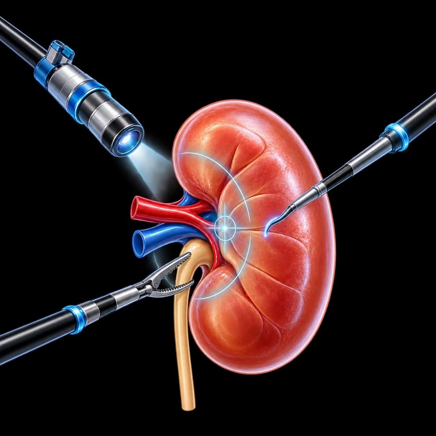
The procedure is performed through several small access points in the abdomen or the retroperitoneal space, using a camera and long laparoscopic instruments that the surgeon directly handles with his own hands. The kidney is mobilised internally and the specimen is removed through an appropriately sized extraction incision.
This is not “scarless surgery” — incisions, even small ones, are always required.
Three-Dimensional (3D) Laparoscopy
A three-dimensional laparoscopic camera can offer improved depth perception, anatomical orientation, and precision during vessel dissection, compared with conventional two-dimensional laparoscopy.
The 3D image alone does not guarantee a better oncological outcome.
The instruments used in 3D laparoscopy remain conventional laparoscopic instruments — the surgeon moves them directly by hand, without a console or robotic arm.
Laparoscopic ≠ Robotic
Laparoscopic surgery
The surgeon directly handles the laparoscopic instruments with his own hands.
Robotic surgery
The surgeon controls articulated robotic instruments through a console.
Both can use minimally invasive access, but they are not the same technology. See also robotic nephrectomy.
Comparing Laparoscopic & Robotic Nephrectomy
| Laparoscopic | Robotic | |
|---|---|---|
| Minimally invasive | Yes | Yes |
| Instrument control | Direct laparoscopy | Robotic console |
| Articulation | Standard laparoscopic | Wristed/robotic |
| 3D vision | If the system supports it | Standard robotic architecture |
| Radical nephrectomy | Established | Feasible |
| Oncologic objective | Same | Same |
| Recovery | Broadly comparable in many settings | Broadly comparable in many settings |
| Best approach | Case/team dependent | Case/team dependent |
Neither is presented as the “winner” approach.
What the Evidence Shows
For standard radical nephrectomy, comparative data on blood loss, operative time, hospital stay, complications, conversion, and cost between laparoscopic and robotic approaches vary by study and centre.
We avoid statements such as “robotic is always better”, “laparoscopy is always faster”, or “robotic always causes less bleeding”.
For routine radical nephrectomy, both are minimally invasive approaches and major clinical outcomes depend strongly on patient selection and surgical expertise.
Laparoscopic vs Open
Laparoscopic
Small incisions, minimally invasive, selected localised tumours, generally faster postoperative functional recovery.
Open
Larger incision, important for selected complex/invasive disease, major venous tumour thrombus, multivisceral resection, difficult oncological anatomy.
We do not characterise open surgery as outdated.
Transperitoneal vs Retroperitoneal Approach
Transperitoneal
Access through the peritoneal cavity, offering a wider working space and familiar orientation anatomy.
Retroperitoneal
Direct access through the retroperitoneal space without entering the peritoneal cavity — may be preferred in patients with multiple previous abdominal operations.
Selection depends on:
- Tumour location.
- Previous abdominal surgery.
- Tumour size.
- Anatomy.
- Surgeon experience.
- Need for other concurrent procedures.
Neither approach is presented as universally better.
Right vs Left Nephrectomy
Right side
Anatomical relationships with the liver, duodenum, inferior vena cava, and renal vessels.
Left side
Anatomical relationships with the spleen, pancreas, colon, aorta, and renal vessels.
This description remains at a general level — it is not an operative manual.
How the Procedure Is Performed
This description is deliberately kept at a general level — it is not a surgical manual with vessel-control sequence, port coordinates, or device details.
Specimen Removal
The kidney/tumour is placed inside an appropriate retrieval bag; the incision may need modest enlargement for intact removal of the whole specimen.
Especially in malignant disease: preserving specimen integrity and avoiding tumour rupture/spillage are important. Routine morcellation is not suggested for a suspected malignant renal tumour.
This is not “scarless” surgery. We do not promise a tiny incision regardless of tumour size.
Is the Adrenal Gland Always Removed?
No.
Routine ipsilateral adrenalectomy is not required when:
- The adrenal gland is radiologically normal.
- There is no direct tumour involvement/suspicion.
- There is no otherwise suspicious finding.
according to current RCC guidance. Adrenalectomy may be indicated with direct invasion, a suspicious adrenal lesion, or another specific oncological reason.
Are Lymph Nodes Always Removed?
No.
Routine extended lymph-node dissection (LND) in organ-confined, clinically node-negative RCC has not shown universal therapeutic benefit.
Suspicious/enlarged nodes may be removed for staging, local control, or appropriate oncological management, according to current guidance. Not every radical nephrectomy includes lymphadenectomy.
What Happens to the Ureter
In standard radical nephrectomy for RCC, the ureter and bladder cuff are not normally removed en bloc, as they would be in nephroureterectomy for UTUC. Only the necessary ureteral segment/structures are managed according to the surgery — this distinction must be very clear.
Preoperative Assessment
- Contrast CT/MRI.
- Kidney function.
- Contralateral kidney.
- Full blood count.
- Biochemical/metabolic profile.
- Urinalysis as appropriate.
- Medication review.
- Anticoagulants/antiplatelets.
- Anaesthetic evaluation.
- Comorbidities.
- Cardiopulmonary status.
- Prior abdominal surgery.
- Staging as appropriate.
Anticoagulants & Antiplatelets
The patient must not stop anticoagulants or antiplatelets on their own.
The perioperative plan must balance bleeding risk, thrombotic risk, the specific drug, indication, and the surgical plan.
Anaesthesia
Laparoscopic nephrectomy requires general anaesthesia. It is not an office or local-anaesthetic procedure.
How Long Does the Procedure Take
There is no universal operating time. It depends on the tumour, side, anatomy, obesity, adhesions, prior surgery, vascular anatomy, oncological extent, tumour thrombus, and additional procedures. If a time range is shown elsewhere, it should be treated only as approximate.
Hospital Stay
Length of stay is variable and depends on patient health, the surgery performed, oral intake, mobilisation, pain, kidney function, complications, and centre protocol. We do not guarantee a fixed number of days.
Recovery
Covers mobilisation, oral intake, wound care, fatigue, pain, kidney-function monitoring, activity, driving, return to work, and exercise.
We prefer healing-based, individualised advice over rigid timetables unless a current local protocol is verified.
Recovery Timeline
Immediate postoperative phase
Clinical monitoring, pain control, mobilisation, urine output, creatinine/eGFR.
Early home recovery
Wound healing, progressive walking/activity, fatigue may gradually improve.
Pathology review
Tumour type, stage, grade, margins.
Long term
Renal-function monitoring, blood pressure, and risk-adapted oncological surveillance.
Discomfort may relate to the incisions, the abdominal wall, the operative field, or temporary referred shoulder-tip discomfort after transperitoneal CO₂ insufflation. This is not a painless procedure.
Return to Work & Exercise
Depends on the type of work, wound healing, fatigue, pain, and any complications. Desk-based work is not the same as heavy manual work or driving heavy vehicles — there is no universal day.
Return to exercise/weightlifting happens gradually, based on abdominal wall healing, symptoms, and medical review. We avoid a rigid one-size-fits-all week threshold unless verified.
Can I Live With One Kidney?
Most patients with a normally functioning contralateral kidney can live with one kidney.
What matters:
- Blood pressure.
- Kidney function.
- Diabetes control.
- Avoidance of unnecessary nephrotoxic exposures/medications.
- Long-term medical follow-up.
We do not promise unchanged kidney function for everyone. CKD risk is individual.
Kidney Function (GFR) After Nephrectomy
Removal of one kidney reduces total nephron mass. The remaining kidney may undergo compensatory adaptation.
Final eGFR depends on baseline function, the contralateral kidney, age, diabetes, hypertension, vascular disease, and pre-existing CKD.
Might I Need Dialysis?
Usually not when the other kidney functions satisfactorily.
The risk can be higher with advanced baseline CKD, a solitary/poorly functioning contralateral kidney, or significant comorbidity. This is why preoperative renal assessment is important. We do not give an absolute guarantee.
Do I Need a Special Diet?
A special renal diet is not automatically required simply because one kidney was removed.
Advice depends on eGFR, albuminuria, blood pressure, diabetes, and other metabolic factors. We do not prescribe universal protein/potassium/fluid restrictions without individualised assessment.
Possible Complications
Bleeding
Uncommon but significant, due to proximity to major vessels.
Transfusion
Rare, depending on the course.
Vascular injury
Rare, serious complication.
Injury to adjacent organs
Bowel, spleen, liver, pancreas depending on side — rare.
Infection
Requires assessment and targeted treatment.
Thromboembolic events
Rare, depending on risk factors.
Respiratory/anaesthetic complications
Rare, depending on the patient.
Conversion to open surgery
A safety decision, see the relevant section.
Postoperative renal impairment
Monitored with creatinine/eGFR.
Port-site hernia
Rare with a minimally invasive approach.
Lymphatic complications
If nodal surgery is added.
Rare adjacent-organ injury
Depends on anatomy and side.
We do not use unsourced incidence rates. We do not promise “bloodless surgery” or zero risk of transfusion.
Conversion to Open Surgery
Conversion to open surgery is a safety decision, not a failure.
It may be necessary due to bleeding, unexpected anatomy, adhesions, tumour invasion, oncological concerns, or patient safety.
What Does the Histology Show
The final specimen can establish tumour type, RCC subtype, grade where applicable, tumour size, pathological stage, margins, venous invasion, and lymph-node findings if sampled, using current WHO terminology.
Common RCC subtypes include clear-cell, papillary, and chromophobe RCC, as well as other less common renal tumours. Grade does not equal stage.
Some radiologically suspicious renal masses prove benign after removal — for example, oncocytoma or fat-poor angiomyolipoma.
This does not mean imaging is generally unreliable.
Adjuvant Therapy After Nephrectomy
In selected high-risk clear-cell RCC after nephrectomy, current adjuvant systemic therapy may be considered, according to the latest risk criteria/approval status and current EAU/ESMO/NCCN guidance. The decision is made in consultation with an oncologist, individualised per patient. We do not imply that every high-risk RCC needs postoperative systemic therapy.
Follow-Up After Nephrectomy
Follow-up is risk-adapted and depends on pathology, stage, grade, subtype, margins, kidney function, and comorbidity. It may include clinical review, creatinine/eGFR, blood pressure, and chest/abdominal imaging according to oncological risk.
Renal follow-up
Kidney function, proteinuria where relevant, blood pressure, cardiovascular risk factors — selected patients may need nephrology input.
Oncological follow-up
Depends on recurrence risk. The same surveillance protocol is not used for low-risk pT1 and high-risk locally advanced RCC.
There is no universal CT schedule for everyone.
Prognosis & Surveillance
Nephrectomy does not eliminate all risk of recurrence. Outcome depends on tumour stage, biology, histology, grade, venous/nodal involvement, and metastatic status — not on whether access was laparoscopic, robotic, or open, provided appropriate oncological surgery is achieved.
We avoid generic cure percentages. Prognosis is determined primarily by pathological stage, grade, subtype, and nodal/metastatic disease.
The Laparoscopic Approach Does Not Change Tumour Biology
Laparoscopy affects surgical access and recovery, not the biology of the cancer.
Oncological quality depends on correct indication, complete surgery, tumour stage, surgical expertise, and appropriate multidisciplinary care — not by itself on the surgical access.
The Patient Journey
Key Questions in Plain Language
I Have a Kidney Tumour – Does the Whole Kidney Need to Be Removed?
Not necessarily. First it is assessed whether partial nephrectomy can achieve safe tumour removal while preserving kidney tissue.
When Is Radical Nephrectomy Needed?
When nephron-sparing surgery is not appropriate, when complete tumour removal cannot reasonably be achieved by partial surgery, or in selected advanced/complex situations — not size alone.
Laparoscopic or Robotic Nephrectomy?
Both are established minimally invasive approaches. For straightforward radical nephrectomy, team experience, anatomy, and correct indication are often more important than the platform.
What Is the Difference Between 3D Laparoscopy and the Robot?
3D laparoscopy is a stereoscopic camera with conventional laparoscopic instruments directly handled by the surgeon. Robotic surgery offers 3D vision and articulated instruments through a console. They are not the same technology.
Can I Live With One Kidney?
Usually yes, provided the remaining kidney is healthy. Kidney function monitoring is needed.
Will I Need Dialysis?
Usually not with satisfactory function of the contralateral kidney, but the risk must be individually assessed.
Is the Adrenal Gland Removed Too?
Not routinely. Only if indicated by tumour location/involvement or suspicious adrenal findings.
Are Lymph Nodes Removed?
Not in every clinically node-negative localised kidney cancer. Suspicious nodes may be removed according to oncological context.
Frequently Asked Questions (FAQ)
What is laparoscopic radical nephrectomy?
It is the surgical removal of the entire affected kidney through small incisions, with the help of a laparoscopic camera and instruments, when preserving the kidney with partial nephrectomy is not technically or oncologically appropriate.
Does every kidney tumour need to be removed with the whole kidney?
No. When technically feasible and oncologically safe, partial nephrectomy has an important role because it preserves more functioning kidney tissue.
What is the difference between partial and radical nephrectomy?
Partial nephrectomy removes the tumour with a small margin of healthy kidney, preserving the remaining tissue. Radical nephrectomy removes the entire kidney. The choice depends on anatomy, complexity, kidney function, and team experience — not size alone.
When is partial nephrectomy possible?
In localised kidney tumours, particularly when it is technically and oncologically feasible, nephron-sparing surgery should be considered before deciding on radical nephrectomy.
When is radical nephrectomy needed?
When the tumour is too large/complex for safe partial nephrectomy, its location makes nephron-sparing surgery technically unsuitable, there is locally advanced disease, or oncological clearance cannot reasonably be achieved with partial nephrectomy.
Does tumour size alone determine the decision?
No. Size, location, depth, hilar involvement, complexity, kidney function, the contralateral kidney, comorbidities, and surgeon expertise are all taken into account.
Is a biopsy needed before surgery?
Not always. Renal mass biopsy has an important role in selected patients, particularly when it could change management, active surveillance or ablation is being considered, or the imaging diagnosis is uncertain.
What is the difference between laparoscopic and robotic nephrectomy?
Both are established minimally invasive approaches. For straightforward radical nephrectomy, team experience and correct patient selection are often more important than the platform.
What is 3D laparoscopy?
It is a high-definition stereoscopic camera that gives the surgeon a three-dimensional sense of depth, while the instruments remain conventional laparoscopic instruments — it is not a robotic system.
When is open surgery needed?
With extensive local invasion, a very large/complex tumour, a significant venous thrombus, or need for multivisceral resection, the open approach may be necessary or preferred.
Transperitoneal or retroperitoneal approach?
The choice depends on tumour location, previous abdominal surgery, size, anatomy, and surgeon experience. Neither is universally better.
Is the adrenal gland always removed?
No. Routine ipsilateral adrenalectomy is not required when the adrenal gland is radiologically normal and there is no direct tumour involvement/suspicion.
Are lymph nodes always removed?
No. Routine extended lymph-node dissection in organ-confined, clinically node-negative kidney cancer has not shown a universal therapeutic benefit. Suspicious/enlarged nodes may be removed for staging or local control.
What happens if there is a renal vein or IVC tumour thrombus?
RCC can, in selected cases, extend as a venous tumour thrombus. This significantly changes operative complexity and surgical planning, and may require specialised, multidisciplinary management — it is not equivalent to a standard laparoscopic radical nephrectomy.
How long is the hospital stay?
It is variable, depending on complexity, kidney function, any complications, and the recovery course.
When can I return to work?
It depends on the type of work, healing course, and the surgery performed. Desk-based work usually allows a faster return than manual work.
Can I live with one kidney?
Most patients with a normally functioning contralateral kidney can live with one kidney. Long-term medical follow-up of blood pressure and kidney function is important.
Will my eGFR drop after surgery?
Removal of one kidney reduces total nephron mass. The remaining kidney may undergo compensatory adaptation, but the final eGFR depends on baseline function, the contralateral kidney, age, diabetes, hypertension, and vascular disease.
Might I need dialysis?
Usually not, when the other kidney functions satisfactorily. The risk can be higher with advanced pre-existing chronic kidney disease, a solitary/poorly functioning contralateral kidney, or significant comorbidity.
What are the possible complications?
These include bleeding, transfusion, vascular injury, injury to adjacent organs, infection, thromboembolic events, respiratory/anaesthetic complications, conversion to open surgery, postoperative renal impairment, and wound hernia.
What does the histology show?
It establishes tumour type, RCC subtype, grade where applicable, tumour size, pathological stage, margins, venous invasion, and lymph-node findings if sampled.
What follow-up is needed after nephrectomy?
It depends on pathology, stage, grade, subtype, margins, kidney function, and comorbidity. There is no single universal CT schedule for everyone.
Related Topics
Evaluation for Laparoscopic Radical Nephrectomy in Rhodes
The key question before a radical nephrectomy is not only whether the procedure can be done laparoscopically or robotically, but whether it is truly necessary to remove the entire kidney. Tumour anatomy, oncological extent, and the function of both kidneys should be assessed before choosing partial or radical nephrectomy and the appropriate surgical approach.
Scientific References
- Ljungberg B, et al. European Association of Urology Guidelines on Renal Cell Carcinoma. Eur Urol. 2022;82(4):399–410.
- Marconi L, Kuusk T, Hora M, et al. Hospital Volume as a Determinant of Outcomes After Partial Nephrectomy: A Systematic Review by the EAU Renal Cell Carcinoma Guidelines Panel. Eur Urol Oncol. 2025;8(3):616–622.
- Choueiri TK, et al. Adjuvant Pembrolizumab after Nephrectomy in Renal-Cell Carcinoma (KEYNOTE-564). N Engl J Med. 2021;385(8):683–694.
- Choueiri TK, et al. Overall Survival with Adjuvant Pembrolizumab in Renal-Cell Carcinoma. N Engl J Med. 2024;390(15):1359–1371.
Meet the Doctor

Dr. Marinos Vasilas, Urologist – Andrologist
Dr. Marinos Vasilas specialises in robotic and 3D laparoscopic urological oncology, with emphasis on clearly informing every patient about the choice between partial and radical nephrectomy and the real approach options available.
Full ProfileSurgical Team
Laparoscopic nephrectomy is performed in collaboration with a specialised surgical centre and urology team, ensuring access to modern minimally invasive infrastructure.
Meet Our TeamThe content on this page is informational and does not replace individualised urological/oncological assessment. Renal mass treatment is individualised. Tumour size alone does not determine radical nephrectomy. The feasibility of partial nephrectomy requires specialist evaluation. Kidney function must be assessed before surgery. Tumour-thrombus surgery is specialised. Follow-up depends on final pathology.


