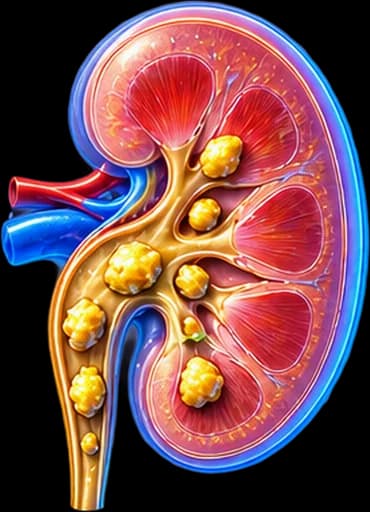
1. My Clinical Experience with Kidney Stones
Kidney stones are among the most common conditions that bring patients to my urology practice in Rhodes — often urgently, in the middle of a dramatic night with the typical renal colic. Renal colic pain is internationally described as one of the most intense pains in medicine, comparable only to labour.
My Approach
There is a truth I emphasise to every patient in daily clinical practice: removing the stone is only half the job. The other half — often more important — is identifying why the stone formed, to prevent recurrence. Without proper metabolic evaluation, recurrence rates reach 50% at 5 years and 75% at 20 years.
My approach follows closely the EAU Guidelines on Urolithiasis and the AUA Guideline:
- Low-dose NCCT for accurate diagnosis and stone mapping (sensitivity >95%, specificity >96%).
- A urine culture is mandatory before every endoscopic procedure (to prevent urosepsis).
- Emergency decompression (JJ stent or nephrostomy) when infection or obstruction coexists — antibiotics alone are not enough.
- Individualised choice of technique (SWL / RIRS / PCNL / mini-PCNL) based on size, location, composition, anatomy, and patient preference.
- Chemical composition analysis of every stone removed, and metabolic evaluation in high-risk patients.
- Modern technologies: Thulium Fiber Laser (TFL), suction-enabled URS, ultrasound-guided PCNL puncture.
My goal is a complete care plan: emergency relief, definitive stone treatment with the appropriate technique, and — equally important — long-term recurrence prevention.
2. What Is Nephrolithiasis — Pathogenesis & Classification
Nephrolithiasis is the aggregation of crystals within urine, forming solid stones inside the urinary tract. The formation mechanism follows a sequence: urine supersaturation with lithogenic substances (calcium, oxalate, uric acid, cystine) → crystal nucleation → growth and aggregation → formation of a macroscopic stone, often on Randall’s plaques.
2.1 Classification by Location
Kidney Stones (Pyelocaliceal System)
Form within the calyces or renal pelvis. Many remain asymptomatic while located there — becoming symptomatic once they move into the ureter.
Ureteral Stones
Cause the typical renal colic. The EAU distinguishes three locations: proximal (UPJ), mid-ureter (near the iliac vessels), and distal (UVJ). Location significantly affects treatment choice.
Staghorn Calculus
A large stone filling part or all of the pyelocaliceal system, shaped like a stag’s horn. Usually struvite, forming with chronic infection by urease-producing organisms. Practically always requires PCNL as first choice.
2.2 Clarifications
Nephrolithiasis should not be confused with:
- Nephrocalcinosis (diffuse calcium deposition within the renal parenchyma).
- Simple renal cysts (Bosniak I).
- Cholelithiasis (gallstones) — an entirely different condition.
3. Epidemiology & Risk Factors
Nephrolithiasis affects 10–15% of the general population, with a clear predominance in men (ratio ~2:1) and peak incidence between 30–60 years. In Mediterranean countries, including Greece and particularly the islands such as Rhodes, incidence is among the highest in Europe — a consequence of the hot, dry climate, increased fluid loss through sweating, and Mediterranean dietary habits.
Risk Factors
| Factor | Mechanism |
|---|---|
| Low fluid intake | The most important modifiable factor. Urine output <1.5 L/day quadruples the risk. |
| Diet rich in sodium and animal protein | Increases calcium and uric acid excretion, reduces citrate. |
| Obesity & metabolic syndrome | Associated with hypercalciuria, hyperuricaemia, and low urine pH. |
| Type 2 diabetes | Insulin resistance leads to low urine pH (uric acid stones). |
| Family history | Risk 2–3× higher in first-degree relatives. |
| Genetic factors | Cystinuria, primary hyperoxaluria, Dent disease. |
| Anatomical variants | UPJ obstruction, medullary sponge kidney, horseshoe kidney. |
| Systemic conditions | Primary hyperparathyroidism, gout, Crohn’s disease, chronic UTIs. |
| Medications | Indinavir, sulfonamides, topiramate, high-dose calcium/vitamin D. |
| Climate factors | Hot/dry climate — a relevant factor for Rhodes. |
4. Symptoms & Renal Colic
Many stones, especially small intrarenal ones, remain asymptomatic and are found incidentally on ultrasound or CT. Symptoms begin once a stone migrates into the ureter and causes acute obstruction.
4.1 Renal Colic — The Clinical “Signature”
Intense, cramping pain in the loin or flank that radiates towards:
- The groin.
- The testicle (in men) or labia majora (in women).
- The lower abdomen as the stone descends.
Pathognomonic clinical sign: the patient cannot stay still and constantly changes position seeking relief — a characteristic distinction from peritonitis, where the patient remains still.
4.2 Associated Symptoms
- Haematuria (present in ~85% of cases — microscopic or visible). Its absence does not rule out the diagnosis.
- Nausea and vomiting — from reflex vagal stimulation.
- Frequency and urgency — when the stone is in the distal ureter, near the bladder.
- Dysuria (burning during urination).
Red Flags — Immediate Specialised Evaluation
- Fever >38°C with chills and flank pain → stone + infection = emergency (risk of urosepsis).
- Persistent vomiting preventing hydration and oral medication.
- Pain resistant to analgesia for >6 hours.
- Oliguria or anuria (suspicion of bilateral obstruction or obstruction of a solitary kidney).
- A stone in pregnancy, a solitary kidney, or a patient with renal impairment.
- Clinical signs of sepsis (qSOFA ≥2: tachycardia, tachypnoea, confusion).
5. Diagnostic Algorithm
5.1 Low-Dose Non-Contrast CT (NCCT)
It is the gold standard according to the EAU Guidelines:
- Sensitivity >95%, specificity >96% for all stone types.
- Accurate measurement of size, location, and density in Hounsfield Units (HU) — HU density predicts response to different treatments.
- Assessment of hydronephrosis grade and complications (perinephric urine leak).
- Low radiation dose (≤3 mSv) — comparable to traditional IVU radiography.
| HU density | Treatment implications |
|---|---|
| <800 HU | Usually favours SWL/ESWL — softer stone |
| 800–1000 HU | Intermediate category — choice is individualised |
| >1000 HU | Hard stone — laser (RIRS or PCNL) preferred |
5.2 Other Imaging Modalities
Kidney & Bladder Ultrasound
First choice in pregnancy, children, and very young patients. Detects hydronephrosis and renal stones, but often misses mid-ureteral stones.
Plain Abdominal X-Ray (KUB)
Useful for monitoring radiopaque (calcium) stones after treatment. Does not show radiolucent uric acid or cystine stones.
CT Urography with Contrast
When another anatomical abnormality is suspected or findings are unclear — precise mapping before interventional treatment.
5.3 Laboratory Work-Up
- Urinalysis + culture — mandatory before any intervention.
- Complete blood count, CRP — screening for leukocytosis/sepsis.
- Creatinine, eGFR, electrolytes — assessment of renal function.
- Serum calcium, uric acid, PTH when primary hyperparathyroidism is suspected.
6. Stone Composition & Metabolic Workup
Chemical composition analysis of every stone removed is mandatory per EAU. Composition guides both treatment choice and prevention strategy.
6.1 Main Stone Types
| Type | Frequency | Characteristics | Strategy |
|---|---|---|---|
| Calcium (oxalate/phosphate) | 75–80% | Hard, radiopaque | Often laser, thiazides for hypercalciuria |
| Uric acid | 10% | Radiolucent, at low urine pH (<5.5) | Chemical dissolution with alkalinisation + allopurinol |
| Struvite (magnesium ammonium phosphate) | 5–10% | From chronic infection (Proteus, Klebsiella) | Complete removal with PCNL + targeted antibiotics |
| Cystine | 1% | Hereditary cystinuria, hard | High hydration (>3 L), alkalinisation, thiola |
| Rare (xanthine, indinavir) | <1% | Drug-related or enzymatic disorders | Drug discontinuation, specialised care |
6.2 Genetic Testing
The newer EAU guideline added a section on genetic factors. Genetic testing is recommended in:
- Early onset (children, adolescents, young adults <25 years).
- A strong family history.
- Cystine or xanthine stones.
- Primary hyperoxaluria (PH1, PH2, PH3) — new siRNA therapies (lumasiran/nedosiran) have changed the prognosis.
- Dent disease or Bartter syndrome.
6.3 24-Hour Urine Metabolic Testing
In high-risk patients (recurrent stone formers, solitary kidney, children, family history, non-calcium stones):
- Two 24-hour urine collections (on separate days, on a usual diet).
- Parameters: volume, calcium, oxalate, citrate, uric acid, sodium, magnesium, creatinine, pH.
- Findings guide individualised pharmacological prevention (thiazides, potassium citrate, allopurinol, pyridoxine).
7. Modern Treatment Options
7.1 Conservative Management & Medical Expulsive Therapy (MET)
Stones ≤10 mm in the distal ureter without infection or significant obstruction can be managed conservatively:
- Analgesia: NSAIDs (diclofenac or ketoprofen) first line. Paracetamol or tramadol as alternatives.
- Hydration (not “forced diuresis” — simply adequate).
- Tamsulosin 0.4 mg/day (Medical Expulsive Therapy — MET): relaxes the distal ureter and increases spontaneous passage by approximately 20% in stones 5–10 mm. Strong recommendation.
- Re-evaluation in 2–4 weeks with NCCT or low-dose CT.
| Stone Size | Probability of Spontaneous Passage |
|---|---|
| ≤4 mm | 80–90% |
| 5–6 mm | 60% |
| 7–9 mm | 35–50% |
| ≥10 mm | <25% |
7.2 Extracorporeal Shock Wave Lithotripsy (SWL/ESWL)
A non-invasive method for:
- Renal stones ≤20 mm with density <1000 HU.
- Stones in the pelvis or upper/mid calyx (lower pole has lower stone-free rates).
- Selected ureteral stones.
Uses shock waves to fragment the stone into small pieces that pass naturally. Usually 1–3 sessions.
Contraindications: pregnancy, bleeding disorders, uncontrolled UTI, obstruction distal to the stone, nearby aneurysms, extreme obesity.
7.3 Ureteroscopy with Laser (URS / RIRS) — The Modern “First Choice”
Flexible ureteroscopy (Retrograde Intrarenal Surgery — RIRS) with laser has become the technique of choice for most kidney and ureteral stones.
Holmium:YAG vs Thulium Fiber Laser (TFL)
The major technological development of 2024–2026 is the Thulium Fiber Laser (TFL). According to the AUA Guideline and the meta-analysis by Uleri et al. (Eur Urol 2024):
| Parameter | Holmium:YAG | Thulium Fiber Laser (TFL) |
|---|---|---|
| Mechanism | Photothermal | Photothermal with higher water absorption |
| Effectiveness | Gold standard | Comparable or superior, faster dusting |
| Fibre diameter | 200–365 μm | Down to 150 μm — allows smaller ureteroscopes |
| Heat generation | High — requires pauses | Lower per pulse but cumulative |
EAU/AUA conclusion: Both lasers are acceptable. The choice depends on availability and operator experience.
Lithotripsy Strategies
- Fragmenting + basketing: breaking into larger pieces and extracting with a basket.
- Dusting: fragmenting into fine dust that passes naturally — preferred for larger stones.
- Pop-dusting: combination of high frequency and low energy.
Suction-Enabled Ureteroscopy — Newer Technology
Direct In-Scope Suction (DISS) and suction-enabled ureteral access sheaths are among the most significant developments. They allow continuous aspiration of fragments during lithotripsy, with results including:
- Higher stone-free rates in larger stones.
- Reduced intrarenal pressure → less bacterial spread.
- Shorter operative time.
- Reduced postoperative sepsis.
7.4 Percutaneous Nephrolithotomy (PCNL) & Variants
First choice for:
- Renal stones >20 mm.
- Staghorn calculi.
- Multiple lower-pole stones.
- Stones resistant to SWL/RIRS.
Modern PCNL Variants
| Technique | Diameter | Indications |
|---|---|---|
| Standard PCNL | 24–30 Fr | Stones >25 mm, staghorn |
| Mini-PCNL | 14–22 Fr | Stones 15–25 mm, less bleeding |
| SuperMini-PCNL | 10–14 Fr | Stones <20 mm, very safe |
| Micro-PCNL | 4.8 Fr | Very small stones, paediatric cases |
Recent Developments
- Supine vs prone: the supine position allows simultaneous RIRS access (ECIRS — Endoscopic Combined Intra-Renal Surgery) for complex stones, with a similar stone-free rate and shorter procedure time.
- Ultrasound-guided puncture: equally safe as fluoroscopy, with significantly reduced radiation exposure for patient and surgeon.
7.5 Emergency Decompression: JJ Stent or Percutaneous Nephrostomy
In sepsis with an obstructed kidney, definitive stone treatment is postponed. The first priority is decompression within hours:
- Retrograde JJ stent (cystoscopically) — less invasive.
- Percutaneous nephrostomy (ultrasound-guided) — preferred in severe sepsis or when retrograde access is not possible.
Antibiotics are given at the same time, guided by culture. Definitive stone treatment is performed 2–4 weeks later, once sepsis has resolved.
8. Indications for Interventional Treatment
According to the EAU and AUA guidelines, indications for interventional treatment include:
- Stone >10 mm (spontaneous passage unlikely).
- MET failure after 4–6 weeks of observation.
- Persistent pain uncontrolled with analgesia.
- Renal impairment due to obstruction.
- Coexisting infection — an emergency.
- A solitary kidney or bilateral disease.
- A stone during pregnancy (usually URS, safely).
- Asymptomatic stones >15 mm (discussed with the patient).
- Occupational reasons: pilots, professional drivers, seafarers.
Technique Selection by Size & Location
| Stone | 1st Choice | 2nd Choice |
|---|---|---|
| Kidney <10 mm | Observation or SWL | RIRS |
| Kidney 10–20 mm | RIRS | SWL |
| Kidney >20 mm | PCNL / mini-PCNL | RIRS in selected cases |
| Upper ureter | RIRS or SWL | Antegrade URS |
| Lower ureter | URS with laser | SWL |
| Staghorn | PCNL | Combined (ECIRS) |
9. Emergency Management: Sepsis & Obstructed Kidney
Urosepsis with an obstructed kidney is one of the most dangerous conditions in urology and requires immediate intervention.
Diagnostic Criteria
- History of renal colic plus fever/chills.
- Clinical signs of sepsis (qSOFA ≥2).
- Leukocytosis, high CRP, elevated procalcitonin.
- NCCT: stone + hydronephrosis + possible renal parenchymal inflammation.
Management Algorithm
- Immediate hydration + broad-spectrum antibiotics (e.g., piperacillin/tazobactam, a carbapenem in severe sepsis).
- Blood cultures obtained before antibiotics.
- Emergency decompression within hours — JJ stent or percutaneous nephrostomy.
- ICU monitoring in septic shock.
- Definitive stone treatment after 2–4 weeks.
10. Recurrence Prevention & Follow-Up
Nephrolithiasis is a chronic condition. Without prevention, recurrence risk reaches 50% at 5 years. With appropriate measures, it drops to 10–15%.
10.1 General Recommendations (All Patients)
- Urine output ≥2.5 L/day (fluid intake ~3 L) — the most important factor.
- Moderate animal protein intake (≤1 g/kg/day).
- Sodium restriction (≤5 g salt/day).
- Adequate dietary calcium intake (1000–1200 mg/day). Calcium supplements increase risk and should be avoided.
- Citrate (lemon, orange, citrus fruits) — a practical, non-prescriptive dietary role: it alkalinises urine and directly inhibits crystallisation. Lemon does not “dissolve” an existing stone.
- Reducing high-oxalate foods (spinach, beetroot, chocolate, nuts) in hyperoxaluria.
- Healthy body weight and regular physical activity.
- Smoking cessation and moderate alcohol consumption.
10.2 Targeted Pharmacological Prevention
| Metabolic Finding | Pharmacological Intervention |
|---|---|
| Hypercalciuria | Thiazides (HCT 25 mg or chlorthalidone) |
| Hypocitraturia | Potassium citrate 30–60 mEq/day |
| Uric acid stones | Alkalinisation + allopurinol in hyperuricaemia |
| Hyperoxaluria | Pyridoxine (B6) 5–10 mg/kg, dietary oxalate restriction |
| Struvite | Complete removal + targeted antibiotics + culture monitoring |
| Cystinuria | High hydration (>3 L), alkalinisation, thiola/captopril |
| Primary hyperoxaluria | Lumasiran/nedosiran (siRNA) — newer genetic therapy |
10.3 Follow-Up Schedule
- 4–6 weeks after a procedure: NCCT or US + KUB to confirm stone-free status. Removal of the JJ stent if present.
- High-risk patients: annual US + 24-hour urine every 1–2 years.
- Low-risk patients: US every 2–3 years.
- Children and adolescents: more intensive follow-up + genetic testing.
11. Renal Pain vs Musculoskeletal Pain
Flank or back pain is not always caused by a stone. It can also originate from:
- The musculoskeletal system (muscle strain, spinal dysfunction).
- Gastrointestinal causes (e.g., appendicitis, diverticulitis, depending on side).
- Other urological conditions (e.g., pyelonephritis, UPJ obstruction).
Diagnosis is not based on pain location alone — it requires clinical examination and, when needed, imaging for a safe distinction.
12. Hydronephrosis
A stone in the ureter can obstruct urine flow and cause dilation of the collecting system (hydronephrosis).
- Hydronephrosis is not always caused by a stone — it can also result from a stricture or another obstructive cause.
- Its severity should be assessed together with renal function, symptoms, and any coexisting infection — not in isolation.
13. ESWL vs RIRS vs PCNL — Comparison Table
| Feature | ESWL | RIRS | PCNL |
|---|---|---|---|
| Access | External | Via urethra/ureter | Percutaneous |
| Mechanism | Shock waves | Laser fragmentation/dusting | Direct fragmentation/removal |
| Typical stone size | Depends on composition/density | Wide range of selected stones | Large stone burden |
| Anaesthesia | Per protocol/light sedation | Usually general anaesthesia | General anaesthesia |
| Invasiveness | Lower | Endoscopic | More invasive |
| Stone-free probability | Depends on stone factors | High in suitable cases | Very high for large burden |
| Repeat procedure | May be needed | May be needed | May also be needed |
| Hospitalisation | Variable, usually minimal | Variable | Usually longer |
14. The Treatment Journey
- 1
Kidney stone — size, location, symptoms, CT characteristics
Initial assessment with NCCT/ultrasound.
- 2
Is there infection or dangerous obstruction?
If YES → emergency decompression (JJ stent/nephrostomy) and infection management first.
- 3
Small, asymptomatic, or low-risk stone
Observation in selected patients.
- 4
Suitable stone for ESWL
Extracorporeal lithotripsy when size/density/location favour it.
- 5
Suitable endoscopic treatment
RIRS with laser for a wide range of kidney/ureteral stones.
- 6
Large or complex stone burden
PCNL/mini-PCNL, possibly combined (ECIRS).
- 7
After treatment
Stone analysis, individualised recurrence prevention, scheduled follow-up.
The final choice is always individualised — the journey above is not a rigid, size-only algorithm.
15. Nephrologist or Urologist?
Urologist
Mainly for stone diagnosis, obstruction, renal colic, and interventional/endoscopic treatment (ESWL, RIRS, PCNL, JJ stent, nephrostomy).
Nephrologist / Metabolic Specialist
May be involved in complex metabolic disorders, chronic kidney disease, or recurrent metabolic stone disease, depending on the case.
16. Treatments for Kidney Stones
Extracorporeal Shock Wave Lithotripsy (ESWL)
Non-invasive stone fragmentation with shock waves, generally for suitable stones of lower density.
Learn moreRIRS — Flexible Intrarenal Surgery
Endoscopic laser lithotripsy via a natural route, without an external incision in typical cases.
Learn morePCNL — Percutaneous Nephrolithotomy
Percutaneous management of a large or complex stone burden, including staghorn calculi.
Learn more17. Frequently Asked Questions
How do I distinguish renal colic from other abdominal pain?
Renal colic is intense, cramping pain in the flank or loin that radiates to the groin or genitalia. The patient cannot find a position of relief and keeps moving, in contrast to peritonitis, where stillness helps. It is often accompanied by nausea, vomiting, and haematuria. Definitive distinction is made with low-dose non-contrast CT (NCCT).
Which is the most reliable diagnostic test?
According to the EAU Guidelines on Urolithiasis, low-dose non-contrast CT (NCCT) is the gold standard, with sensitivity >95% and specificity >96%. It identifies almost all stones and measures size, density in Hounsfield Units, and the degree of obstruction. Ultrasound remains the first choice in pregnancy and children.
Will the stone pass on its own?
The probability depends mainly on size: stones ≤4 mm pass in 80–90% of cases, 5–6 mm in around 60%, 7–9 mm in 35–50%. Stones >10 mm rarely pass spontaneously. Tamsulosin 0.4 mg/day (medical expulsive therapy) increases the probability by around 20% for distal ureteral stones 5–10 mm.
When is emergency treatment needed?
A stone combined with infection (fever, chills, leukocytosis) or obstruction of a solitary kidney is a vital emergency. Immediate decompression within hours by JJ stent or percutaneous nephrostomy is required, regardless of stone size. Antibiotics alone are not sufficient.
Which treatment is appropriate for each stone?
The choice is individualised based on size, location, composition, and anatomy: SWL/ESWL for renal stones ≤20 mm with lower density, RIRS with Holmium or Thulium Fiber Laser for kidney stones 10–20 mm and most ureteral stones, PCNL or mini-PCNL for stones >20 mm or staghorn calculi, and a combined approach (ECIRS) for complex stones.
What is Thulium Fiber Laser (TFL), and is it better than Holmium?
TFL is the newer generation of laser used in urological lithotripsy since 2020. According to a meta-analysis by Uleri et al. (Eur Urol 2024), it is comparable or slightly superior to Holmium:YAG in dusting speed, with the advantage of using thinner fibres (down to 150 μm). The AUA 2025 Guideline considers both equally acceptable.
What is a staghorn calculus?
It is a large stone that fills part or all of the pyelocaliceal system, taking the shape of a coral/stag horn. It is usually struvite, forming in chronic infections with urease-producing organisms (Proteus, Klebsiella). It can gradually damage kidney function and practically always requires PCNL as first-line treatment.
How can I prevent recurrence?
Three fundamental pillars: 1) Adequate hydration (>2.5 L/day intake, urine output target ≥2 L). 2) Dietary changes: moderate restriction of animal protein and sodium, adequate dietary calcium (not supplements), increased citrate intake (lemon). 3) Targeted medication (citrate salts, thiazides, allopurinol) based on 24-hour urine metabolic testing results.
Is metabolic workup needed after a stone?
Yes in high-risk patients: children, recurrent stone formers, renal impairment, solitary kidney, family history, non-calcium stones (uric acid, struvite, cystine). It includes stone composition analysis, two 24-hour urine collections, blood biochemistry, and — where indicated — genetic testing (per the newer EAU guideline).
Is stone treatment safe during pregnancy?
Yes, but with particular caution. First line: JJ stent or nephrostomy placement for drainage. Ureteroscopy with laser is safe in selected cases. SWL is absolutely contraindicated. Definitive treatment is often postponed until after delivery.
How long is recovery after each procedure?
SWL/ESWL: return to activities in 1–2 days. URS/RIRS: 2–5 days, with a JJ stent remaining for 1–2 weeks. PCNL/mini-PCNL: hospital stay 1–3 days, full recovery 2–3 weeks.
Do diabetes or obesity affect kidney stone risk?
Clearly. Patients with type 2 diabetes have an increased incidence of uric acid stones due to low urine pH. Obesity is associated with hypercalciuria and hyperuricosuria. Weight reduction and glycaemic control are an integral part of prevention.
Ultrasound or CT for kidney stones?
Ultrasound avoids radiation and is useful for detecting hydronephrosis and larger renal stones, particularly in pregnancy and children, but it can miss small ureteral stones. Low-dose non-contrast CT has much higher sensitivity and is the preferred test in most other adults with suspected obstructing stones.
Is a JJ stent always needed after stone treatment?
No. The need for a JJ stent depends on the technique, stone size/burden, pre-existing ureteral anatomy, and whether a prior stent was already in place. In selected RIRS cases, no stent is needed, or it can remain for a shorter interval.
How much water should I drink to avoid another stone?
There is no single fixed “2 litres for everyone” target — the goal is adequate urine output (practically ≥2–2.5 L/24h for most recurrent formers), spread throughout the day. Practical needs increase with heat, exercise, and sweating, and may need individualisation in select cardiovascular/renal conditions.
Related Topics
Book an Appointment in Rhodes
The right management of a kidney stone does not depend only on its size. Location, composition, kidney anatomy, the presence of obstruction or infection, and patient characteristics determine whether observation, ESWL, RIRS, or PCNL is needed.
References
- EAU Guidelines on Urolithiasis. European Association of Urology — uroweb.org
- Skolarikos A, Geraghty R, Somani B, et al. European Association of Urology Guidelines on the Diagnosis and Treatment of Urolithiasis. Eur Urol 2025;88:64–75.
- AUA Guideline on Surgical Management of Kidney and Ureteral Stones. American Urological Association — auanet.org
- Pearle MS, et al. Medical Management of Kidney Stones: AUA Guideline. J Urol 2014;192:316.
- Uleri A, Farr\u00e9 A, et al. Thulium Fiber Laser Versus Holmium:Yttrium Aluminum Garnet for Lithotripsy: A Systematic Review and Meta-analysis. Eur Urol 2024;85(6):529–540.
- Chen R, et al. Efficacy and safety of thulium fiber laser versus holmium:YAG laser in lithotripsy for urolithiasis: a systematic review and meta-analysis. Urolithiasis 2025;53:33.
- Geraghty R, Pietropaolo A, Tzelves L, et al. Which measure of stone burden is the best predictor of interventional outcomes in urolithiasis. Eur Urol Open Sci 2025;71:22–30.
- Hollingsworth JM, et al. Alpha blockers for treatment of ureteric stones: systematic review and meta-analysis. BMJ 2016;355:i6112.
- NIDDK — National Institute of Diabetes and Digestive and Kidney Diseases: Kidney Stones — niddk.nih.gov
The content of this page is for informational purposes only and does not replace an individualised urological evaluation. Treatment is not decided by stone size alone. Analgesics, medical expulsive therapy, and antibiotics should not be taken as a universal self-treatment. Suspected sepsis or obstruction requires immediate medical evaluation.
Medical Review

Dr. Marinos Vasilas, Urologist – Andrologist
Dr. Marinos Vasilas maintains private urology practices in Rhodes and Athens, with particular expertise in the endoscopic and percutaneous management of nephrolithiasis (RIRS with Holmium/Thulium laser, mini-PCNL, suction-enabled URS) and in emergency management of the obstructed and septic kidney. He applies the EAU Guidelines on Urolithiasis.
Full Profile


